. 2009 Sep 4;5(3):298-309.
doi: 10.1016/j.stem.2009.06.001.

Autocrine and paracrine angiopoietin 1/Tie-2 signaling promotes muscle satellite cell self-renewal

Affiliations

Autocrine and paracrine angiopoietin 1/Tie-2 signaling promotes muscle satellite cell self-renewal

Rana Abou-Khalil et al. Cell Stem Cell. .

Abstract

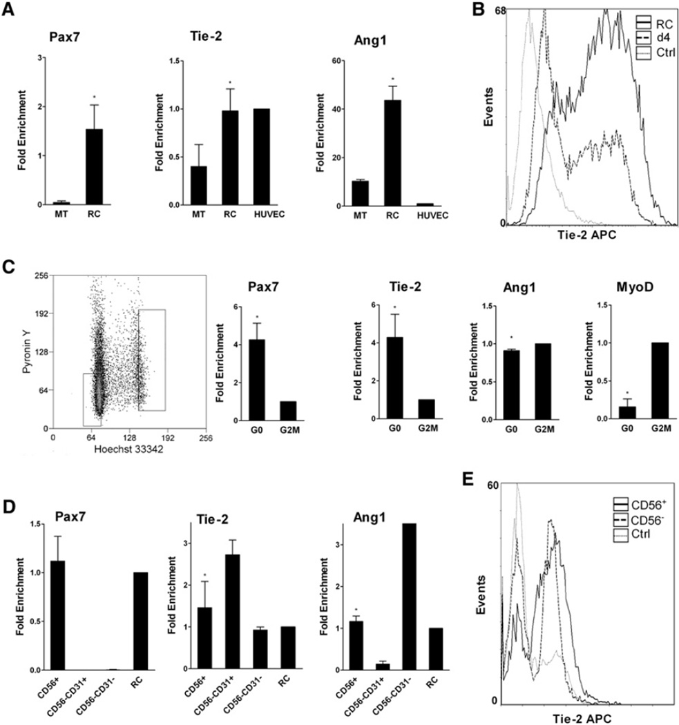
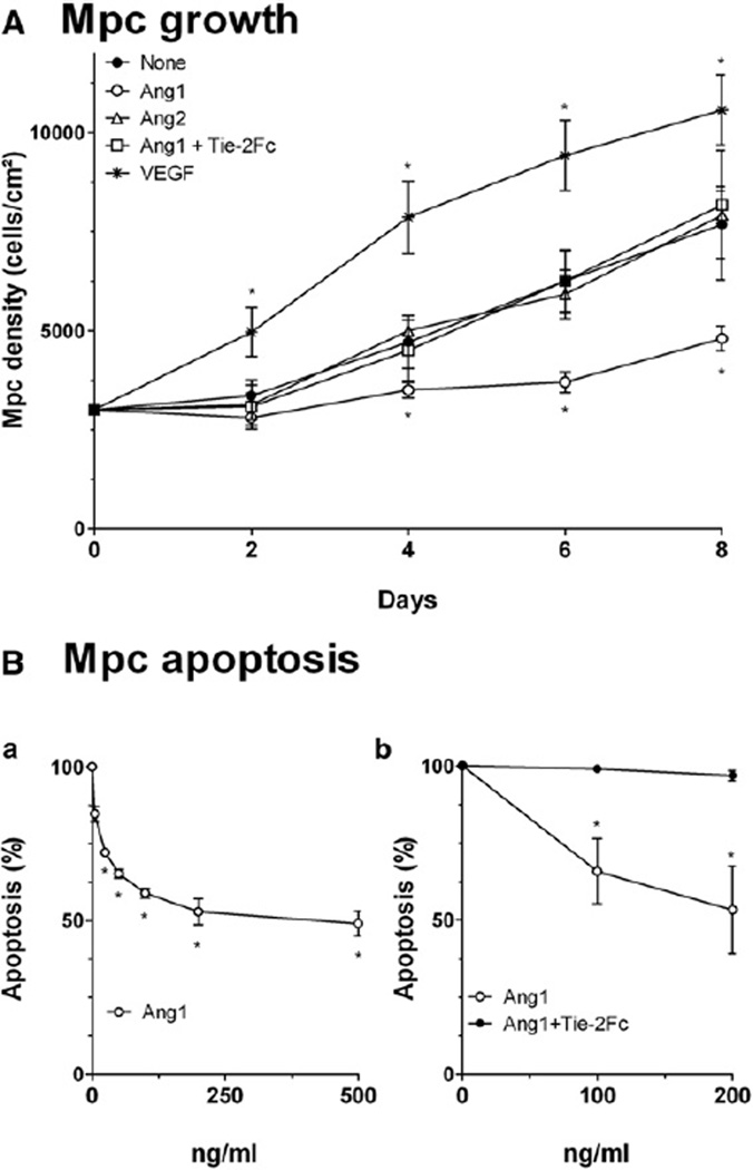
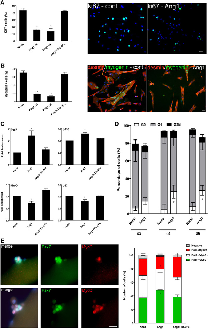
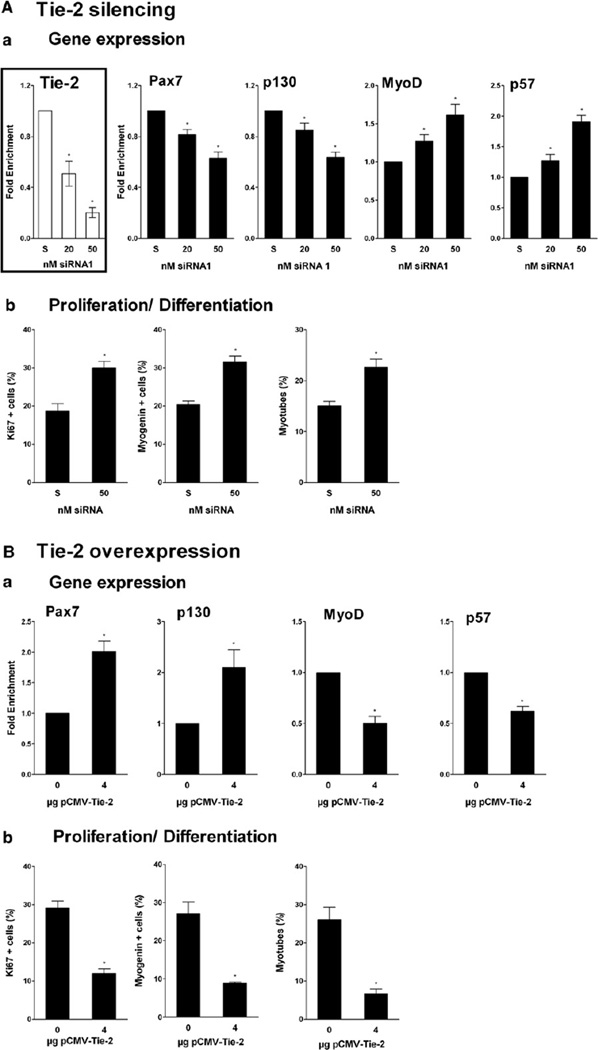
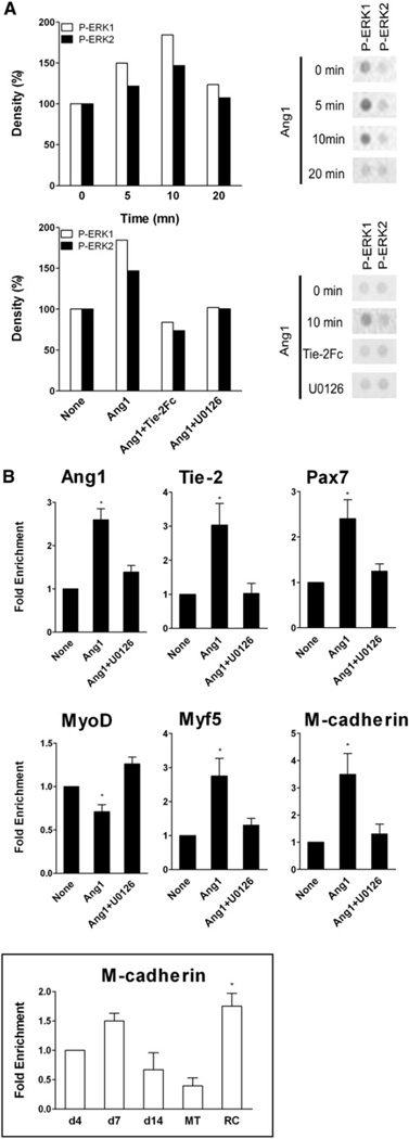
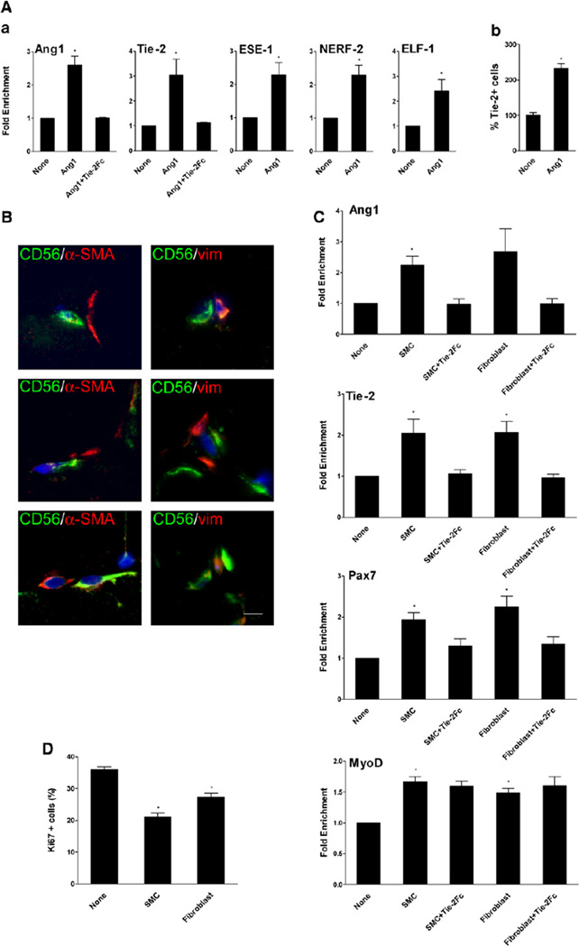
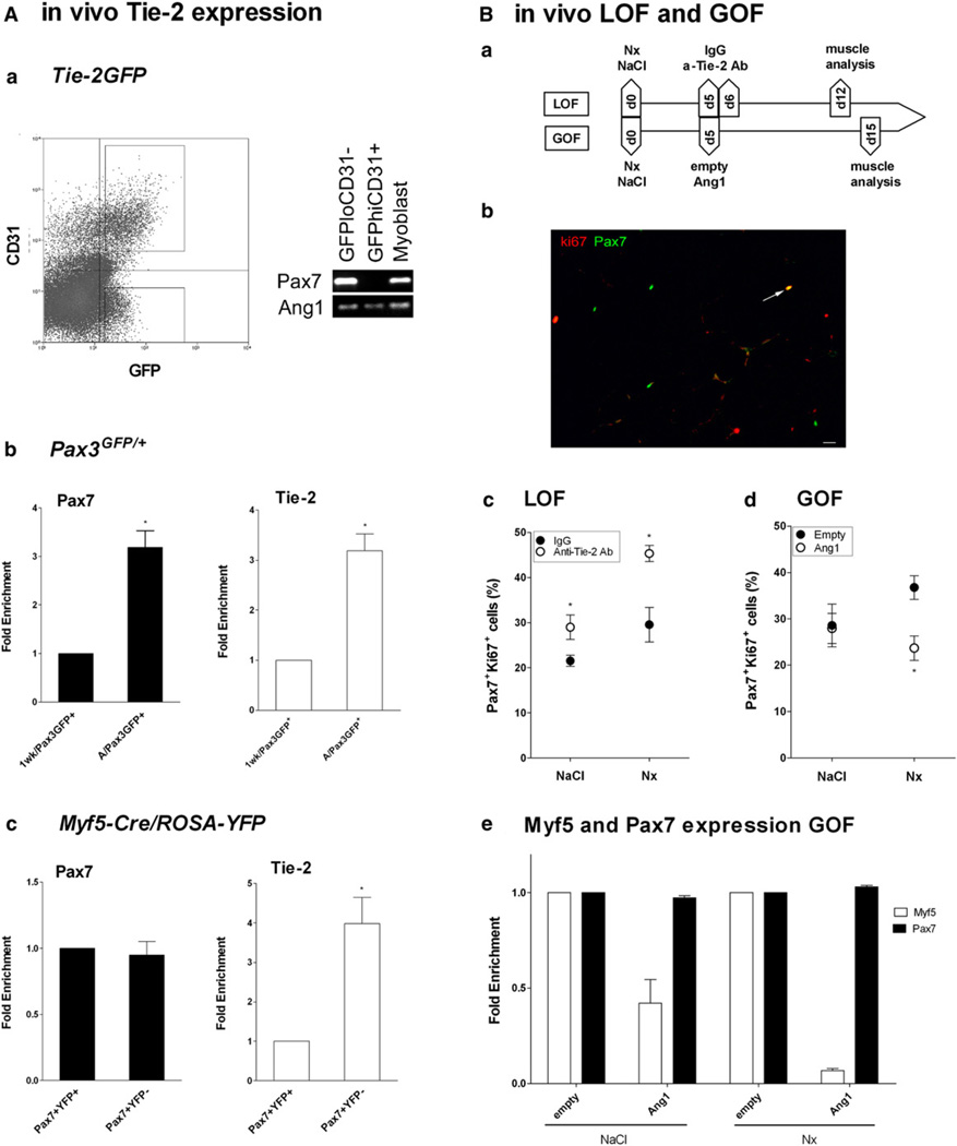
Mechanisms governing muscle satellite cell withdrawal from cell cycle to enter into quiescence remain poorly understood. We studied the role of angiopoietin 1 (Ang1) and its receptor Tie-2 in the regulation of myogenic precursor cell (mpc) fate. In human and mouse, Tie-2 was preferentially expressed by quiescent satellite cells in vivo and reserve cells (RCs) in vitro. Ang1/Tie-2 signaling, through ERK1/2 pathway, decreased mpc proliferation and differentiation, increased the number of cells in G0, increased expression of RC-associated markers (p130, Pax7, Myf-5, M-cadherin), and downregulated expression of differentiation-associated markers. Silencing Tie-2 had opposite effects. Cells located in the satellite cell neighborhood (smooth muscle cells, fibroblasts) upregulated RC-associated markers by secreting Ang1 in vitro. In vivo, Tie-2 blockade and Ang1 overexpression increased the number of cycling and quiescent satellite cells, respectively. We propose that Ang1/Tie-2 signaling regulates mpc self-renewal by controlling the return to quiescence of a subset of satellite cells.


Figures

Figure 1
Figure 1. Expression of Tie-2 and Ang1 by Human Myogenic Cells
(A) RT-qPCR analysis of Pax7, Tie-2, and Ang1 expression in human RCs, MTs, and HUVECs. Results are means ± SEM. (B) Flow cytometry analysis of Tie-2 expression in day 4 mpcs and RCs. (C) G0 and G2/M cell-cycle fractions were sorted from day 4 mpcs and were analyzed for expression of various genes by RT-qPCR. Results are means ± SD. (D) Freshly isolated human skeletal muscle-derived CD56+, CD56 CD31, and CD56CD31+ cell fractions were analyzed for Pax7, Tie-2, and Ang1 expression by RT-qPCR and compared to human RCs. Results are means ± SEM. (E) Flow cytometry analysis of Tie-2 expression in freshly isolated human skeletal muscle-derived CD56+ and CD56 cell fractions.
Figure 2
Figure 2. Mpc Growth and Apoptosis
(A) Mpcs were grown with or without Ang1 ± Tie-2Fc, Ang2, or VEGF, and cell density was estimated every 2 days. (B) Myogenic cell death was induced by staurosporine, mpcs were incubated with Ang1 (Ba) or with Ang1 ± Tie-2Fc (Bb), and apoptosis was evaluated. Results are means ± SEM.
Figure 3
Figure 3. Effects of Ang1 on Myogenic Cells
(A and B) Mpcs were incubated with Ang1 ± Tie-2Fc from day 0 or from day 4 and then analyzed. (A) Mpc proliferation was evaluated after Ki67 immunolabeling. (B) The number of myogenin+ cells was evaluated after myogenin/desmin immunolabeling. (C) Mpcs were incubated with Ang1 ± Tie-2Fc from day 0 and then analyzed for Pax7, p130, MyoD, and p57 expression by RT-qPCR. (D) Mpcs were incubated with Ang1 ± Tie-2Fc from day 0, stained with Pyronin Y and Hoechst, and sorted for their cell-cycle status at days 2, 4, and 6. The number of cells in each cell-cycle phase is given as percentage of total events. (E) Isolated mouse myofibers were incubated with Ang1 ± Tie-2Fc and stained for Pax7 (green) and MyoD (red). The number of Pax7+ and MyoD+ cells was counted in the clusters derived from satellite cells and reported as percentages. Pictures show examples of labeling observed in myofiber-associated cell clusters. Results are means ± SEM except in (D), where they are means ± SD. Bars, 10 µm.
Figure 4
Figure 4. Effects of Tie-2 Silencing and Tie-2 Overexpression on Mpcs
(A) Mpcs were incubated with Tie-2 siRNAs and scrambled control siRNA (S) and analyzed. (Aa) Tie-2, Pax7, p130, MyoD, and p57 expression were evaluated by RT-qPCR. (Ab) Mpc proliferation and differentiation were evaluated after Ki67 and myogenin/desmin immunolabeling, respectively. (B) Mpcs were incubated with Tie-2-overexpressing plasmid and analyzed. (Ba) Pax7, p130, MyoD, and p57 expression were evaluated by RT-qPCR. (Bb) Mpc proliferation and differentiation were evaluated as in (Ab). Results are means ± SEM.
Figure 5
Figure 5. Ang1/Tie-2 Intracellular Signaling
(A) Mpcs were incubated with Ang1 during increasing times or with Ang1 ± Tie-2Fc or U0126 for 10 min. Blot density on phosphosignaling membrane array (example is shown on right panel) was expressed as percentages of signal density at time 0 or in untreated cells, respectively. (B) Mpcs were incubated with Ang1 from day 4 ± U0126. Expression of various genes was evaluated by RT-qPCR. Insert: M-cadherin expression during in vitro myogenesis was evaluated by RT-qPCR. Results are means ± SEM.
Figure 6
Figure 6. Paracrine Effect of Neighboring Cells
(A) Mpcs were incubated with or without Ang1 ± Tie-2Fc, and (Aa) expression of various genes, including Ang1, Tie-2, and transcription factors involved in the regulation of Ang1 and Tie-2 expression, was evaluated by RT-qPCR. (Ab) Expression of Tie-2 was analyzed by flow cytometry. (B) Normal human muscle sections were immunolabeled with anti-α-SMA (red) or anti-vimentin (red) and anti-CD56 (green) antibodies. Bar, 5 µm. (C and D) Mpcs were incubated with conditioned medium from SMCs and fibroblasts ± Tie-2Fc. Expression of several genes was evaluated by RT-qPCR (C), and mpc proliferation was evaluated after Ki67 immunolabeling (D). Results are means ± SEM.
Figure 7
Figure 7. In Vivo Association between Ang1/Tie-2 System and Satellite Cell Quiescence
(A) Satellite cells were sorted from skeletal muscle of various mouse strains and analyzed for Pax7 and Tie-2 expression by RT-PCR and RT-qPCR. (Aa) CD31 GFPlo and CD31+GFPhi cell populations were sorted from Tg:Tie-2-GFP mice. (Ab) GFP+ cells were sorted from 1-week-old (1 wk/Pax3GFP+) and adult normal (A/Pax3GFP+) Pax3GFP/+ mice. (Ac) YFP+ and YFP cells were sorted from adult normal Myf5-Cre/ROSA-YFP mice. Results are means ± SEM. (B) Loss-of-function (LOF) and gain-of-function (GOF) studies were performed as shown on the scheme (Ba): muscle was injured with notexin (Nx) or not (Nacl). In LOF, at days 5 and 6, blocking anti-Tie-2 antibodies (a-Tie-2Ab or IgG) were i.m. injected, and muscle was analyzed at day 12. In GOF, at day 5, Ang1 plasmid (Ang1) (or empty plasmid) (empty) was electroporated, and was muscle analyzed at day 15. (Bb) Example of muscle-section labeling for Pax7 (green) and ki67 (red) (arrow shows double-positive satellite cell). Bar, 10 µm. (Bc) LOF and (Bd) GOF analysis of satellite cell staining. (Be) Whole-muscle expression of Pax7and Myf5 genes by RT-qPCR in GOF experiments. Results are means ± SEM.

Comment in

References

    1. Arai F, Hirao A, Ohmura M, Sato H, Matsuoka S, Takubo K, Ito K, Koh GY, Suda T. Tie2/angiopoietin-1 signaling regulates hematopoietic stem cell quiescence in the bone marrow niche. Cell. 2004;118:149–161. - PubMed
    1. Arnold L, Henry A, Poron F, Baba-Am Y, van Rooijen N, Plonquet A, Gherardi RK, Chazaud B. Inflammatory monocytes recruited after skeletal muscle injury switch into antiinflammatory macrophages to support myogenesis. J. Exp. Med. 2007;204:1071–1081. - PMC - PubMed
    1. Beauchamp JR, Heslop L, Yu DS, Tajbakhsh S, Kelly RG, Wernig A, Buckingham ME, Partridge TA, Zammit PS. Expression of CD34 and Myf5 defines the majority of quiescent adult skeletal muscle satellite cells. J. Cell Biol. 2000;151:1221–1234. - PMC - PubMed
    1. Black EJ, Clark W, Gillespie DA. Transient deactivation of ERK signalling is sufficient for stable entry into G0 in primary avian fibroblasts. Curr. Biol. 2000;10:1119–1122. - PubMed
    1. Brack AS, Conboy IM, Conboy MJ, Shen J, Rando TA. A temporal switch from notch to wnt signaling in muscle stem cells is necessary for normal adult myogenesis. Cell Stem Cell. 2008;2:50–59. - PubMed

Publication types

MeSH terms

Substances