Review
. 2009 Dec;126(11-12):925-41.
doi: 10.1016/j.mod.2009.08.004. Epub 2009 Sep 4.

Spemann's organizer and the self-regulation of embryonic fields

Affiliations
Review

Spemann's organizer and the self-regulation of embryonic fields

E M De Robertis. Mech Dev. 2009 Dec.

Abstract

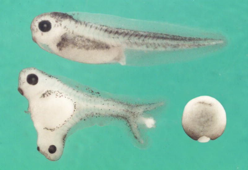
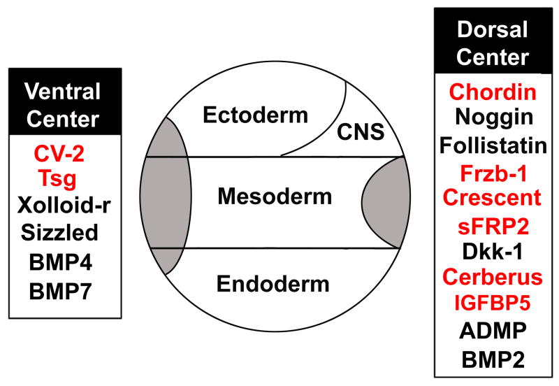
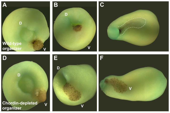
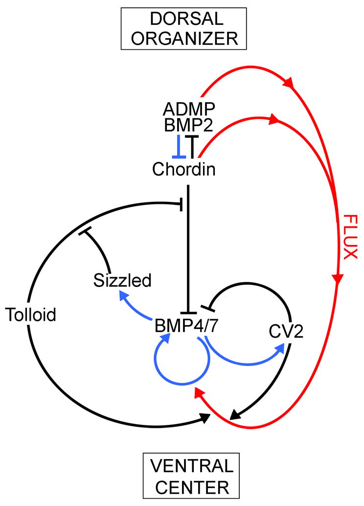
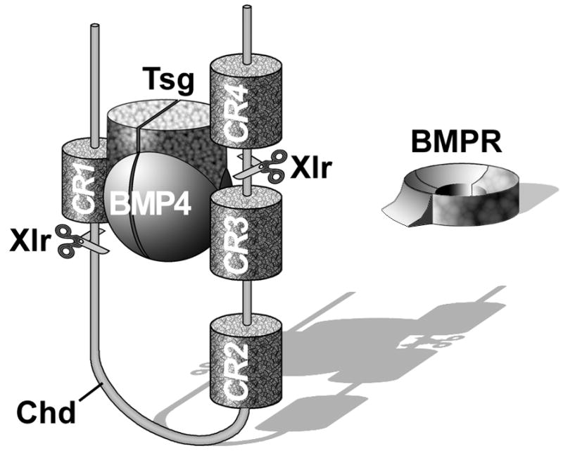
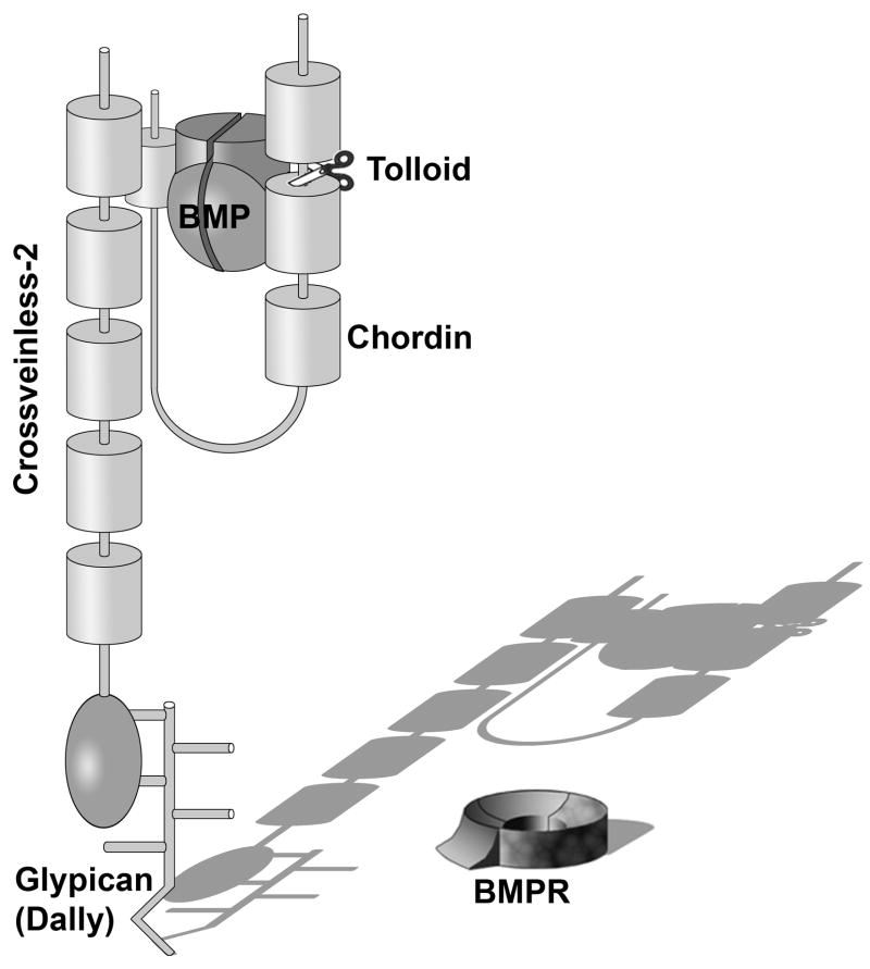
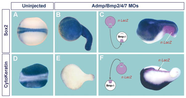
Embryos and developing organs have the remarkable ability of self-regenerating after experimental manipulations. In the Xenopus blastula half-embryos can regenerate the missing part, producing identical twins. Studies on the molecular nature of Spemann's organizer have revealed that self-regulation results from the battle between two signaling centers under reciprocal transcriptional control. Long-range communication between the dorsal and ventral sides is mediated by the action of growth factor antagonists - such as the BMP antagonist Chordin - that regulate the flow of BMPs within the embryonic morphogenetic field. BMPs secreted by the dorsal Spemann organizer tissue are released by metalloproteinases of the Tolloid family, which cleave Chordin at a distance of where they were produced. The dorsal center secretes Chordin, Noggin, BMP2 and ADMP. The ventral center of the embryo secretes BMP4, BMP7, Sizzled, Crossveinless-2 and Tolloid-related. Crossveinless-2 binds Chordin/BMP complexes, facilitating their flow towards the ventral side, where BMPs are released by Tolloid allowing peak BMP signaling. Self-regulation occurs because transcription of ventral genes is induced by BMP while transcription of dorsal genes is repressed by BMP signals. This assures that for each action of Spemann's organizer there is a reaction in the ventral side of the embryo. Because both dorsal and ventral centers express proteins of similar biochemical activities, they can compensate for each other. A novel biochemical pathway of extracellular growth factor signaling regulation has emerged from these studies in Xenopus. This remarkable dorsal-ventral positional information network has been conserved in evolution and is ancestral to all bilateral animals.


Figures

Fig. 1
Fig. 1
Separation of the first four blastomeres of a sea urchin embryo can give rise to four well-formed pluteus larvae. This powerful regulation was first reported by H. Driesch in 1891, marking the beginning of experimental embryology. It now appears that the self-regulation of embryonic fragments had been reported even earlier, in 1869, by Ernst Haeckel in cnidarian embryos (Sanchez-Alvarado, 2008). The experiment shown here is from Hörstadius and Wolsky, 1936, W. Roux. Arch. Entw. Mech. Org. 135, 69–113, reproduced with permission.
Fig. 2
Fig. 2
In Xenopus, the blastula constitutes a self-differentiating morphogenetic field, in which cells are able to communicate over long distances. When the blastula is bisected with a scalpel blade, identical twins can be obtained, provided that both fragments retain Spemann’s organizer tissue. Thus a half-embryo can regenerate the missing half. In humans, identical twins are found in three out of 1000 live births, and usually arise from the spontaneous separation of the inner cell mass of the blastocyst into two. A normal tadpole is shown on top, and two identical twins derived from the same blastula below, all at the same magnification. Reproduced from De Robertis, 2006, with permission of Nature Reviews.
Fig. 3
Fig. 3
Organ-fields identified by experimental embryologists in the amphibian neurula. The concept of self-regulating morphogenetic fields arose from a transplantation experiment by R. Harrison (1918) using the forelimb field. Reproduced from Huxley and de Beer, 1934, with permission of Cambridge University Press.
Fig. 4
Fig. 4
The Spemann-Mangold experiment reproduced in Xenopus laevis. A graft of albino dorsal lip was transplanted into the ventral side of the gastrula (bottom right). Signals emanating from this small graft were able to divide the embryonic morphogenetic field of the host into two almost equal parts, which formed a Siamese twin. Note that the D-V and A-P axes are perfectly integrated; this can be seen, for example, in the perfect alignment of somites (segments) of the duplicated axes. Reprinted, with permission, from the Annual Review of Cell and Developmental Biology, Volume 20 (c) 2004 by Annual Reviews www.annualreviews.org.
Fig. 5
Fig. 5
Sand (SiO2) particles serve as heterologous neural inducers in ectodermal explants of the American salamander Ambystoma maculatum. (A) A single grain of sand sandwiched between two ectodermal explants induces neural tissue marked by Sox3 mRNA. (B) Multiple sand particles cause patches of Cytokeratin-negative cells, which correspond to neural tissue. (C) Ectodermal explants cultured without sand particles, showing that the normal fate of these cells is to form Cytokeratin-epidermal positive cells. Reproduced from Hurtado and De Robertis, 2007, with permission.
Fig. 6
Fig. 6
CNS differentiations induced by culturing Ambystoma maculatum ectoderm attached to a glass surface (in Holtfreter’s saline solution) can be blocked by addition of UO126, a chemical inhibitor of the MAPK/Erk pathway. (A) Ectoderm cultured attached to glass can develop extensive neural differentiations. After the initial induction of CNS tissue, differentiations of secondary fields also take place, giving rise to olfactory placodes, retina, retinal pigmented epithelium, and lens (of which an enlargement is shown). (B) In the presence of UO126 CNS differentiations are blocked. The explants develop as atypical epidermis (which is called atypical because it contains small cavities containing keratinized cells). (C) Section of a sibling embryo at the same stage of development (9 days) to illustrate the normal histological appearance of CNS tissues. (D) Outside view of Ambystoma maculatum 9-day larva indicating the plane of section. Abbreviations: ba, balancer; CNS, central nervous system; ey, eye; g, gills; gm, gray matter; le, lens; me, mesencephalon; op, olfactory placodes; re, retina; rpe, retinal pigmented epithelium; te, telencephalon; v, ventricle; vm, white matter. Reproduced from Hurtado and De Robertis, 2007, with permission.
Fig. 7
Fig. 7
Secreted proteins that have been cloned from the dorsal lip or the ventral center of the Xenopus gastrula. Many laboratories contributed to this effort; genes first isolated by our group are shown in red. See text for further description.
Fig. 8
Fig. 8
Chordin is required for the activity of Spemann organizer grafts. Shown here are transplants of pigmented organizers into albino hosts. (A–C) Transplant of a wild-type organizer followed for a few hours, showing how it involutes through the ventral blastopore until it is barely seen by transparency below the ectoderm (dotted line). (D–F) Depletion of Chordin in the organizer graft (Oelgeschläger et al., 2003) prevents all inductive activity, and the transplanted cells remain in the surface of the embryo, becoming epidermis. D, dorsal: V, ventral. Transplantation experiment by E.M.D.R., photographs by J.L. Plouhinec
Fig. 9
Fig. 9
An extracellular biochemical network of interacting proteins explains self-regulation of the Xenopus embryonic field. The dorsal organizer and the ventral center communicate with each other through secreted proteins that bind to each other, inhibiting or activating the BMP gradient. All these protein-protein interactions (shown in black) were determined in our laboratory using biochemical measurements of affinity constants. Blue arrows indicate transcriptional regulation by Smad1/5/8 signaling, which activates ventral center genes and represses dorsal center genes. The two centers self-adjust to signaling changes in one another because of this opposite transcriptional control by BMPs. For example, when BMP levels increase, this causes an increase in Sizzled expression, which is an inhibitor of the Tolloid metalloproteinase that degrades Chordin. Thus, when Sizzled increases, Chordin levels increase, inhibiting BMP signaling and restoring the gradient. Red arrows denote the flux of Chordin/ADMP/BMP2 complexes from dorsal to more ventral regions. Mathematical modeling suggests that this Chordin-mediated flow of BMP is essential for the resilience of the gradient.
Fig. 10
Fig. 10
Chordin (Chd) forms a ternary complex with BMP4 and Twisted gastrulation (Tsg), which prevents binding to BMPR and allows the complex to diffuse within the embryo. The inhibition of BMP signaling is reversed by a ventral enzyme called Xolloid-related (Xlr) which is able to cleave Chordin at two specific sites (indicated by scissors), releasing BMPs for signaling through BMPR. Note that Chordin has four BMP-binding Cysteine-rich modules called CRs. CR domains function as regulators of BMP or TGF-β signaling in many extracellular proteins (De Robertis and Kuroda, 2004).
Fig. 11
Fig. 11
Crossveinless-2 (CV2) serves as a molecular sink that concentrates Chordin/Tsg/BMP complexes on the ventral side of the embryo. Once there, BMPs secreted by more dorsal regions of the embryo can be released by Tolloid enzymes and signal through BMP receptors (BMPR). CV2 is a secreted protein but does not diffuse far from the cells that secrete it, because it remains anchored to the cell surface by GPI-anchored glypicans, such as Dally in Drosophila, via its COOH-terminal vWF-d (von Willebrand Factor-D) domain.
Fig. 12
Fig. 12
The self-adjusting nature of the D-V BMP gradient can be revealed by lowering or increasing BMP signaling. In this experiment, BMP signaling was lowered by Chordin or increased by BMP proteins microinjected into the blastula cavity at stage 8. A molecular see-saw explains self-regulation of the gradient. When BMP signaling is inhibited, transcription of ADMP (a BMP) increases and restores the gradient. When BMP4 signaling is increased, Sizzled transcription is elevated and Sizzled protein inhibits tolloid proteinases, indirectly increasing levels of the BMP antagonist Chordin. ODC, Ornithine Decarboxylase, serves as an mRNA loading control in these RT-PCR reactions. From Reversade and De Robertis, 2005, reproduced with permission.
Fig. 13
Fig. 13
Simultaneous depletion of four BMPs causes ubiquitous CNS differentiation, which can be restored by transplantation of either a wild-type ventral center or a dorsal organizer. (A) Control Xenopus embryo showing normal Sox2 mRNA expression in the CNS. (B) Sibling depleted of ADMP, BMP2, 4 and 7 with antisense morpholinos; note that the entire embryonic surface is covered by CNS tissue. (C) Transplantation of a wild-type ventral center (labeled with nuclear LacZ lineage tracer) into BMP-depleted embryos restores formation of a neural plate with epidermis ventrally to it. (D) Cytokeratin mRNA is abundantly expressed in epidermis. (E) Cytokeratin expression is eliminated in BMP-depleted embryos (because epidermis is replaced by CNS). (F) Transplantation of a wild-type dorsal organizer rescues BMP depletion. Epidermis is induced, but at a considerable distance from the transplanted tissue (which gives rise to notochord). BMP does not signal close to the graft because it is inhibited by Chordin. These experiments show, first, that BMP inhibition causes ubiquitous neural induction and, second, that the embryo has dorsal and ventral sources of BMP signals. Experiments from Reversade and De Robertis (2005), reproduced with permission.
Fig. 14
Fig. 14
The common ancestor of bilateral animals had a D-V axis patterned by the Chd/BMP/Tsg/Tolloid/CV2 pathway. Shown here are the two branches of bilateral animals, which underwent a D-V inversion of the CNS. The protostomes (proto, first; stomo, mouth) have the nerve cord ventral to the gut. The deuterostomes (deutero, second) have the CNS dorsal to the gut. Urbilateria is the last common ancestor of all bilateral animals. Evo-Devo studies suggest that Urbilateria was a highly complex animal (De Robertis, 2008a). The blastopore of Urbilateria is shown as an elongated slit that gives rise both to the mouth and anus (a situation called amphistomy); recent findings showing that hemichordates (acorn worms) have not yet undergone D-V inversion of the CNS (Benito-Gutiérrez and Arendt, 2009) imply that the urbilaterian CNS likely resembled that of protostomes. In the diagram, Urbilateria is depicted as a segmented bottom-dwelling (benthic) animal. While a common ancestry of animal segmentation mechanisms is the subject of debate, two recent studies favor this idea: in the cockroach Notch pathway genes cycle rhythmically as in vertebrate segmentation (Pueyo et al., 2008), and Smad1/5/8 and Mad are required for segmentation in Xenopus and Drosophila (Eivers et al., 2009). Urbilateria probably had a life-cycle including a marine free-swimming (pelagic) primary larval stage, shown here with trochophore-like beating cilia. Many extant phyla have such larvae – annelids, mollusks, hemichordates and echinoderms – although this phase of the life-cycle has been repeatedly lost during evolution (Jägersten, 1972; Nielsen, 1998). Both the D-V (Chd/BMP/Tsg/Tld/CV2) and A-P (Hox genes) patterning systems were utilized by the urbilaterian ancestor to generate pattern. The use of these ancestral gene networks must have placed important developmental constraints in the evolution of animal body plans. Ectoderm is shown in green, CNS in blue, eye in black, and endoderm in red, with its openings in yellow. Reproduced, with permission, from De Robertis 2008b.
Fig. 15
Fig. 15
The Hox complexes have been conserved between Drosophila and mammals, down to level of micro RNAs (miRs) that repress the translation of more anterior genes. (A) Vertebrates have four Hox complexes and Drosophila only one (which became separated into two segments). Vertebrates underwent two rounds of whole-genome duplication when they evolved from a simpler chordate ancestor. These whole-genome duplications may explain the evolutionary success of the vertebrates. The ancestral chordate Hox complex had 13 genes, but some paralogues have been lost in mammals. (B) Hox-C6 protein is detected in eight thoracic segments of mouse embryos. The inset shows that Hox-C6 mRNA is expressed all the way to the tail. Hox-C6 protein is not detected posteriorly probably because of translational repression by miR196 inhibition. Redrawn from De Robertis, 2008a, reproduced with permission.

References

    1. Akiyama-Oda Y, Oda H. Axis specification in the spider embryo: dpp is required for radial-to-axial symmetry transformation and sog for ventral patterning. Development. 2006;133:2347–2357. - PubMed
    1. Ambrosio AL, Taelman VF, Lee HX, Metzinger CA, Coffinier C, De Robertis EM. Crossveinless-2 is a BMP feedback inhibitor that binds Chordin/BMP to regulate Xenopus embryonic patterning. Dev Cell. 2008;15:248–260. - PMC - PubMed
    1. Appel TA. The Cuvier-Geoffroy Debate. Oxford University Press; Oxford: 1987.
    1. Bachiller D, et al. The organizer secreted factors Chordin and Noggin are required for forebrain development in the mouse. Nature. 2000;403:658–661. - PubMed
    1. Barth LG. Neural diffferentiation without organizer. J Exp Zool. 1941;87:371–384.

Publication types

MeSH terms