. 2009 Oct;46(16):3224-31.
doi: 10.1016/j.molimm.2009.08.015. Epub 2009 Sep 5.

The Sts proteins target tyrosine phosphorylated, ubiquitinated proteins within TCR signaling pathways

Affiliations

The Sts proteins target tyrosine phosphorylated, ubiquitinated proteins within TCR signaling pathways

Nick Carpino et al. Mol Immunol. 2009 Oct.

Abstract

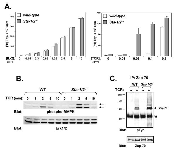
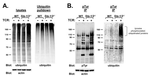
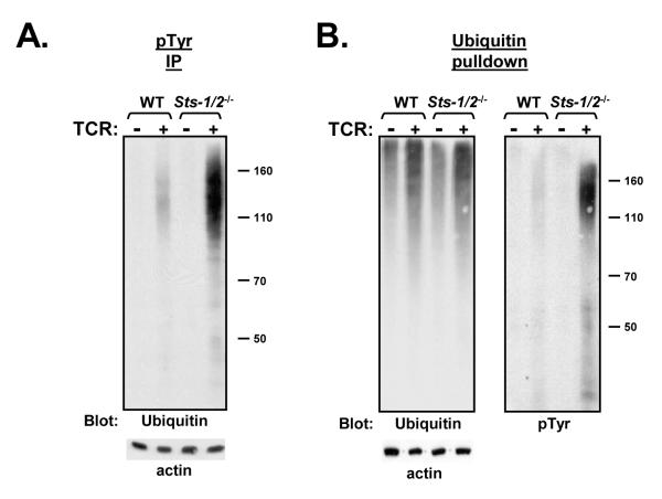
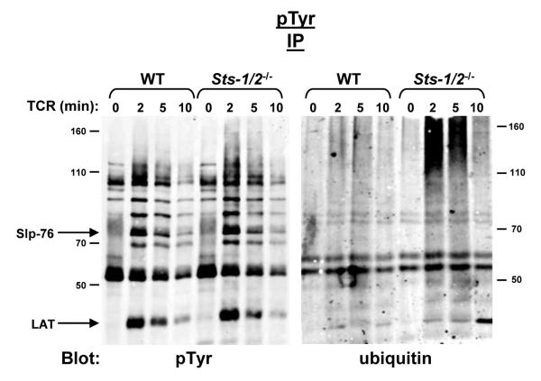
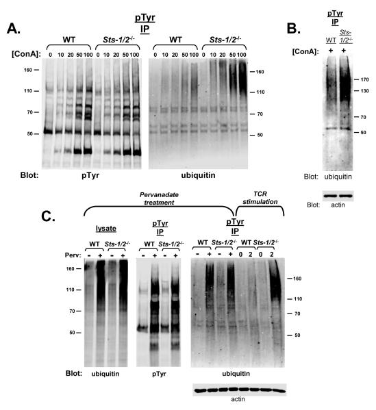
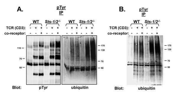
The T cell receptor (TCR) detects the presence of infectious pathogens and activates numerous intracellular signaling pathways. Protein tyrosine phosphorylation and ubiquitination serve as key regulatory mechanisms downstream of the TCR. Negative regulation of TCR signaling pathways is important in controlling the immune response, and the Suppressor of TCR Signaling proteins (Sts-1 and Sts-2) have been shown to function as critical negative regulators of TCR signaling. Although their mechanism of action has yet to be fully uncovered, it is known that the Sts proteins possess intrinsic phosphatase activity. Here, we demonstrate that Sts-1 and Sts-2 are instrumental in down-modulating proteins that are dually modified by both protein tyrosine phosphorylation and ubiquitination. Specifically, both naïve and activated T cells derived from genetically engineered mice that lack the Sts proteins display strikingly elevated levels of tyrosine phosphorylated, ubiquitinated proteins following TCR stimulation. The accumulation of the dually modified proteins is transient, and in activated T cells but not naïve T cells is significantly enhanced by co-receptor engagement. Our observations hint at a novel regulatory mechanism downstream of the T cell receptor.


Figures

Figure 1
Figure 1. Hyper-responsiveness of Sts-1/2-/- activated T cells to TCR engagement
(A) The response of wild-type and Sts-1/2-/- activated T cells to either IL-2 or TCR stimulation was evaluated by [3H]-thymidine incorporation assay. Sts-1/2-/- cells display a hyper-proliferative phenotype in response to TCR stimulation. Each assay was conducted in triplicate, and data representative of over three separate experiments are displayed. (B) Elevated MAPK activation in Sts-1/2-/- activated, cultured T cells. Stimulated T cells were lysed at the indicated time points. Whole-cell lysates were separated by SDS-PAGE and subjected to Western analysis with anti-phospho-MAPK antibodies (pMAPK indicated with arrows). The same blot was reprobed with anti-Erk1/2 antibodies, as a loading control (see lower blot). (C) Hyper-phosphorylation of Zap-70 in Sts-1/2-/- activated T cells. Cells were stimulated for two minutes, lysed, and processed for IP/Western analysis. Half of the precipitated Zap-70 was assessed for levels of tyrosine phosphorylation and half was assessed for levels of Zap-70. Arrow indicates tyrosine phosphorylated Zap-70. This data is representative of over five experiments.
Figure 2
Figure 2. Stimulation — induced ubiquitination in wild-type and Sts-1/2-/- activated T cells
(A) Wild-type and Sts-1/2-/- cultured T cells demonstrate similar levels of activation-induced ubiquitination. Resting (−) or TCR-stimulated (+) cells were lysed and levels of ubiquitination were assessed by western analysis. Total lysates (left panel) or proteins binding to a poly-ubiquitin affinity column (right panel) were separated by SDS-PAGE and ubiquitin levels were assessed by anti-ubiquitin western analysis. (B) Tyrosine phosphorylated proteins in resting or TCR-stimulated activated T cells were isolated by immuno-precipitation. The pool of precipitated proteins was split in two, with half analyzed by anti-pTyr western analysis and half analyzed by anti-ubiquitin western analysis. An anti-actin immunoblot serves as a loading control. The dotted bracket indicates increased levels of proteins dually modified by tyrosine phosphorylation and ubiquitination that appear in stimulated Sts-1/2-/- activated T cells.
Figure 3
Figure 3. Appearance of hyper-phosphorylated, ubiquitinated proteins in Sts-1/2-/- naïve T cells
(A) Freshly isolated wild-type and Sts-1/2-/- splenic T cells were stimulated through the TCR, phosphorylated proteins were isolated by anti-pTyr immunoprecipitation and levels of ubiquitination were assessed by anti-ubiquitin western analysis. An anti-actin immunoblot demonstrates equivalent levels of protein in each lysates. (B) Dually modified proteins in stimulated Sts-1/2-/- naïve T cells. Poly-ubiquitinated proteins from lysates of resting or stimulated naïve T cells were isolated by poly-ubiquitin affinity pull-down analysis. Levels of ubiquitinated proteins were assessed by anti-ubiquitin western analysis (left panel), following which the gel was stripped and reprobed with anti-pTyr antibodies (right panel). An anti-actin immunoblot was used as a loading control.
Figure 4
Figure 4. Transient appearance of hyper-phosphorylated, ubiquitinated proteins in stimulated Sts-1/2-/- T cells
IP/Western analysis with the indicated antibodies was used to detect the appearance and disappearance of hyper-phosphorylated, ubiquitinated proteins in Sts-1/2-/- cultured T cells along a time-course of TCR stimulation from 0 to 10 minutes.
Figure 5
Figure 5. Indirect stimulation of T cells suggests the Sts proteins regulate a TCR-dependent pathway
(A) Wild-type and Sts-1/2-/- cultured T cells were incubated for five minutes with the indicated concentrations (μg/ml) of Concanavalin A, a lectin that binds surface glycoproteins and is thought to indirectly activate the TCR. The appearance of tyrosine phosphorylated, ubiquitinated proteins, as detected by IP/Western analysis with the indicated antibodies, was only evident in Sts-1/2-/- cells. (B) Wild-type and Sts-1/2-/- purified splenic T cells were incubated for five minutes with 100 ug/ml ConA. IP/Western analysis was utilized to assess the appearance of dually modified proteins in Sts-1/2-/- naïve T cells. (C) Wild-type and Sts-1/2-/- cultured T cells were treated for five minutes with pervanadate to inactivate intracellular tyrosine phosphatases. An equivalent level of overall treatment-induced protein tyrosine phosphorylation was evident in both samples (left panel). In addition, levels of phosphorylation-dependent ubiquitination were similar (middle panel), as assessed by anti-ubiquitin blotting of cell lysates. Both wild-type and Sts-1/2-/- pervanadate-treated cells displayed robust levels of tyrosine phosphorylated, ubiquitinated proteins (right panel). However, the SDS-PAGE migration pattern of the latter proteins differed significantly from the dually-modified proteins that appear in TCR-stimulated Sts-1/2-/- cells (see right panel), suggesting either the two modified protein populations are different or arise by different signaling mechanisms.
Figure 6
Figure 6. Regulation of a co-receptor — dependent pathway by the Sts proteins
(A) Co-receptor engagement stimulates increased levels of dually modified proteins in Sts-1/2-/- activated T cells. Wild-type and Sts-1/2-/- cultured T cells were stimulated with anti-TCR antibody or anti-TCR antibody plus antibodies to the CD8 co-receptor, as indicated. The level of intracellular tyrosine phosphorylation proteins and the level of dually modified proteins were assessed by IP/western analysis with the indicated antibodies. (B) Co-receptor engagement has little effect on the level of dually modified protein in naïve cells. Wild-type and Sts-1/2-/- purified splenic T cells were stimulated with anti-TCR antibody or anti-TCR antibody plus antibodies to the CD4 and CD8 co-receptors. Levels of tyrosine phosphorylated, ubiquitinated proteins were assessed by IP/western analysis with the indicated antibodies.

References

    1. Acuto O, Bartolo VD, Michel F. Tailoring T-cell receptor signals by proximal negative feedback mechanisms. Nat. Rev. Immunol. 2008;8:699–712. - PubMed
    1. Agrawal R, Carpino N, Tsygankov A. TULA proteins regulate activity of the protein tyrosine kinase Syk. J. Cell. Biochem. 2008;104:953–964. - PubMed
    1. Balagopalan L, Barr VA, Sommers CL, Barda-Saad M, Goyal A, Isakowitz MS, Samelson LE. c-Cbl-mediated regulation of LAT-nucleated signaling complexes. Mol. Cell. Biol. 2007;27:8622–8636. - PMC - PubMed
    1. Carpino N, Turner S, Mekala D, Takahashi Y, Zang H, Geiger TL, Doherty P, Ihle JN. Regulation of Zap-70 activation and TCR signaling by two related proteins, Sts-1 and Sts-2. Immunity. 2004;20:37–46. - PubMed
    1. Cenciarelli C, Hou D, Hsu KC, Rellahan BL, Wiest DL, Smith HT, Fried VA, Weissman AM. Activation-induced ubiquitination of the T cell antigen receptor. Science. 1992;257:795–797. - PubMed

Publication types