. 2009 Nov 6;284(45):31097-108.
doi: 10.1074/jbc.M109.015800. Epub 2009 Sep 4.

A UV-responsive internal ribosome entry site enhances serine hydroxymethyltransferase 1 expression for DNA damage repair

Affiliations

A UV-responsive internal ribosome entry site enhances serine hydroxymethyltransferase 1 expression for DNA damage repair

Jennifer T Fox et al. J Biol Chem. .

Abstract

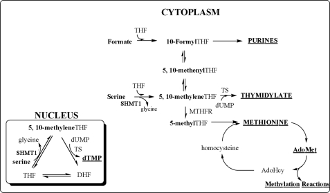
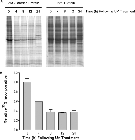
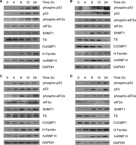
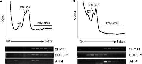
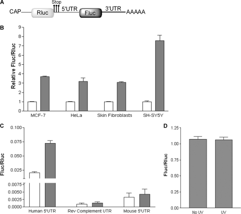
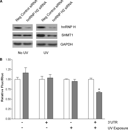
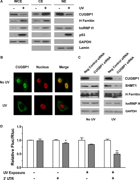
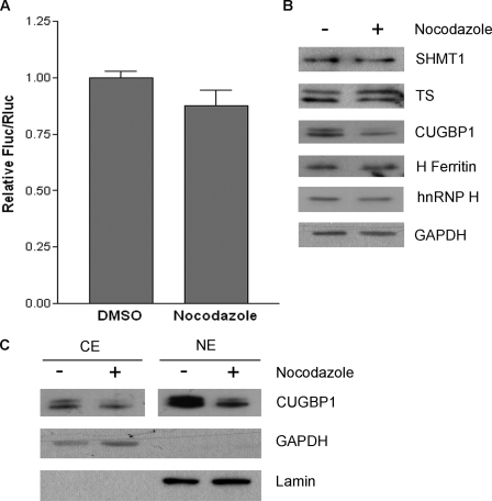
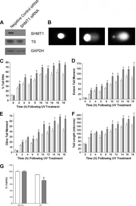
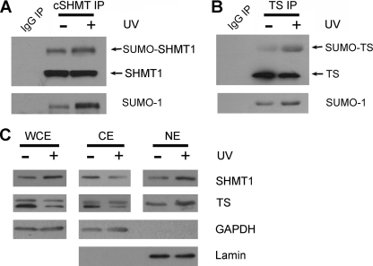
Thymidine nucleotides are required for faithful DNA synthesis and repair, and their de novo biosynthesis is regulated by serine hydroxymethyltransferase 1 (SHMT1). The SHMT1 transcript contains a heavy chain ferritin, heterogeneous nuclear ribonucleoprotein H2, and CUG-binding protein 1-responsive internal ribosome entry site (IRES) that regulates SHMT1 translation. In this study a non-lethal dose of UVC is shown to increase SHMT1 IRES activity and protein levels in four different cell lines. The mechanism for the UV-induced activation of the SHMT1 IRES involves an increase in heavy chain ferritin and heterogeneous nuclear ribonucleoprotein H2 expression and the translocation of CUG-binding protein 1 from the nucleus to the cytoplasm. The UV-induced increase in SHMT1 translation is accompanied by an increase in the small ubiquitin-like modifier-dependent nuclear localization of the de novo thymidylate biosynthesis pathway and a decrease in DNA strand breaks, indicating a role for SHMT1 and nuclear folate metabolism in DNA repair.


Figures

FIGURE 1.
FIGURE 1.
Folate-mediated one-carbon metabolism. Folate-mediated one-carbon metabolism is required for the de novo synthesis of purines and thymidylate and for the remethylation of homocysteine to methionine. Mitochondrial-derived formate can enter the cytoplasm and function as a one-carbon donor through the conversion of THF to 10-formyl-THF. 5,10-Methylene-THF, which can be generated through the reduction of 10-formyl-THF or through the catalytic activity of SHMT1, provides the one-carbon units for the TS-catalyzed conversion of dUMP to thymidylate. It also serves as a substrate for methylene-THF reductase (MTHFR), which reduces 5,10-methylene-THF to ultimately form S-adenosylmethionine (AdoMet), the one-carbon donor in numerous cellular methylation reactions. De novo thymidylate biosynthesis also occurs in the nucleus through the SUMO-dependent import of the thymidylate synthesis pathway during S-phase. Hcy, homocysteine; DHF, dihydrofolate.
FIGURE 2.
FIGURE 2.
Nascent protein synthesis decreases after UV treatment. A, MCF-7 cells were treated with 10,000 μJ/cm2 UVC (254 nm). At the indicated times after UV treatment, the cells were pulse-labeled with [35S]Met/Cys, and total cell extracts were resolved by SDS-PAGE. The gel was then stained with Coomassie Blue (right panel) to visualize total proteins followed by autoradiography (left panel) to detect newly synthesized proteins. B, equal amounts of protein from A were precipitated with trichloroacetic acid, and 35S incorporation was quantified in a scintillation counter. The counts per min recorded for the 0 h sample was given a value of 1.0. The data represent the average of three independent trichloroacetic acid precipitations ±S.E.
FIGURE 3.
FIGURE 3.
Effect of UVC on protein levels. MCF-7 cells (A), HeLa cells (B), transformed skin fibroblasts (C), and SH-SY5Y cells (D) were treated with 10,000 μJ/cm2 UVC (254 nm). At the indicated times after UV treatment, total protein lysates were prepared and resolved by SDS-PAGE. Protein levels were determined by immunoblotting using antibodies against phosphorylated p53, p53, phosphorylated eIF2α, eIF2α, SHMT1, TS, CUGBP1, H ferritin, and hnRNP H. GAPDH served as a control for equal protein loading.
FIGURE 4.
FIGURE 4.
SHMT1 mRNA remains associated with polysomes after UV exposure. The polysome profile of untreated (A) and UV-irradiated MCF-7 cells (B) was recorded 22 h after UV treatment. The ribosomal species, as determined by the optical density (OD) at 254 nm, are indicated. Total RNA was extracted from each fraction, and SHMT1 mRNA was detected by reverse transcription PCR. CUGBP1 mRNA, which is not known to contain an IRES, and ATF4 mRNA, which is known to be translated during conditions where cap-dependent translation is reduced by the phosphorylation of eIF2α (52–54), are shown for comparison.
FIGURE 5.
FIGURE 5.
UV treatment results in an increase in SHMT1 IRES activity. A, shown is the bicistronic construct used to quantify SHMT1 IRES activity. It consists of (in the 5′ to 3′ direction) a cap analog, the Rluc reporter gene followed by three sequential in-frame stop codons, the human SHMT1 5′-UTR, which contains the IRES element, the Fluc reporter gene, the human SHMT1 3′-UTR, which was shown to stimulate SHMT1 IRES activity (26), and a 30-nucleotide poly(A) tail. B, untreated (light bars) and UV-treated (dark bars) cells were transiently transfected with the bicistronic construct in A. 22 h after treatment Fluc and Rluc activities were quantified. The relative ratio of total Fluc activity divided by total Rluc activity in untreated cells was given a value of 1.0. The data represent the average of three independent experiments ±S.E. C, untreated (light bars) and UV-treated (dark bars) MCF-7 cells were transiently transfected with either the bicistronic construct containing the human SHMT1 5′-UTR or bicistronic constructs where the human SHMT1 5′-UTR was replaced with either the reverse (Rev) complement of the human SHMT1 5′-UTR or the mouse SHMT1 5′-UTR. None of the bicistronic constructs used in this experiment contained the SHMT1 3′-UTR. IRES activity is reported as the ratio of total Fluc activity divided by total Rluc activity as measured 22 h after UV treatment. The data represent the average of three independent experiments ±S.E. D, MCF-7 cells were treated with 10,000 μJ/cm2 UVC and transiently transfected with the bicistronic mRNA in A. 22 h after UV treatment, total RNA was extracted from the cells and reverse-transcribed into cDNA. Rluc and Fluc mRNA levels were determined by real time PCR. The data represent the average of three independent experiments ±S.E.
FIGURE 6.
FIGURE 6.
hnRNP H2 is involved in the UV-induced activation of the SHMT1 IRES. A, MCF-7 cells were transfected with negative control siRNA or hnRNP H2 siRNA, treated with UV, and then subjected to Western blot analysis using antibodies against hnRNP H and SHMT1. GAPDH serves as a control for equal protein loading. B, control (white bars) and hnRNP H2 siRNA-treated cells (dark bars) were transiently transfected with bicistronic mRNAs with and without the SHMT1 3′-UTR and in the presence and absence of UVC exposure. The relative ratio of Fluc/Rluc for each bicistronic mRNA in the control cells was given a value of 1.0. The data represent the average of three independent experiments ±S.E. The asterisk represents statistical significance (p = 0.001) as determined by Student's t test.
FIGURE 7.
FIGURE 7.
Exposure to UV radiation results in the cytoplasmic accumulation of CUGBP1. A, nuclear (NE) and cytoplasmic (CE) extracts were isolated from untreated and UV-treated MCF-7 cells 22 h after UV exposure. Whole cells extracts (WCE) were also obtained from the same samples. All extracts were run side-by-side on an SDS gel and subjected to immunoblotting using an anti-CUGBP1, anti-hnRNP H, or an anti-H ferritin antibody. p53 is shown as a control for UV treatment. GAPDH is shown as a control to demonstrate that the nuclear fractions are free of cytoplasmic contamination. Lamin A is shown as a control to demonstrate that the cytoplasmic fractions are free of nuclear contamination. Both GAPDH and Lamin A serve as controls for equal protein loading. B, immunofluorescence was used to determine CUGBP1 localization in untreated and UV-treated MCF-7 cells 22 h after UV exposure. CUGBP1 was visualized with Alexa Fluor 488 (green), and the nucleus was visualized with DRAQ5 (red). The right column is a merge of the green and red channels. C, MCF-7 cells were transfected with negative control siRNA or CUGBP1 siRNA and then treated with UV. 22 h after UV treatment, CUGBP1, SHMT1, hnRNP H, and H ferritin protein levels were visualized by Western blotting. GAPDH serves as a control for equal protein loading. D, the activity of the SHMT1 IRES was quantified as the Fluc/Rluc ratio in MCF-7 cells treated with negative control siRNA (light bars) and cells treated with CUGBP1 siRNA (dark bars). Cells were transfected with the bicistronic mRNA described in Fig. 5A containing or lacking the SHMT1 3′-UTR in the presence and absence of UVC exposure. For each experimental condition, values obtained from negative control siRNA-treated cells were given a relative value of 1.0. The data represent the average of three independent experiments ±S.E. The single and double asterisks represent statistical significance (p = 0.04 and p = 0.002, respectively) as determined by Student's t test.
FIGURE 8.
FIGURE 8.
Nocodazole treatment does not produce the same effects as UV radiation. A, MCF-7 cells were treated with either vehicle (DMSO) or 60 ng/ml nocodazole for 24 h and then transiently transfected with the bicistronic mRNA in Fig. 5A. The ratio of Fluc/Rluc in DMSO-treated cells was given a value of 1.0. The data represent the average of three independent experiments ±S.E. B, total protein lysates were prepared from untreated and nocodazole-treated cells and resolved by SDS-PAGE. Protein levels were determined by immunoblotting using antibodies against SHMT1, TS, CUGBP1, and H ferritin. GAPDH served as a control for equal protein loading. C, nuclear (NE) and cytoplasmic (CE) fractions were also isolated from these cells and subjected to immunoblotting using an anti-CUGBP1 antibody. GAPDH is shown as a control to demonstrate that the nuclear fractions are free of cytoplasmic contamination. Lamin A is shown as a control to demonstrate that the cytoplasmic fractions are free of nuclear contamination. Both GAPDH and Lamin A serve as controls for equal protein loading.
FIGURE 9.
FIGURE 9.
SHMT1 is involved in the repair of UV-induced DNA damage. MCF-7 cells were treated with negative control siRNA (light bars) or SHMT1 siRNA (dark bars) for 55 h and then exposed to UV radiation. At the indicated times after UV treatment, the cells were harvested and divided into two samples. One sample was used for immunoblotting to ensure the knockdown of SHMT1, and the other sample was used in the comet assay to determine DNA damage or in the MTT assay to determine cell viability. A, shown is a representative Western blot showing SHMT1 and TS protein levels in cells treated with either the negative control siRNA or the SHMT1 siRNA. GAPDH serves as a control for equal protein loading. B, for the comet assay, single cells were embedded in agarose, lysed, and subjected to electrophoresis. The DNA content of each cell was visualized using SYBR Gold. The panels are representative images of (from left to right) a cell with little DNA damage, a cell with an intermediate amount of DNA damage, and a cell with extensive DNA damage. C–F, the DNA content of the cells was quantified using Komet 5.5 software. The data represent the average of three independent experiments ±S.E. Each experiment was performed in duplicate, and 75 cells were analyzed per experiment per time point. The asterisk represents statistical significance (p < 0.05) at a given time point as determined by Student's t test. C, % tail DNA = the proportion of DNA that has migrated from the nucleoid core. D, extent of tail moment = % tail DNA × tail length/100. E, olive tail moment = (tail center of gravity − head center of gravity) × % tail DNA/100. F, tail length = the distance (microns) of DNA migration from the nucleoid core. G, for the MTT assay, the production of formazan by living mitochondria was measured in negative control siRNA-treated cells (light bars) and SHMT1 siRNA-treated cells (dark bars) 24 h after UV treatment by recording the absorbance at 550 nm. The absorbance from non-irradiated, negative control siRNA-treated cells was given a value of 100%. The results represent the average of four independent experiments ±S.E. The asterisk indicates statistical significance (p = 0.0002) as determined by Student's t test.
FIGURE 10.
FIGURE 10.
SHMT1 and TS SUMOylation and nuclear localization increase in response to UV treatment. SHMT1 (A) and TS (B) were immunoprecipitated (IP) from untreated and UV-treated MCF-7 whole cell extracts 22 h after UV exposure. Immunoblotting was performed on the immunoprecipitates using antibodies against SUMO-1, SHMT1, and TS. The IgG immunoprecipitate (lane 1) serves as control for nonspecific binding. C, whole cell (WCE), nuclear (NE), and cytoplasmic (CE) fractions were isolated from untreated and UV-treated MCF-7 cells 22 h after UV exposure. All extracts were resolved by SDS-PAGE and subjected to immunoblotting using an anti-SHMT1 or anti-TS antibody. GAPDH is shown as a control to demonstrate that the nuclear fractions are free of cytoplasmic contamination. Lamin A is shown as a control to demonstrate that the cytoplasmic fractions are free of nuclear contamination. Both GAPDH and Lamin A serve as controls for equal protein loading.

Similar articles

Cited by

References

    1. Ravanat J. L., Douki T., Cadet J. (2001) J. Photochem. Photobiol. B 63, 88–102 - PubMed
    1. Tornaletti S., Hanawalt P. C. (1999) Biochimie 81, 139–146 - PubMed
    1. de Laat W. L., Jaspers N. G., Hoeijmakers J. H. (1999) Genes Dev. 13, 768–785 - PubMed
    1. Hori T., Ayusawa D., Shimizu K., Koyama H., Seno T. (1984) Cancer Res. 44, 703–709 - PubMed
    1. Scott J. M., Weir D. G. (1981) Lancet 2, 337–340 - PubMed

Publication types

MeSH terms

Substances

LinkOut - more resources