. 2009 Oct 30;284(44):30643-51.
doi: 10.1074/jbc.M109.044099. Epub 2009 Sep 4.

Identification of amino acids in the N-terminal domain of atypical methanogenic-type Seryl-tRNA synthetase critical for tRNA recognition

Affiliations

Identification of amino acids in the N-terminal domain of atypical methanogenic-type Seryl-tRNA synthetase critical for tRNA recognition

Jelena Jaric et al. J Biol Chem. .

Abstract

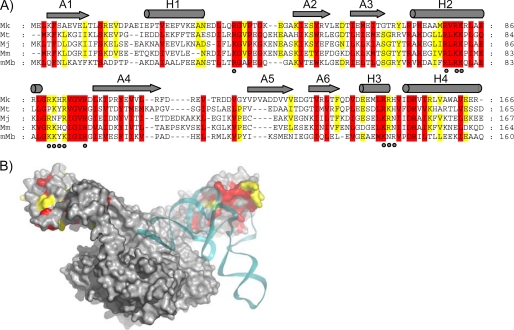
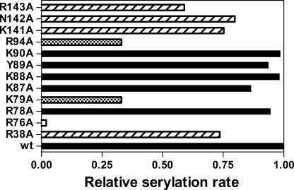
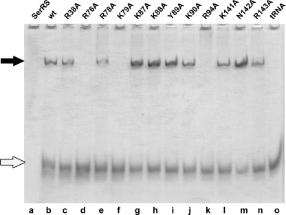
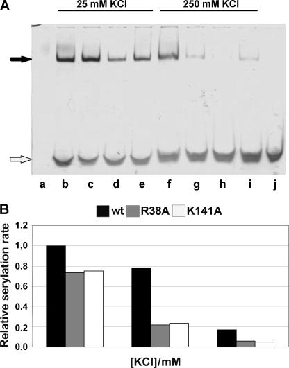
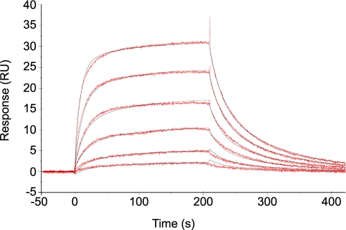
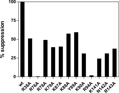
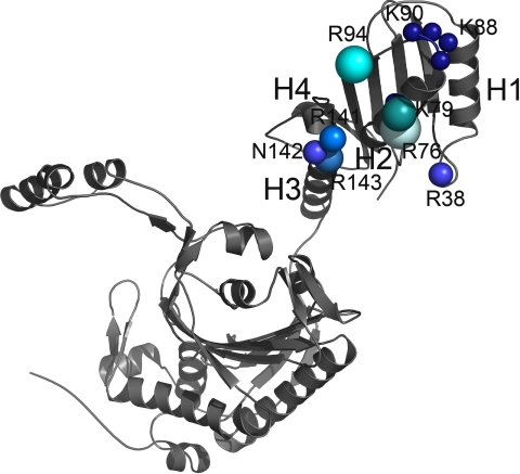
Seryl-tRNA synthetase (SerRS) from methanogenic archaeon Methanosarcina barkeri, contains an idiosyncratic N-terminal domain, composed of an antiparallel beta-sheet capped by a helical bundle, connected to the catalytic core by a short linker peptide. It is very different from the coiled-coil tRNA binding domain in bacterial-type SerRS. Because the crystal structure of the methanogenic-type SerRSxtRNA complex has not been obtained, a docking model was produced, which indicated that highly conserved helices H2 and H3 of the N-terminal domain may be important for recognition of the extra arm of tRNA(Ser). Based on structural information and the docking model, we have mutated various positions within the N-terminal region and probed their involvement in tRNA binding and serylation. Total loss of activity and inability of the R76A variant to form the complex with cognate tRNA identifies Arg(76) located in helix H2 as a crucial tRNA-interacting residue. Alteration of Lys(79) positioned in helix H2 and Arg(94) in the loop between helix H2 and beta-strand A4 have a pronounced effect on SerRSxtRNA(Ser) complex formation and dissociation constants (K(D)) determined by surface plasmon resonance. The replacement of residues Arg(38) (located in the loop between helix H1 and beta-strand A2), Lys(141) and Asn(142) (from H3), and Arg(143) (between H3 and H4) moderately affect both the serylation activity and the K(D) values. Furthermore, we have obtained a striking correlation between these results and in vivo effects of these mutations by quantifying the efficiency of suppression of bacterial amber mutations, after coexpression of the genes for M. barkeri suppressor tRNA(Ser) and a set of mMbSerRS variants in Escherichia coli.


Figures

FIGURE 1.
FIGURE 1.
Design of mMbSerRS variants based on the conserved amino acid residues in the N-terminal domain of methanogenic-type SerRSs and their proximity to tRNA in mMbSerRS·tRNASer docking model. A, the structure-based sequence alignment of the mMbSerRS N-terminal domain with selected SerRS sequences derived from methanogenic archaea (Mk, Methanopyrus kandleri; Mt, Methanothermobacter thermoautotrophicus; Mj, Methanococcus jannaschii; Mm, Methanococcus maripaludis; mMb, Methanosarcina barkeri). The sequence alignment was generated using the program MUSCLE (22). Amino acids that are completely conserved are in red, whereas those with 80% conservation are in yellow. Secondary structural elements are indicated above the alignment with gray cylinders and arrows for helices and β-sheets, respectively. Mutated residues are marked with dots. B, mMbSerRS·tRNASer docking model. The SerRS dimer is shown in gray and tRNASer is colored cyan. Conserved amino acids are indicated by the same color code as in A.
FIGURE 2.
FIGURE 2.
Aminoacylation activity of mMbSerRS variants. Relative serylation rates of in vitro transcribed MbtRNASer by mMbSerRS variants are presented by horizontal bars. Mutated enzymes that retain the serylation ability comparable with the WT enzyme are colored black, mutants with the serylation rate lowered to 59–80% of the WT are designated with traverse lines, variants with a significant drop in the serylation rate (33% of the WT serylation rate) are marked with a black and white square pattern, and one which completely loses the serylation ability is highlighted white.
FIGURE 3.
FIGURE 3.
Gel mobility shift assay of the non-covalent complexes between MbtRNASer and various mMbSerRS mutants. MbtRNASer was incubated with different mMbSerRS variants (final concentration of tRNA and enzymes was 1.1 and 0.6 μm, respectively) and subjected to PAGE under native conditions: WT, lane b; R38A, lane c; R76A, lane d; R78A, lane e; K79A, lane f; K87A, lane g; K88A, lane h; Y89A, lane i; K90A, lane j; R94A, lane k; K141A, lane l; N142A, lane m; and R143A, lane n. Non-complexed mMbSerRS (8.3 pmol) and MbtRNASer (14.8 pmol) were loaded on the gel as electrophoretic mobility markers (lanes a and o, respectively). Non-covalent complexes and non-complexed tRNA are marked with black and white arrows, respectively.
FIGURE 4.
FIGURE 4.
Effect of different salt concentrations on non-covalent complex formation and serylation propensity. A, non-covalent complexes were made as described under “Experimental Procedures” and in the legend to Fig. 3, except that in the reaction mixture the concentration of KCl was varied as indicated, and then subjected to PAGE under native conditions. Non-complexed WT mMbSerRS is visible in lane a, whereas non-covalent complexes between tRNA and mMbSerRS variants in the presence of 25 mm KCl were loaded into following lanes: WT, lane b; R38A, lane c; K141A, lane d; and R143A, lane e; non-covalent complexes between tRNA and mMbSerRS variants in the presence of 250 mm KCl were loaded into following lanes: WT, lane f; R38A, lane g; K141A, lane h; and R143A, lane i; non-complexed tRNA is in lane j. Non-covalent complexes and non-complexed tRNA are marked with black and white arrows, respectively. B, relative serylation rate of the enzymes was tested at three different KCl concentrations (25, 125, and 250 mm). Activity of the WT enzyme at 25 mm KCl was taken as the reference point (100% activity). Contrary to the WT enzyme, a significant drop in the serylation rate was detected for both mutants after raising the KCl concentration from 25 to 125 mm.
FIGURE 5.
FIGURE 5.
Kinetic analysis of tRNA binding to immobilized mMbSerRS R143A monitored by a biosensor. Sensorgrams (red) were obtained for the binding of different concentrations of tRNASer (70.3–2250 nm) in 30 mm Hepes, pH 7.0, 6 mm MgCl2, and 5 mm DTT to R143A mMbSerRS. Data were fit to the two-state conformational change model (black).
FIGURE 6.
FIGURE 6.
Suppression efficiency of M. barkeri SerRS variants. Suppression efficiency was determined by measuring the β-galactosidase activity in E. coli strain XAC-A24. 100% corresponds to the β-galactosidase activity of strain XAC-A24 co-transformed with the pET15b plasmid, carrying the gene for a wild type mMbSerRS, and pTech vector, carrying the M. barkeri suppressor tRNASer sequence. Results were reported as the percentage of mutant enzyme suppression activity relative to that of the wild type enzyme.
FIGURE 7.
FIGURE 7.
Contribution of the individual amino acids in tRNA binding. One subunit of the mMbSerRS dimer is shown. The size and the brightness of the spheres designate the significance of the particular residue in tRNA binding according to the cumulative influence on biochemical properties of mMbSerRS. The largest and the brightest sphere (arginine 76) annotates the most important side chain in tRNA recognition.

Similar articles

Cited by

References

    1. Ibba M., Soll D. (2000) Annu. Rev. Biochem. 69, 617–650 - PubMed
    1. Eriani G., Delarue M., Poch O., Gangloff J., Moras D. (1990) Nature 347, 203–206 - PubMed
    1. Cusack S., Berthet-Colominas C., Härtlein M., Nassar N., Leberman R. (1990) Nature 347, 249–255 - PubMed
    1. Ibba M., Morgan S., Curnow A. W., Pridmore D. R., Vothknecht U. C., Gardner W., Lin W., Woese C. R., Söll D. (1997) Science 278, 1119–1122 - PubMed
    1. Zhang C. M., Perona J. J., Ryu K., Francklyn C., Hou Y. M. (2006) J. Mol. Biol. 361, 300–311 - PubMed

Publication types

LinkOut - more resources