. 2009 Oct;37(19):6540-9.
doi: 10.1093/nar/gkp685. Epub 2009 Sep 4.

SrmB, a DEAD-box helicase involved in Escherichia coli ribosome assembly, is specifically targeted to 23S rRNA in vivo

Affiliations

SrmB, a DEAD-box helicase involved in Escherichia coli ribosome assembly, is specifically targeted to 23S rRNA in vivo

Dmitrii Trubetskoy et al. Nucleic Acids Res. 2009 Oct.

Abstract

DEAD-box proteins play specific roles in remodeling RNA or ribonucleoprotein complexes. Yet, in vitro, they generally behave as nonspecific RNA-dependent ATPases, raising the question of what determines their specificity in vivo. SrmB, one of the five Escherichia coli DEAD-box proteins, participates in the assembly of the large ribosomal subunit. Moreover, when overexpressed, it compensates for a mutation in L24, the ribosomal protein (r-protein) thought to initiate assembly. Here, using the tandem affinity purification (TAP) procedure, we show that SrmB forms a complex with r-proteins L4, L24 and a region near the 5'-end of 23S rRNA that binds these proteins. In vitro reconstitution experiments show that the stability of this complex reflects cooperative interactions of SrmB with L4, L24 and rRNA. These observations are consistent with an early role of SrmB in assembly and explain the genetic link between SrmB and L24. Besides its catalytic core, SrmB possesses a nonconserved C-terminal extension that, we show, is not essential for SrmB function and specificity. In this regard, SrmB differs from DbpA, another DEAD-box protein involved in ribosome assembly.


Figures
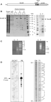
Figure 1.
Figure 1.
Identification of SrmB partners by TAP purification. (A) Schematic representation of the SrmB-TAP fusion protein. The TAP tag consists of a calmodulin-binding peptide (CBP), a TEV cleavage site (TEV) and the IgG-binding domain of protein A (ProtA). (B) Visualization of the proteins interacting with SrmB. (Left) Proteins from crude extracts (‘Extract’, 1/1000 of the sample), after the first IgG column (‘IgG’, 1/50 of the sample) and from three TAP eluate fractions (whole sample) were resolved by denaturing PAGE and stained with Coomassie. C and T stand for Control (empty vector) and TAP (vector expressing SrmB-TAP protein) strains, respectively. Left and right panels correspond to two experiments yielding different protein patterns (see text). (C) Analysis of SrmB-interacting RNA species. RNA from the same elution fractions as in (B) was separated on a 1% agarose gel and stained with ethidium bromide. The left and right panels correspond to the same two experiments as in (B). (D) The ∼0.5-kb RNA species co-eluting with SrmB [(C), left panel] corresponds to domain I of 23S rRNA. Left, Primer extension (PE) analysis of the ∼0.5-kb RNA. A sequencing ladder (GCAT) obtained with the same primer from plasmid pNO2680, which carries the rrnB operon, was run in parallel. The 5′-end of mature 23S rRNA (23S 5′-end) is indicated. Right, Partial digestion of the ∼0.5-kb RNA with RNase T1, which cleaves 3′ to guanine residues. The digest (T1), together with a sample of the same RNA after limited alkaline hydrolysis (OH), was analyzed by urea–PAGE. The sequence of 23S rRNA from nucleotide 15 (bottom) to 50 (top) is shown on the right, with guanine residues (G) facing the corresponding RNase T1 fragments. In this and other figures, dotted, vertical lines mean that lanes from the same gel have been brought together at this position.
Figure 2.
Figure 2.
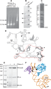
Characterization of a minimal SrmB complex. (A) The SrmB complex was treated at 0°C with different RNase A concentrations [from 0 (−) to 0.5 µg/ml] and further purified on a calmodulin column. The RNA from elution fractions was analyzed as in Figure 1C. (B) The purified ∼0.2-kb fragment (0.05 µg/ml RNase A treatment) was run on a denaturing polyacrylamide gel, transferred to a nylon membrane and probed with the 5′-end-labeled oligonucleotides a-e (see below). Hybridization signals are indicated by arrows. An in vitro transcript corresponding to domain I of 23S rRNA was used as a positive control. (C) The ∼0.2-kb RNA species was labeled at its 5′ end and digested with RNase T1. The sequence of 23S rRNA from nt 263 (bottom) to nt 291 (top) is shown on the left of the gel, with guanine residues (G) facing the corresponding RNase T1 fragments. Note that the sequence at position 264 (marked [G]) is highly heterogeneous in E. coli. We assume that G predominates in the E. coli B strain used here, like in E. coli 0157:H7 but unlike in E. coli K12. (D) Secondary structure of E. coli 23S rRNA domain I. Regions complementary to the probes a–e used in the northern blot analysis (B) are highlighted. The fragment resulting from mild RNase A treatment of the SrmB complex is surrounded in red. Nucleotides that, based on biochemical evidence (16), interact with L4 (319–322) and L24 (298–301 and 337–338) are shown as circles and triangles, respectively. (E) Protein content of the SrmB complex after mild RNase treatment. (+) 0.05 µg/ml RNase A. (−) control without RNase. The previously identified bands 1–3 (Figure 1B) are indicated. (F) Part of the structure of the E. coli ribosome (PDB2aw4) showing nt 200–400 of 23S rRNA (yellow) together with L4 (blue) and L24 (purple).
Figure 3.
Figure 3.
Reconstitution of the SrmB complex in vitro. (A–C) Proteins SrmB-CBP, L4 and L24 were mixed together with a 215 nt rRNA fragment encompassing the L4 and L24 binding sites (spe-RNA) or a control fragment of similar size (ctrl-RNA). SrmB-CBP was then affinity purified, and the eluate was analysed by PAGE. The ‘Input’ samples (upper panels) correspond to aliquots (20%) that were withdrawn prior to RNA addition, whereas ‘elution’ samples (lower) correspond to 50% of the eluates. The composition of each mix is shown above the corresponding lane. Positions of SrmB-CBP (SrmB), L4 and L24 are shown by arrows. Blue and brownish backgrounds in (A) and (C) correspond to Coomassie or silver staining of the same gels. The stars shown in (B) pinpoint traces of L4 and L24 that are recovered in the control lacking SrmB-CBP. (D) Fluorescence anisotropy assays. The fluorescent anisotropy of Alexa-SrmB is plotted versus the concentrations of (I+II) or 16S RNA, with or without added r-proteins L4, L24 or L20 (‘RNP’ or ‘RNA’, respectively).
Figure 4.
Figure 4.
The C-terminal extension of SrmB is dispensable for ribosome assembly. (A) SrmB-specific residues are essentially located within the helicase core. Upper frame: alignment of the E. coli SrmB (444 amino acids) with orthologs from representatives of four distant orders of gamma-proteobacteria. Vertical bars indicate identical residues. Besides E. coli K12 (Enterobacteriales), selected representatives were Vibrio vulnificus CMCP6 (Vibrionales), Pseudoalteromonas haloplanktis TAC125 (Alteromonadales), Aeromonas hydrophila ATCC7966 (Aeromonadales) and Haemophilus influenzae Rd KW20 (Pasteurellales) (the pattern remains largely invariant upon changing representatives within the same orders). Orthologs were identified by their high BLAST scores and aligned with CLUSTALW to the E. coli protein. Shown above the panel is the helicase DEAD-box core, assumed to extend down to amino acid 368, after a predicted alpha-helix that is conserved in known structures of DEAD-box proteins. The start of the ΔC truncation is indicated by an open triangle. Lower frame: same as upper frame, except that SrmB was aligned with the four other E. coli DEAD-box proteins: identity in this case is largely restricted to the DEAD-box motifs [horizontal bars; see (1)]. (B) Same as [(A), upper frame], except that orthologs of DbpA were aligned. The bacteria used for alignment are the same as in (A), except that Haemophilus influenzae, which lacks a DbpA ortholog (8) was replaced by Pseudomonas aeruginosa PAO1 (Pseudomonadales). The DbpA orthlogs used here (457–462 amino acids) are much closer in length than the SrmB orthologs (407–444 residues), due to the homogeneous size of their C-terminal extensions. (C) ΔsrmB cells transformed with either pCL1920 or its derivatives carrying the wild-type or srmB ΔC-TAP gene, were grown at 30°C. Ribosome profiles show the 30S and 50S subunits, 70S ribosomes (free couples and monosomes), polysomes, as well as the aberrant 40S particles. The ΔsrmB ribosome assembly defect (deficit of 50S subunits and accumulation of 40S particles) is fully corrected by the wild-type and ΔC proteins. (D) Deletion of the SrmB C-terminal extension weakens the SrmB complex. (a) Proteins co-eluting with wild-type and truncated SrmB during TAP purification were analysed as in Figure 1B. Previously identified bands 1–3 (see Figure 1B) are indicated. (b) Crude extracts and eluates from cells expressing wild-type or truncated SrmB were analyzed by western blotting using L4 and L24 antibodies. (c) RNA co-eluting with wild-type and truncated SrmB was visualized as in Figure 1C, except that a 6% polyacrylamide-urea gel stained was used.

Similar articles

Cited by

References

    1. Cordin O, Banroques J, Tanner NK, Linder P. The DEAD-box protein family of RNA helicases. Gene. 2006;367:17–37. - PubMed
    1. Jankowsky E, Fairman ME. RNA helicases–one fold for many functions. Curr. Opin. Struct. Biol. 2007;17:316–324. - PubMed
    1. Diges CM, Uhlenbeck OC. Escherichia coli DbpA is an RNA helicase that requires hairpin 92 of 23S rRNA. EMBO J. 2001;20:5503–5512. - PMC - PubMed
    1. Nicol SM, Fuller-Pace FV. The “DEAD box” protein DbpA interacts specifically with the peptidyltransferase center in 23S rRNA. Proc. Natl Acad. Sci. USA. 1995;92:11681–11685. - PMC - PubMed
    1. Tsu CA, Kossen K, Uhlenbeck OC. The Escherichia coli DEAD protein DbpA recognizes a small RNA hairpin in 23S rRNA. RNA. 2001;7:702–709. - PMC - PubMed

Publication types

MeSH terms