. 2009 Nov;297(5):H1697-710.
doi: 10.1152/ajpheart.00595.2009. Epub 2009 Sep 4.

Regulation of VASP phosphorylation in cardiac myocytes: differential regulation by cyclic nucleotides and modulation of protein expression in diabetic and hypertrophic heart

Affiliations

Regulation of VASP phosphorylation in cardiac myocytes: differential regulation by cyclic nucleotides and modulation of protein expression in diabetic and hypertrophic heart

Juliano L Sartoretto et al. Am J Physiol Heart Circ Physiol. 2009 Nov.

Abstract

Vasodilator-stimulated phosphoprotein (VASP) is a major substrate for cyclic nucleotide-dependent kinases that has been implicated in cardiac pathology, yet many aspects of VASP's molecular regulation in cardiomyocytes are incompletely understood. In these studies, we explored the role of VASP, both in signaling pathways in isolated murine myocytes, as well as in a model of cardiac hypertrophy in VASP(null) mice. We found that the beta-adrenergic agonist isoproterenol promotes the rapid and reversible phosphorylation of VASP at Ser157 and Ser239. Forskolin and the cAMP analog 8-(4-chlorophenylthio)-cAMP promote a similar pattern of VASP phosphorylation at both sites. The effects of isoproterenol are blocked by atenolol and by compound H-89, an inhibitor of the cAMP-dependent protein kinase. By contrast, phosphorylation of VASP only at Ser239 is seen following activation of particulate guanylate cyclase by atrial natriuretic peptide, or following activation of soluble guanylate cyclase by sodium nitroprusside, or following treatment of myocytes with cGMP analog. We found that basal and isoproterenol-induced VASP phosphorylation is entirely unchanged in cardiomyocytes isolated from either endothelial or neuronal nitric oxide synthase knockout mice. In cardiomyocytes isolated from diabetic mice, only basal VASP phosphorylation is increased, whereas, in cells isolated from mice subjected to ascending aortic constriction (AAC), we found a significant increase in basal VASP expression, along with an increase in VASP phosphorylation, compared with cardiac myocytes isolated from sham-operated mice. Moreover, there is further increase in VASP phosphorylation in cells isolated from hypertrophic hearts following isoproterenol treatment. Finally, we found that VASP(null) mice subjected to transverse aortic constriction develop cardiac hypertrophy with a pattern similar to VASP(+/+) mice. Our findings establish differential receptor-modulated regulation of VASP phosphorylation in cardiomyocytes by cyclic nucleotides. Furthermore, these studies demonstrate for the first time that VASP expression is upregulated in hypertrophied heart.


Figures
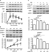
Fig. 1.
Fig. 1.
Time-concentration responses for isoproterenol (ISO)-mediated vasodilator-stimulated phosphoprotein (VASP) phosphorylation and β1-adrenergic receptor blockade effect on ISO-induced VASP phosphorylation. Shown are the results of immunoblots analyzed in lysates prepared from adult murine cardiac myocytes treated with ISO (100 nM) for the indicated times (A) or concentrations (B; 5-min incubation with ISO), and treated with the β1-adrenergic antagonist atenolol (ATE) at the indicated concentrations and then treated with ISO (C; 100 nM for 5 min). Cell lysates were analyzed in immunoblots probed using antibodies directed against phospho-VASP (pVASP) Ser157, pVASP Ser239, total VASP, Akt, and GAPDH, as indicated. The experiment shown is representative of three independent experiments that yielded similar results. Below each immunoblot are the results of densitometric analyses from pooled data, showing the fold increase in VASP phosphorylation (in arbitrary units) in cardiac myocytes treated with ISO at the indicated times (A), ISO concentrations (B), or ATE concentrations (D) plotted relative to the signals present in unstimulated cells. Each data point represents the mean ± SE derived from three independent experiments. The results are significant at the P < 0.05 level. *P < 0.05, **P < 0.01, and ***P < 0.001 for VASP phosphorylation vs. unstimulated cells (ANOVA).
Fig. 2.
Fig. 2.
Effects of the adenylate cyclase/cAMP/PKA pathway on VASP phosphorylation in cardiac myocytes. A, C, E, and G: results of immunoblots analyzed in lysates prepared from cardiac myocytes isolated from adult mice treated with ISO (100 nM, 5 min), forskolin (10 μM, 5 min), 8-(4-chlorophenylthio)adenosine-3′,5′-cyclic monophosphate acetoxymethyl ester (8-CPT-cAMP-AM; 10 μM, 5 min), PKA inhibitor H-89 (10 μM, 30 min), sildenafil (SIL; 20 μM, 30 min), or 3-isobutyl-1-methylxanthine (IBMX; 1 mM, 30 min). Cell lysates were analyzed in immunoblots probed using antibodies directed against pVASP Ser157, pVASP Ser239, total VASP, Akt, or GAPDH, as indicated. The experiment shown is representative of three independent experiments each that yielded similar results. B, D, F, and H: results of densitometric analyses from pooled data, plotting the fold increase in VASP phosphorylation (in arbitrary units) relative to the signals present in unstimulated cells. Each data point represents mean ± SE derived from three independent experiments. The results are significant at the P < 0.05 level (ANOVA). eNOS, endothelial nitric oxide synthase (NOS).
Fig. 3.
Fig. 3.
Time concentration response for ISO-mediated eNOS phosphorylation at Ser1177 and the effects of NOS or guanylate cyclase inhibitor on ISO-induced VASP phosphorylation. This figure shows the results of time course (treated with 100 nM isoproterenol; A) and dose-response (treated with ISO for 5 min; B) experiments for ISO-stimulated eNOS Ser1177 phosphorylation in murine cardiac myocytes. C and E: results from cardiac myocytes incubated either with NG-nitro-l-arginine methyl ester (l-NAME; 1 mM, 30 min) or with soluble guanylate cyclase inhibitor 1H-[1,2,4]oxadiazolo[4,3-a]quinoxalin-1-one (ODQ; 10 μM, 30 min), respectively, and then treated with ISO (100 nM, 5 min). Cell lysates were resolved by SDS-PAGE and probed using antibodies directed against phospho-eNOS (peNOS) Ser1177, total eNOS, pVASP Ser157, pVASP Ser239, total VASP, Akt, and GAPDH, as indicated. The experiment shown is representative of three independent experiments that yielded similar results. Below each immunoblot is shown the results of densitometric analyses from pooled data, plotting the fold increase of the degree of phosphorylation (in arbitrary units) of eNOS at the times (A) and concentrations (B) indicated, relative to the signals present in unstimulated cardiac myocytes. Each data point represents the mean ± SE derived from three independent experiments. *P < 0.05 and **P < 0.01 for eNOS phosphorylation vs. unstimulated cells (ANOVA). D and F: results of densitometric analyses from pooled data, plotting the fold increase in VASP phosphorylation relative to the signals present in the untreated cells. Each data point represents the mean ± SE derived from three independent experiments (ANOVA).
Fig. 4.
Fig. 4.
Isoproterenol-induced VASP phosphorylation in cardiac myocytes isolated from eNOS or neuronal NOS (nNOS) knockout mice. This figure shows results of immunoblots analyzed in lysates prepared from cardiac myocytes isolated from wild-type, endothelial (eNOSnull), or neuronal (nNOSnull) NOS knockout adult mice and treated with ISO (100 nM, 5 min). Cell lysates were resolved by SDS-PAGE and probed using antibodies directed against pVASP Ser157, pVASP Ser239, total VASP, eNOS, Akt, or GAPDH, as indicated. A and C: experiment shown are representative of three independent experiments each that yielded similar results. B and D: results of densitometric analyses from pooled data, plotting the fold increase in VASP phosphorylation relative to the signals present in the untreated cells. Each data point represents the mean ± SE derived from three independent experiments (ANOVA).
Fig. 5.
Fig. 5.
Sodium nitroprusside (SNP) or atrial natriuretic peptide (ANP) promotes phosphorylation of VASP Ser239 in cardiac myocytes. This figure shows results of immunoblots analyzed in lysates prepared from isolated murine cardiac myocytes treated with SNP (1 μM) for the indicated times. A: cells treated with the soluble guanylate cyclase inhibitor ODQ (10 μM, 30 min) and then incubated with SNP (1 μM, 5 min). C: cells treated with ANP (1 μM) for the indicated times. Results are shown of cells treated with forskolin (10 μM, 5 min; E) or 8-bromo-cGMP (8-Br-cGMP; 200 μM, 15 min; G). Cell lysates were resolved by SDS-PAGE and probed using antibodies directed against pVASP Ser157, pVASP Ser239, or total VASP, as shown. B, D, F, and H: results of densitometric analyses from pooled data, plotting the fold increase in VASP phosphorylation relative to the signals present in unstimulated cells. Each data point represents the mean ± SE derived from three independent experiments. *P < 0.05, **P < 0.01, and ***P < 0.001 for pVASP239 vs. unstimulated cells (ANOVA). The experiment shown is representative of three independent experiments that yielded similar results.
Fig. 6.
Fig. 6.
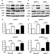
VASP phosphorylation in cardiac myocytes isolated from db/db obese diabetic mice. This figure shows results of immunoblots analyzed in lysates prepared from cardiac myocytes isolated from db/db obese diabetic mice or their wild-type littermates (db/+), lean nondiabetic mice, and treated either with ISO (100 nM for 5 min) or H-89 (10 μM for 30 min). A: cell lysates were resolved by SDS-PAGE and probed using antibodies directed against pVASP Ser157, pVASP Ser239, total VASP, actin, or PKA, as indicated. The experiment is representative of five independent experiments that yielded similar results. B: results of densitometric analyses from pooled data, plotting the total VASP (no treatment) abundance relative to actin abundance. Each data point represents the mean ± SE derived from five independent experiments (t-test). C: the results of densitometric analyses from pooled data, plotting the fold increase in pVASP Ser157 or Ser239 relative to the signals present in untreated cells. D and E: results of immunoblots analyzed in lysates prepared from cardiac myocytes isolated from db/db obese diabetic mice (E) or their wild-type littermates (db/+; D) and treated with ISO (100 nM for 5 min), insulin (INS; 1 μM for 15 min), or ANP (1 μM for 15 min). Each data point represents the mean ± SE derived from five independent experiments (ANOVA). VEH, vehicle.
Fig. 7.
Fig. 7.
MENA (mammalian homolog of Enabled)/VASP expression and VASP phosphorylation in cardiac myocytes isolated from hypertrophic hearts. This figure shows results of immunoblots analyzed in lysates prepared from cardiac myocytes isolated from mice subjected to ascending aortic constriction (AAC) or sham-operated mice. A: the time line from AAC surgery to cardiac myocyte isolation. B and C: echocardiographic analyses from AAC and sham-operated mice, including diastolic interventricular septal thickness (IVS-d; B) and left ventricular fractional shortening (LV-FS; C). D: cell lysates were resolved by SDS-PAGE and probed using antibodies directed against pVASP Ser157, pVASP Ser239, total VASP, MENA, actin, GAPDH, and PKA, as indicated. The experiment is representative of six independent experiments that yielded similar results. E: results of densitometric analyses from pooled data, plotting the total VASP or MENA abundance relative to GAPDH abundance. F: results of densitometric analyses from pooled data, plotting the pVASP Ser157 or Ser239 abundance relative to GAPDH abundance. Each data point represents the mean ± SE derived from six independent experiments (t-test). The number of mice per group is shown inside bars of B and C.
Fig. 8.
Fig. 8.
ISO-induced VASP phosphorylation in cardiac myocytes isolated from hypertrophic hearts. This figure shows results of immunoblots analyzed in lysates prepared from cardiac myocytes isolated from mice subjected to AAC or sham-operated mice treated with ISO (100 nM for 5 min). A: cell lysates were resolved by SDS-PAGE and probed using antibodies directed against pVASP Ser157, pVASP Ser239, total VASP, actin, or PKA, as indicated. The experiment is representative of four independent experiments that yielded similar results. B: results of densitometric analyses from pooled data, plotting the fold increase in pVASP Ser157 or Ser239 relative to the signals present in untreated cells. Each data point represents the mean ± SE derived from four independent experiments (ANOVA).
Fig. 9.
Fig. 9.
Blood pressure, heart rate, and development of cardiac hypertrophy induced by transverse aortic constriction (TAC) in VASP+/+ and VASPnull mice. A and B: basal mean blood pressure and basal heart rate in awake VASP+/+ and VASPnull mice, respectively. C: the time line from TAC surgery to the two-dimensional echocardiogram analyses and cardiac myocyte isolation. D and E: echocardiographic parameters measured in TAC mice 2 wk before surgery (pre); 2 wk postsurgery (post 2 wks); and 5 wk postsurgery (post 5 wks). IVS-d (D) and LV FS (E) are shown. F: the results of immunoblots analyzed in lysates and hematoxylin/eosin staining of representative cross sections of cardiac myocytes from VASP+/+ and VASPnull mice. Cell lysates were resolved by SDS-PAGE and probed using antibodies directed against total VASP and GAPDH, as indicated. Each data point represents the mean ± SE (ANOVA). The number of mice per group is shown inside the bars. BPM, heart rate in beats per minute.

Similar articles

Cited by

References

    1. An D, Rodriguez B. Role of changes in cardiac metabolism in development of diabetic cardiomyopathy. Am J Physiol Heart Circ Physiol 291: H1489–H1506, 2006 - PubMed
    1. Aszodi A, Pfeifer A, Ahmad M, Glauner M, Zhou XH, Ny L, Andersson KE, Kehrel B, Offermanns S, Fassler R. The vasodilator-stimulated phosphoprotein (VASP) is involved in cGMP- and cAMP-mediated inhibition of agonist-induced platelet aggregation, but is dispensable for smooth muscle function. EMBO J 18: 37–48, 1999 - PMC - PubMed
    1. Belhassen L, Feron O, Kaye DM, Michel T, Kelly RA. Regulation by cAMP of post-translational processing and subcellular targeting of endothelial nitric-oxide synthase (type 3) in cardiac myocytes. J Biol Chem 272: 11198–11204, 1997 - PubMed
    1. Blume C, Benz PM, Walter U, Ha J, Kemp BE, Renne T. AMP-activated protein kinase impairs endothelial actin cytoskeleton assembly by phosphorylating vasodilator-stimulated phosphoprotein. J Biol Chem 282: 4601–4612, 2007 - PubMed
    1. Brancaccio M, Hirsch E, Notte A, Selvetella G, Lembo G, Tarone G. Integrin signalling: the tug-of-war in heart hypertrophy. Cardiovasc Res 70: 422–433, 2006 - PubMed

Publication types

MeSH terms

Substances

LinkOut - more resources