Retroviral transfer of human CD20 as a suicide gene for adoptive T-cell therapy
- PMID: 19734426
- PMCID: PMC2738728
- DOI: 10.3324/haematol.2008.001677
Retroviral transfer of human CD20 as a suicide gene for adoptive T-cell therapy
Abstract
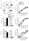
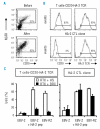
The aim of adoptive T-cell therapy of cancer is to selectively confer immunity against tumor cells. Autoimmune side effects, however, remain a risk, emphasizing the relevance of a suicide mechanism allowing in vivo elimination of infused T cells. We investigated the use of human CD20 as suicide gene in T-lymphocytes. Potential effects of forced CD20 expression on T-cell function were investigated by comparing CD20- and mock-transduced cytomegalovirus (CMV) specific T cells for cytolysis, cytokine release and proliferation. The use of CD20 as suicide gene was investigated in CMV specific T cells and in T cells genetically modified with an antigen specific T-cell receptor. No effect of CD20 on T-cell function was observed. CD20-transduced T cells with and without co-transferred T-cell receptor were efficiently eliminated by complement dependent cytotoxicity induced by therapeutic anti-CD20 antibody rituximab. The data support the broad value of CD20 as safety switch in adoptive T-cell therapy.
Figures


Similar articles
-
The CD20/alphaCD20 'suicide' system: novel vectors with improved safety and expression profiles and efficient elimination of CD20-transgenic T cells.Gene Ther. 2006 May;13(9):789-97. doi: 10.1038/sj.gt.3302705. Gene Ther. 2006. PMID: 16421601
-
Genetic modification of human T cells with CD20: a strategy to purify and lyse transduced cells with anti-CD20 antibodies.Hum Gene Ther. 2000 Mar 1;11(4):611-20. doi: 10.1089/10430340050015798. Hum Gene Ther. 2000. PMID: 10724039
-
Elongation factor 1 (EF1alpha) promoter in a lentiviral backbone improves expression of the CD20 suicide gene in primary T lymphocytes allowing efficient rituximab-mediated lysis.Haematologica. 2004 Jan;89(1):86-95. Haematologica. 2004. PMID: 14754610
-
Anti-CD20 monoclonal antibodies: beyond B-cells.Blood Rev. 2013 Sep;27(5):217-23. doi: 10.1016/j.blre.2013.07.002. Epub 2013 Aug 13. Blood Rev. 2013. PMID: 23953071 Review.
-
Rituximab: a review of its use in non-Hodgkin's lymphoma and chronic lymphocytic leukaemia.Drugs. 2003;63(8):803-43. doi: 10.2165/00003495-200363080-00005. Drugs. 2003. PMID: 12662126 Review.
Cited by
-
Chimeric Antigen Receptor T Cells and Hematopoietic Cell Transplantation: How Not to Put the CART Before the Horse.Biol Blood Marrow Transplant. 2017 Feb;23(2):235-246. doi: 10.1016/j.bbmt.2016.09.002. Epub 2016 Sep 13. Biol Blood Marrow Transplant. 2017. PMID: 27638367 Free PMC article. Review.
-
The Art and Science of Selecting a CD123-Specific Chimeric Antigen Receptor for Clinical Testing.Mol Ther Methods Clin Dev. 2020 Jun 30;18:571-581. doi: 10.1016/j.omtm.2020.06.024. eCollection 2020 Sep 11. Mol Ther Methods Clin Dev. 2020. PMID: 32775492 Free PMC article.
-
CAR-T cell development for Cutaneous T cell Lymphoma: current limitations and potential treatment strategies.Front Immunol. 2022 Aug 18;13:968395. doi: 10.3389/fimmu.2022.968395. eCollection 2022. Front Immunol. 2022. PMID: 36059451 Free PMC article. Review.
-
CA215 and GnRH receptor as targets for cancer therapy.Cancer Immunol Immunother. 2012 Oct;61(10):1805-17. doi: 10.1007/s00262-012-1230-8. Epub 2012 Mar 21. Cancer Immunol Immunother. 2012. PMID: 22430628 Free PMC article.
-
Feasibility of controlling CD38-CAR T cell activity with a Tet-on inducible CAR design.PLoS One. 2018 May 30;13(5):e0197349. doi: 10.1371/journal.pone.0197349. eCollection 2018. PLoS One. 2018. PMID: 29847570 Free PMC article.
References
-
- Bonini C, Ferrari G, Verzeletti S, Servida P, Zappone E, Ruggieri L, et al. HSV-TK gene transfer into donor lymphocytes for control of allogeneic graft-versus-leukemia. Science. 1997;276:1719–24. - PubMed
-
- Bonini C, Bondanza A, Perna SK, Kaneko S, Traversari C, Ciceri F, et al. The suicide gene therapy challenge: how to improve a successful gene therapy approach. Mol Ther. 2007;15:1248–52. - PubMed
-
- Schumacher TN. T-cell-receptor gene therapy. Nat Rev Immunol. 2002;2:512–9. - PubMed
-
- Heemskerk MHM, Hoogeboom M, de Paus RA, Kester MGD, van der Hoorn MAWG, Goulmy E, et al. Redirection of antileukemic reactivity of peripheral T lymphocytes using gene transfer of minor histocompatibility antigen HA-2-specific T-cell receptor complexes expressing a conserved alpha joining region. Blood. 2003;102:3530–40. - PubMed
MeSH terms
Substances
LinkOut - more resources
Full Text Sources
Other Literature Sources

