. 2009 Oct;15(10):1163-9.
doi: 10.1038/nm.2009. Epub 2009 Sep 6.

An integrin alpha(v)beta(3)-c-Src oncogenic unit promotes anchorage-independence and tumor progression

Affiliations

An integrin alpha(v)beta(3)-c-Src oncogenic unit promotes anchorage-independence and tumor progression

Jay S Desgrosellier et al. Nat Med. 2009 Oct.

Abstract

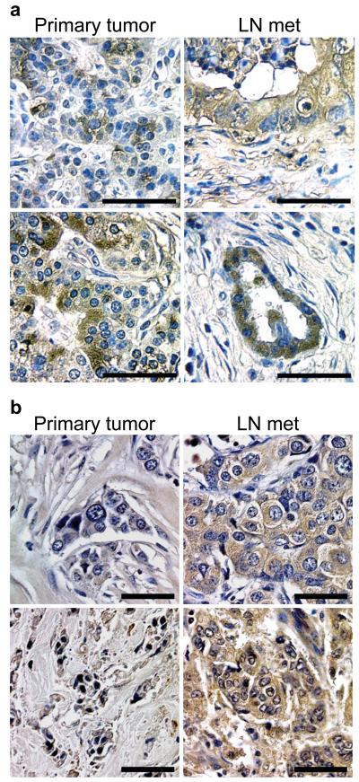
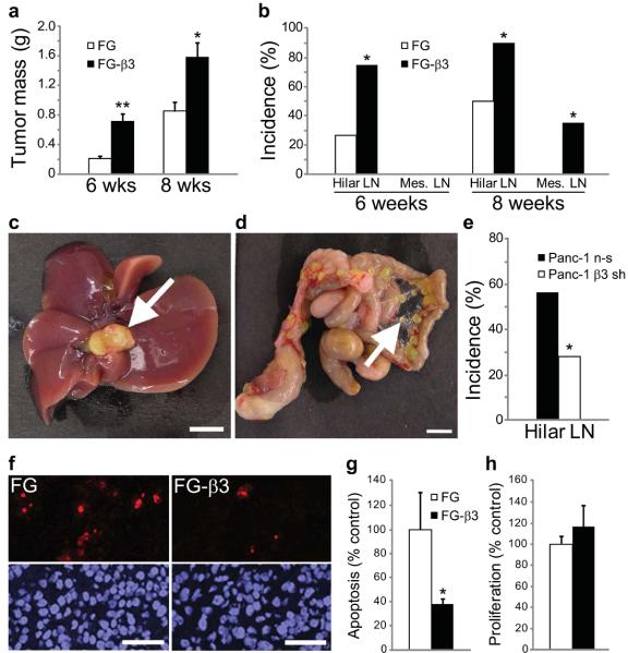
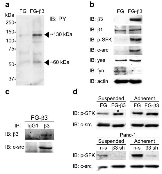
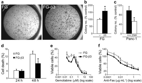
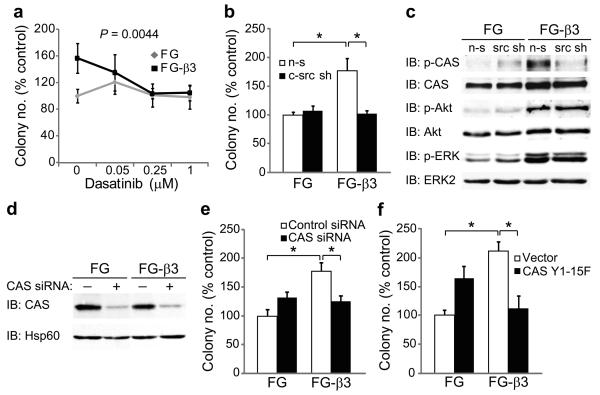
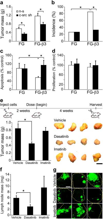
Integrins regulate adhesion-dependent growth, survival and invasion of tumor cells. In particular, expression of integrin alpha(v)beta(3) is associated with progression of a variety of human tumors. Here we reveal a previously undescribed adhesion-independent role for integrin alpha(v)beta(3) in pancreatic cancer and other carcinomas. Specifically, alpha(v)beta(3) expressed in carcinoma cells enhanced anchorage-independent tumor growth in vitro and increased lymph node metastases in vivo. These effects required recruitment of c-Src to the beta(3) integrin cytoplasmic tail, leading to c-Src activation, Crk-associated substrate (CAS) phosphorylation and tumor cell survival that, unexpectedly, was independent of cell adhesion or focal adhesion kinase (FAK) activation. Pharmacological blockade of c-Src kinase activity or decreased expression of endogenous alpha(v)beta(3) integrin or c-Src not only inhibited anchorage-independent growth but also suppressed metastasis in vivo, yet these manipulations did not affect tumor cell migration or invasion. These data define an unexpected role for an integrin as a mediator of anchorage independence, suggesting that an alpha(v)beta(3)-c-Src signaling module may account for the aggressive behavior of integrin alpha(v)beta(3)-expressing tumors in humans.


Figures

Figure 1
Figure 1
Integrin αvβ3 is expressed in a sub-population of human carcinoma cells and correlates with lymph node invasion. (a,b) Representative images of immunohistochemical staining for the integrin β3 subunit in matched pairs of primary tumors (left panels) and lymph node metastases (right panels) from pancreatic (n=7) (a) and breast (n=50) (b) cancer patients. (a) In pancreatic cancer, β3 was expressed heterogeneously throughout all of the primary tumors, however, tumor cells invading lymph nodes from the same patient were primarily β3-positive in 6 of 7 patients. (b) In breast cancer specimens, β3 was expressed in 28 of 50 primary tumors (56%) however, we observed expression of this integrin in 36 of 50 lymph node metastases (72%) in these same patients. Scale bars, 50 μm.
Figure 2
Figure 2
Integrin αvβ3 enhances pancreatic tumor progression and metastasis. (a) Primary tumor mass is enhanced in FG-β3 tumors compared to FG control tumors at both 6 and 8 weeks. (b) At both time-points we also observed enhanced spontaneous metastasis to the hepatic hilar lymph nodes in mice with FG-β3 tumors relative to FG controls. At 8 weeks we observed additional metastasis of FG-β3 cells to the mesenteric lymph nodes. 6 weeks; FG, n=15, FG-β3, n=16. 8 weeks; FG, n=20, FG-β3, n=20. *P<0.05, **P<0.001. (c,d) Representative examples of spontaneous metastases to the hepatic hilar lymph nodes (c) and the mesenteric lymph nodes (d) after orthotopic injection of FG-β3 cells. Scale bar, 5 mm. (e) Knock-down of the β3 integrin subunit in Panc-1 cells (β3 sh) reduced spontaneous metastasis to the liver hilar lymph nodes compared to Panc-1 cells expressing a non-silencing control (n-s). n-s, n=13, β3 sh, n=13. *P<0.05. (f–h) Apoptosis (TUNEL) and proliferation (Ki-67) were assayed in sections from 8 week primary tumors. (f) Representative TUNEL staining results show fewer stained cells in the FG-β3 tumor relative to the FG control. Scale Bar, 50 μm. (g,h) FG-β3 tumors exhibit decreased levels of apoptosis (g) compared to FG controls, with no difference in proliferation (h). FG, n=11; FG-β3, n=10. *P<0.05.
Figure 3
Figure 3
Integrin αvβ3 promotes anchorage-independent activation of c-src. (a) Adhesion of FG-β3 cells to fibronectin enriches for phosphotyrosine bands of 60 and 130 kDa in the triton insoluble lysate relative to FG cells. (b) Immunoblotting for pY416 SFK shows enrichment in the FG-β3 triton insoluble lysate. However, only c-src, and not yes or fyn, exhibited a similar pattern of recruitment as pY416 SFK in FG-β3 cells. (c) Co-immunoprecipitation of c-src with αvβ3 from the triton insoluble fraction of FG-β3 cells plated on the αvβ3 substrate vitronectin. (d). While expression of αvβ3 enhanced adhesion-dependent SFK activation, as expected, αvβ3 unexpectedly increased suspension levels of pY416 SFK in FG-β3 cells (asterisk) while β3 knock-down inhibited suspension SFK activation in Panc-1 cells.
Figure 4
Figure 4
Integrin αvβ3-induces anchorage-independent survival with no affect on survival of adherent cells. (a–c) Expression of αvβ3 in FG cells enhanced colony number in soft agar (a,b) while knock-down of β3 in Panc-1 cells (β3 sh) reduced colony formation compared to cells expressing a non-silencing control (n-s) (c). n=3 independent experiments. *P<0.05. (d) FG and FG-β3 cells were maintained in suspension culture for 24 and 48 h prior to trypan blue staining and counting viable and non-viable cells. Compared to FG control cells, suspension cultures of FG-β3 cells exhibit significantly less cell death. n=4 independent experiments. *P<0.05. (e,f) Ligation of αvβ3 to fibronectin failed to protect FG-β3 cells from apoptosis initiated by either gemcitabine (e) or anti-Fas antibody (CH11) (f). Representative experiments are shown.
Figure 5
Figure 5
Integrin αvβ3-induces anchorage-independence through c-src phosphorylation of CAS (a) To discern a role for c-src activation in αvβ3-induced anchorage-independent growth, FG and FG-β3 cells were treated with dasatinib, a clinically approved SFK inhibitor. Treatment with dasatinib specifically reduced colony number in FG-β3 cells compared to vehicle control (DMSO) while no effect was observed in FG cells. n=3 independent experiments. P=0.0044. (b) Knock-down of c-src (c-src sh) selectively inhibited αvβ3-mediated colony formation relative to a non-silencing control (n-s) with no effect on FG cells. n=3 independent experiments. *P<0.05. (c) Expression of αvβ3 potentiates CAS, Akt and ERK signaling in suspension cultured cells, but only CAS phosphorylation required c-src. (d,e) Knock-down of CAS with siRNA specifically decreased αvβ3-mediated growth in soft agar compared to FG control cells. A representative experiment is shown. n=3 independent experiments. *P<0.05. (f) Stable expression of a CAS mutant lacking c-src phosphorylation sites (Y1-15F) specifically inhibited FG-β3 cell colony formation. A representative experiment is shown. n=3 independent experiments. *P<0.05.
Figure 6
Figure 6
Integrin αvβ3 requires c-src for tumor cell survival and metastasis in vivo, but not for migration in vitro. (a–d) FG and FG-β3 non-silencing (n-s) and c-src knock-down cells (c-src sh) were injected into the pancreas of nude mice and assessed for primary tumor growth, metastasis, apoptosis and proliferation after 8 weeks. (a) Knock-down of c-src decreased primary tumor mass in both FG and FG-β3 tumors relative to non-silencing controls. (b) However, in these same mice, knock-down of c-src selectively inhibited αvβ3-mediated metastasis to the hepatic hilar lymph nodes with no affect on FG cells. (a,b) FG non-silencing, n=15, FG-β3 non-silencing, n=14, FG c-src shRNA, n=22, FG-β3 c-src shRNA, n=21. *P<0.05. (c) In primary tumor sections, knock-down of c-src increased apoptosis in FG-β3 tumors to the level of FG controls. (d) The percentage of proliferating cells in primary tumor sections was unaffected by c-src knock-down. (c,d) FG non-silencing, n=8, FG-β3 non-silencing, n=7, FG c-src shRNA, n=7, FG-β3 c-src shRNA, n=6; *P<0.05. (e–g) Treatment with the SFK inhibitor dasatinib decreased FG-β3 cell primary tumor mass (e) and metastasis to the liver hilar lymph nodes (f,g) while imatinib had no effect. Representative tumors are shown in (e). The three largest lymph node metastases from each group are compared in (g). Vehicle, n=12, Dasatinib, n=12, Imatinib, n=12; *P<0.05. Scale bar, 1 mm.

References

    1. Guo W, Giancotti FG. Integrin signalling during tumour progression. Nat Rev Mol Cell Biol. 2004;5:816–826. - PubMed
    1. Schlaepfer DD, Mitra SK, Ilic D. Control of motile and invasive cell phenotypes by focal adhesion kinase. Biochim Biophys Acta. 2004;1692:77–102. - PubMed
    1. Roberts WG, et al. Antitumor activity and pharmacology of a selective focal adhesion kinase inhibitor, PF-562,271. Cancer research. 2008;68:1935–1944. - PubMed
    1. Pylayeva Y, et al. Ras- and PI3K-dependent breast tumorigenesis in mice and humans requires focal adhesion kinase signaling. The Journal of clinical investigation. 2009;119:252–266. - PMC - PubMed
    1. Lahlou H, et al. Mammary epithelial-specific disruption of the focal adhesion kinase blocks mammary tumor progression. Proc Natl Acad Sci U S A. 2007;104:20302–20307. - PMC - PubMed

Publication types

MeSH terms