

A fast automatic recognition and location algorithm for fetal genital organs in ultrasound images
- PMID: 19735097
- PMCID: PMC2738834
- DOI: 10.1631/jzus.B0930162
A fast automatic recognition and location algorithm for fetal genital organs in ultrasound images
Abstract
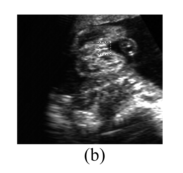
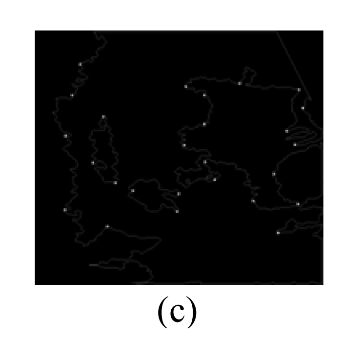
Severe sex ratio imbalance at birth is now becoming an important issue in several Asian countries. Its leading immediate cause is prenatal sex-selective abortion following illegal sex identification by ultrasound scanning. In this paper, a fast automatic recognition and location algorithm for fetal genital organs is proposed as an effective method to help prevent ultrasound technicians from unethically and illegally identifying the sex of the fetus. This automatic recognition algorithm can be divided into two stages. In the 'rough' stage, a few pixels in the image, which are likely to represent the genital organs, are automatically chosen as points of interest (POIs) according to certain salient characteristics of fetal genital organs. In the 'fine' stage, a specifically supervised learning framework, which fuses an effective feature data preprocessing mechanism into the multiple classifier architecture, is applied to every POI. The basic classifiers in the framework are selected from three widely used classifiers: radial basis function network, backpropagation network, and support vector machine. The classification results of all the POIs are then synthesized to determine whether the fetal genital organ is present in the image, and to locate the genital organ within the positive image. Experiments were designed and carried out based on an image dataset comprising 658 positive images (images with fetal genital organs) and 500 negative images (images without fetal genital organs). The experimental results showed true positive (TP) and true negative (TN) results from 80.5% (265 from 329) and 83.0% (415 from 500) of samples, respectively. The average computation time was 453 ms per image.
Figures


Similar articles
-
Detection and measurement of fetal anatomies from ultrasound images using a constrained probabilistic boosting tree.IEEE Trans Med Imaging. 2008 Sep;27(9):1342-55. doi: 10.1109/TMI.2008.928917. IEEE Trans Med Imaging. 2008. PMID: 18753047
-
Automatic segmentation of the cerebellum of fetuses on 3D ultrasound images, using a 3D Point Distribution Model.Annu Int Conf IEEE Eng Med Biol Soc. 2010;2010:4731-4. doi: 10.1109/IEMBS.2010.5626624. Annu Int Conf IEEE Eng Med Biol Soc. 2010. PMID: 21096244
-
The usefulness of ultrasound in the classification of chronic liver disease.Annu Int Conf IEEE Eng Med Biol Soc. 2011;2011:5132-5. doi: 10.1109/IEMBS.2011.6091271. Annu Int Conf IEEE Eng Med Biol Soc. 2011. PMID: 22255494
-
Learning-based prediction of gestational age from ultrasound images of the fetal brain.Med Image Anal. 2015 Apr;21(1):72-86. doi: 10.1016/j.media.2014.12.006. Epub 2015 Jan 3. Med Image Anal. 2015. PMID: 25624045 Free PMC article.
-
Segmentation of 2D fetal ultrasound images by exploiting context information using conditional random fields.Annu Int Conf IEEE Eng Med Biol Soc. 2011;2011:7219-22. doi: 10.1109/IEMBS.2011.6091824. Annu Int Conf IEEE Eng Med Biol Soc. 2011. PMID: 22256004
Cited by
-
Artificial Intelligence in Prenatal Ultrasound Diagnosis.Front Med (Lausanne). 2021 Dec 16;8:729978. doi: 10.3389/fmed.2021.729978. eCollection 2021. Front Med (Lausanne). 2021. PMID: 34977053 Free PMC article. Review.
References
-
- Barandela R, Sanchez JS, Garcia V, Rangel E. Strategies for learning in class imbalance problems. Pattern Recogn. 2003;36(3):849–851. doi: 10.1016/S0031-3203(02)00257-1. - DOI
-
- Batista GE, Prati RC, Monard MC. A study of the behavior of several methods for balancing machine learning training data. ACM SIGKDD Explor Newsl. 2004;6(1):20–29. doi: 10.1145/1007730.1007735. - DOI
-
- Bishop CM. Neural Networks for Pattern Recognition. Oxford: Oxford University Press; 1995.
-
- Blum A, Langley P. Selection of relevant features and examples in machine learning. Artif Intell. 1997;97(1-2):245–271. doi: 10.1016/S0004-3702(97)00063-5. - DOI
Publication types
MeSH terms
LinkOut - more resources
Full Text Sources
Other Literature Sources
