. 2009 Oct;74(1):159-174.
doi: 10.1111/j.1365-2958.2009.06858.x. Epub 2009 Sep 3.

Contribution of AmyA, an extracellular alpha-glucan degrading enzyme, to group A streptococcal host-pathogen interaction

Affiliations

Contribution of AmyA, an extracellular alpha-glucan degrading enzyme, to group A streptococcal host-pathogen interaction

Samuel A Shelburne Iii et al. Mol Microbiol. 2009 Oct.

Abstract

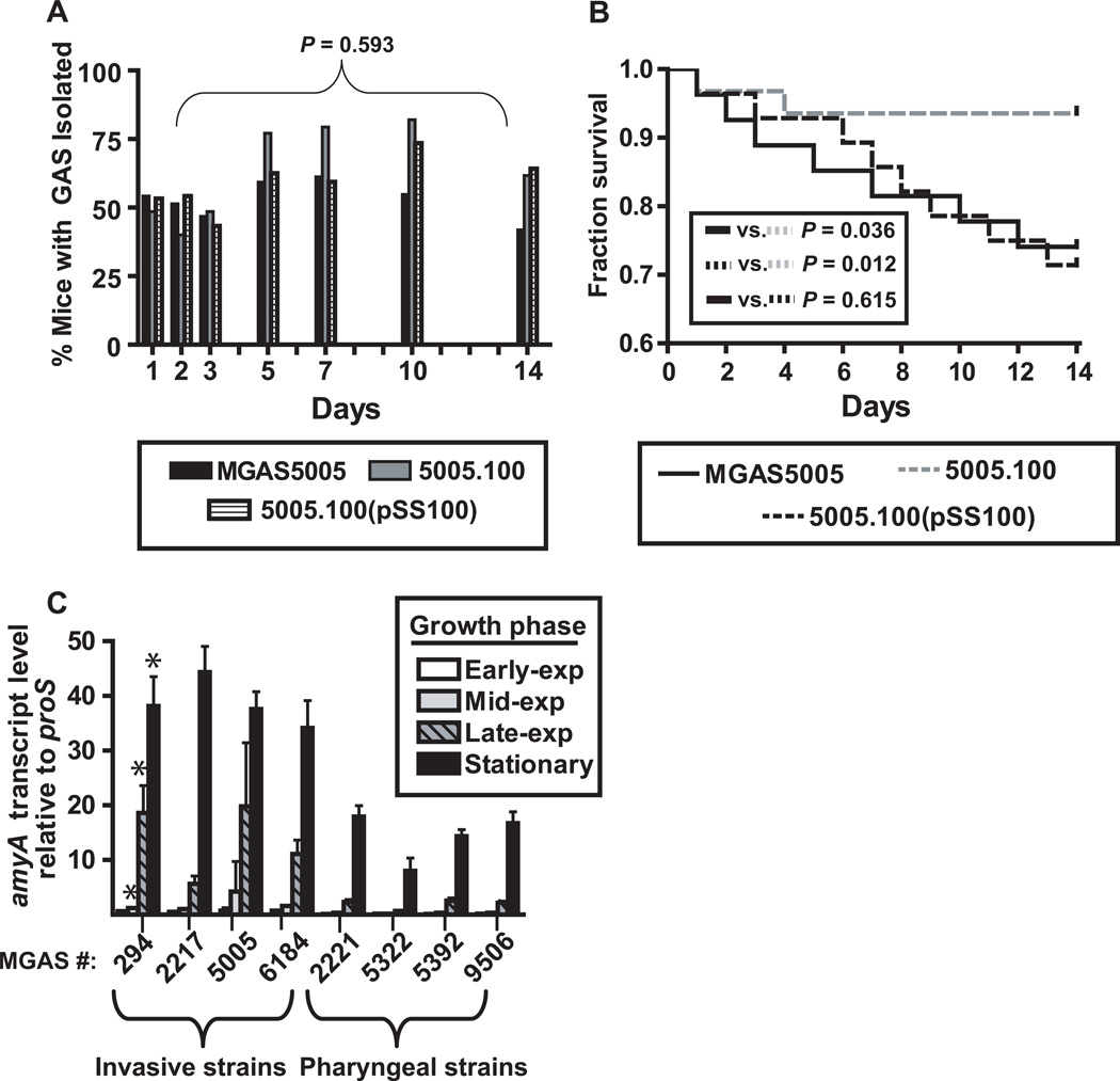
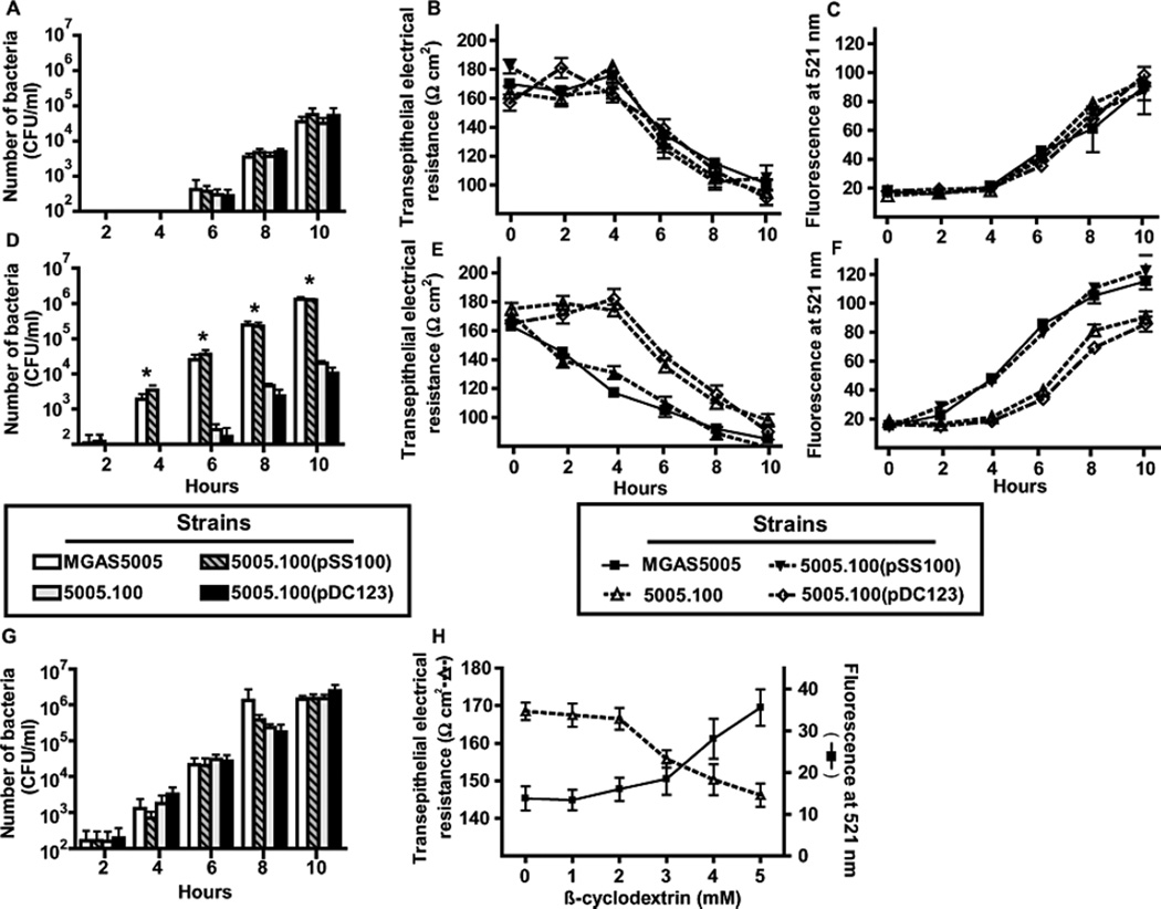
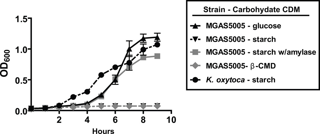
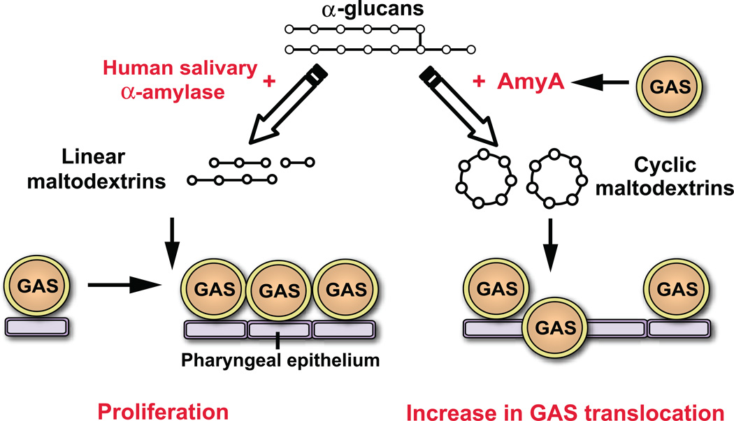
alpha-Glucans such as starch and glycogen are abundant in the human oropharynx, the main site of group A Streptococcus (GAS) infection. However, the role in pathogenesis of GAS extracellular alpha-glucan binding and degrading enzymes is unknown. The serotype M1 GAS genome encodes two extracellular proteins putatively involved in alpha-glucan binding and degradation; pulA encodes a cell wall anchored pullulanase and amyA encodes a freely secreted putative cyclomaltodextrin alpha-glucanotransferase. Genetic inactivation of amyA, but not pulA, abolished GAS alpha-glucan degradation. The DeltaamyA strain had a slower rate of translocation across human pharyngeal epithelial cells. Consistent with this finding, the DeltaamyA strain was less virulent following mouse mucosal challenge. Recombinant AmyA degraded alpha-glucans into beta-cyclomaltodextrins that reduced pharyngeal cell transepithelial resistance, providing a physiologic explanation for the observed transepithelial migration phenotype. Higher amyA transcript levels were present in serotype M1 GAS strains causing invasive infection compared with strains causing pharyngitis. GAS proliferation in a defined alpha-glucan-containing medium was dependent on the presence of human salivary alpha-amylase. These data delineate the molecular mechanisms by which alpha-glucan degradation contributes to GAS host-pathogen interaction, including how GAS uses human salivary alpha-amylase for its own metabolic benefit.


Figures

Fig. 1
Fig. 1
Schematic of the linear maltodextrin, cyclomaltodextrin and pullulanase gene regions in GAS serotype M1 strain MGAS5005 (Sumby et al., 2005). amyA and pulA genes are indicated in white. M5005_spy numbers refer to open reading frame in the serotype M1 strain MGAS5005. ABC = ATP-binding cassette.
Fig. 2
Fig. 2
AmyA α-glucan digestion results in β-cyclomaltodextrin formation. (A) GAS strains of indicated amyA and pulA genotype were grown overnight on THY agar plates supplemented with 0.5% starch. Iodine was added to plates and the presence of clearing was assessed for evidence of starch hydrolysis. (B) Colorimetric analysis of iodine staining following growth in THY supplemented with 1% starch was used to assess starch degrading activity in indicated GAS strains as described in Experimental procedures. (C) β-cyclomaltodextrin production from starch by various strains was assessed using colorimetric analysis as described in Experimental procedures. (D) GAS AmyA was cloned and overexpressed in E. coli and then purified to apparent homogeneity as described in Experimental procedures. Proteins were separated on a 12% polyacrylamide gel. (E) Degradation of starch by purified AmyA with empty pBAD-hisA vector as negative control. (F) Production of B-cyclomaltodextrin by purified AmyA with empty pBAD-hisA vector as negative control. (G) Purified AmyA was run under non-denaturing conditions as described in Experimental procedures and subsequently allowed to diffuse into a 1% starch agar plate. Iodine staining confirmed that starch degradation (bright areas) was limited to the gel locations corresponding to AmyA. Lanes are as follows; 1 = 5 µl purified AmyA, 2 = 5 μl human salivary α-amylase, 3 = 5 µl empty vector. All starch degrading experiments were performed in triplicate on four separate occasions with data graphed being mean and error bars representing standard deviation.
Fig. 3
Fig. 3
Schematic of cyclomaltodextrin (CMD) region genes in various GAS strains. (A) Overview of CMD region in sequenced GAS strains. CMD(+) sequenced strains include serotype M1, M2, M4, and M28 strains (Ferretti et al., 2001, Sumby et al., 2005, Beres et al., 2006, Green et al., 2005). CMD(-) sequenced strains include serotype M3, M5, M6, M12, M18, and M49 strains (Beres et al., 2002, McShan et al., 2008, Nakagawa et al., 2003, Holden et al., 2007, Banks et al., 2004, Smoot et al., 2002, Beres et al., 2006). CMD(+) and CMD(-) strains share CMD flanking region genes and a transposase gene (M5005_spy1068) shown in blue. All GAS strains sequenced to date have a second transposase gene (M5005_spy1862, shown in red) found near the origin of replication that is duplicated in place of the CMD region genes in CMD (-) strains. (B) PCR reactions to determine presence or absence of CMD genes. Lane designations: Serotype M1 strain MGAS5005 with malX primers (1), with CMD region flanking primers (2), with amyA primers (3); serotype M3 strain MGAS315 with malX primers (4), with CMD region flanking primers (5), with amyA primers (6). Small black arrows showing primer locations appear in (A).
Fig. 4
Fig. 4
Analysis of amyA transcript levels. (A) Serotype M1 strain MGAS5005 was grown to early-exponential (exp), mid-exponential, late-exponential, and stationary growth phase in indicated media in quadruplicate biologic replicates on three separate occasions (total of 12 samples at each time point). RNA was isolated, converted to cDNA, and transcript levels for amyA and the endogenous control gene proS were determined using TaqMan real-time PCR. Data graphed are mean +/− standard deviation. (B) amyA transcript levels for 6 patients with GAS pharyngitis were determined by TaqMan real-time PCR. The M serotype of the infecting GAS strain is shown in the circle. Data graphed are mean +/− standard deviation with each experiment done with quadruplicate technical replicates on three separate occasions (total of 12 data points per sample).
Fig. 5
Fig. 5
AmyA does not contribute to GAS colonization of the mouse oropharynx but does increase murine mortality following mucosal challenge. Adult outbred CD-1 mice (n = 35 per group) were intransally inoculated with ∼1.0x10⁷ CFU with indicated GAS strains. Mice oropharynges were swabbed daily and plated onto BSA. Plates were incubated for 24 hr, and β-hemolytic colonies were counted and tested for GAS carbohydrate antigen using latex agglutination. (A) Percentage of mice with GAS isolated by day. P value refers to repeated measures analysis. (B) Percent mice surviving graphed by data with P value referring to χ2 test done on day 14 of experiment. (C) Analysis of amyA transcript levels during growth in THY at indicated growth phase in invasive vs. pharyngeal GAS strains (for strain description see Table 1). * = P < 0.05 at the mid-logarithmic, late-logarithmic, and stationary growth phases when comparing invasive vs. pharyngeal strains. Transcript level analysis was done using quadruplicate biologic replicates on three separate occasions with data graphed being mean +/− standard deviation.
Fig. 6
Fig. 6
AmyA increases GAS transepithelial invasion in the presence of starch. GAS translocation across a D562 epithelial cell monolayer were performed as described in Experimental procedures using indicated strains. For (A), (B), and (C) experiments were performed in standard minimal medium. (A) Number of translocated GAS by indicated strain. (B) and (C) are indicators of epithelial integrity using transepithelial resistance (TEER) and fluorescent-dextran. For (D), (E), and (F) experiments were performed in minimal medium with 0.5% starch added. (G) 5 mM β-cyclomaltodextrins were added to the starch-medium. (H) TEER and fluorescent measurements in the presence indicated concentrations of β-cyclomaltodextrin without GAS. Transepithelial invasion assays were performed in six replicates per strain on four separate occasions. Data graphed are mean with error bars representing standard deviation. Asterisks indicate a P value of < 0.05 using a repeated measures analysis.
Fig. 7
Fig. 7
GAS uses human salivary α-amylase to initiate α-glucan catabolism. Data graphed are growth curves of GAS in various carbohydrate supplemented chemically defined media. Serotype M1 strain MGAS5005 was grown overnight in THY and then added at a 1:100 dilution into designated medium with growth monitored via OD600 readings. Human salivary α-amylase was added at physiologic concentrations when indicated. β-CMD = cyclomaltodextrins. Klebsiella oxytoca, which is capable of growth in starch, is included as a positive control. All growth experiments were performed in triplicate on four separate occasions with mean ± standard deviation graphed.
Fig. 8
Fig. 8
Schematic of hypothesized role for α-glucan degrading enzymes in the pathogenesis of GAS pharyngitis. Degradation of α-glucans (such as starch or glycogen) by human salivary α-amylase results in the production of linear maltodextrins which can be used as an energy source for GAS proliferation. Alternatively, GAS AmyA α-glucan digestions leads to cyclic maltodextrin formation resulting in a decrease in epithelial transepithelial resistance and subsequent GAS translocation.

References

    1. Altschul SF, Gish W, Miller W, Myers EW, Lipman DJ. Basic local alignment search tool. J Mol Biol. 1990;215:403–410. - PubMed
    1. Banks DJ, Porcella SF, Barbian KD, Beres SB, Philips LE, Voyich JM, DeLeo FR, Martin JM, Somerville GA, Musser JM. Progress toward characterization of the group A Streptococcus metagenome: complete genome sequence of a macrolide-resistant serotype M6 strain. Journal of Infectious Diseases. 2004;190:727–738. - PubMed
    1. Beisswenger C, Coyne CB, Shchepetov M, Weiser JN. Role of p38 MAP kinase and transforming growth factor-beta signaling in transepithelial migration of invasive bacterial pathogens. J Biol Chem. 2007;282:28700–28708. - PubMed
    1. Beres SB, Richter EW, Nagiec MJ, Sumby P, Porcella SF, DeLeo FR, Musser JM. Molecular genetic anatomy of inter- and intraserotype variation in the human bacterial pathogen group A Streptococcus . Proc Natl Acad Sci U S A. 2006;103:7059–7064. - PMC - PubMed
    1. Beres SB, Sesso R, Pinto SW, Hoe NP, Porcella SF, Deleo FR, Musser JM. Genome sequence of a Lancefield group C Streptococcus zooepidemicus strain causing epidemic nephritis: new information about an old disease. PLoS ONE. 2008;3:e3026. - PMC - PubMed

Publication types

MeSH terms

LinkOut - more resources