. 2010 Jan;20(1):13-23.
doi: 10.1093/glycob/cwp133. Epub 2009 Sep 7.

Analysis of differential expression of glycosyltransferases in healing corneas by glycogene microarrays

Affiliations

Analysis of differential expression of glycosyltransferases in healing corneas by glycogene microarrays

Chandrassegar Saravanan et al. Glycobiology. 2010 Jan.

Erratum in

Abstract

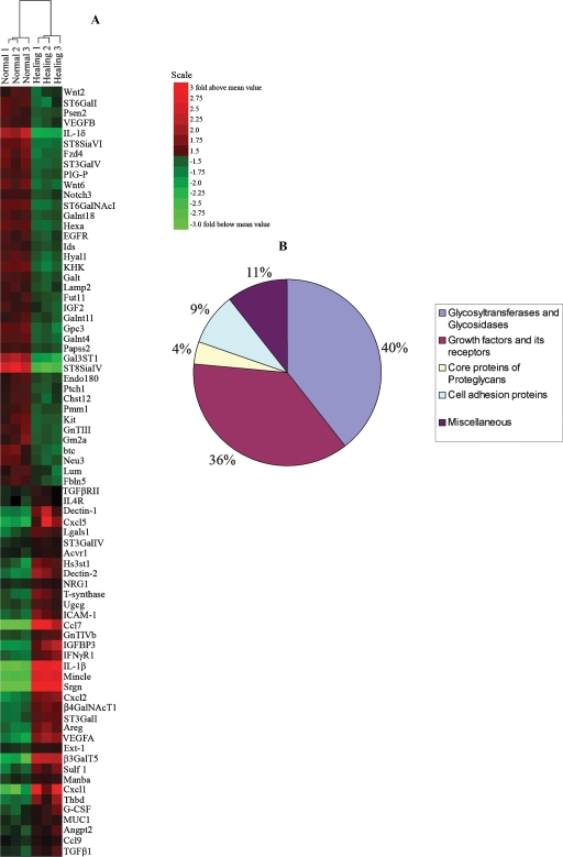
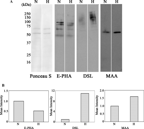
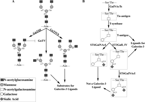
It is generally accepted that the glycans on the cell surface and extracellular matrix proteins play a pivotal role in the events that mediate re-epithelialization of wounds. Yet, the global alteration in the structure and composition of glycans, specifically occurring during corneal wound closure remains unknown. In this study, GLYCOv2 glycogene microarray technology was used for the first time to identify the differentially expressed glycosylation-related genes in healing mouse corneas. Of approximately 2000 glycogenes on the array, the expression of 11 glycosytransferase and glycosidase enzymes was upregulated and that of 19 was downregulated more than 1.5-fold in healing corneas compared with the normal, uninjured corneas. Among them, notably, glycosyltransferases, beta3GalT5, T-synthase, and GnTIVb, were all found to be induced in the corneas in response to injury, whereas, GnTIII and many sialyltransferases were downregulated. Interestingly, it appears that the glycan structures on glycoproteins and glycolipids, expressed in healing corneas as a result of differential regulation of these glycosyltransferases, may serve as specific counter-receptors for galectin-3, a carbohydrate-binding protein, known to play a key role in re-epithelialization of corneal wounds. Additionally, many glycogenes including a proteoglycan, glypican-3, cell adhesion proteins dectin-1 and -2, and mincle, and mucin 1 were identified for the first time to be differentially regulated during corneal wound healing. Results of glycogene microarray data were confirmed by qRT-PCR and lectin blot analyses. The differentially expressed glycogenes identified in the present study have not previously been investigated in the context of wound healing and represent novel factors for investigating the role of carbohydrate-mediated recognition in corneal wound healing.


Figures

Fig. 1
Fig. 1
Dendrogram showing similarities in gene expression profiles within each of the three replicates of healing and normal corneas. Six expression arrays (three healing and three normal) were hierarchically clustered with the algorithm BRB ArrayTools 3.2.2 and displayed with TreeView. The individual samples were clustered in branches of the dendrogram based on overall similarity in patterns of gene expression. Note that all normal corneal samples clustered together on one side of the dendrogram, whereas all three samples of healing corneas clustered on the other side of the dendrogram.
Fig. 2
Fig. 2
(A) Heat map of 75 differentially expressed genes (change >1.5-fold; P < 0.01). All signals are compared to a median value, and fold change from the median is visually represented by color assignment (see scale on the right side). Healing and normal corneas showed visibly distinct profiles of gene expression. (B) Pie diagram showing the classification of differentially expressed genes based on molecular functions. Categories ascribed to genes were determined using DAVID, Gene Cards, and Entrez gene. Note that 40% of the differentially expressed genes are glycosyltransferases and glycosidases.
Fig. 3
Fig. 3
(A) Lectin blots demonstrating that alteration in transcript levels of glycosyltransferases in healing corneas is associated with corresponding changes in glycan structures of glycoproteins. Protein extracts (5 μg) of normal (N) and healing (H) corneas were electrophoresed on 10% SDS–polyacrylamide gels; protein blots of the gels were stained with Ponceau S to ensure equal loading of samples and were then probed with biotinylated E-PHA, DSL, and MAA that recognize glycan products of GnTIII, GnTIV, and α2,3-sialyltransferases, respectively. Note that consistent with the increased mRNA levels of GnTIVb and α2,3-sialyltransferases (ST3GalI and ST3GalIV) and the reduced mRNA level of GnTIII detected in the healing corneas by microarrays (Table I) and qRT-PCR (Table II), the increased expression of DSL- and MAA-reactive glycoproteins and the reduced expression of E-PHA-reactive glycoproteins is detected in healing corneas compared to normal, uninjured corneas. (B) The intensity of lectin-reactive bands from the lanes of normal and healing corneal samples from each lectin blot was quantified by densitometry. A value of 1.0 was assigned to the intensity value of the lectin-reactive components of normal corneas. The intensity values of the lectin-reactive components of healing corneas are expressed as a change in the intensity value with respect to normal corneas. E-PHA, Phaseolus vulgaris erythroagglutinin; DSL, Datura Stromium lectin; MAA, Maakia Amurensis agglutinin.
Fig. 4
Fig. 4
Schematic diagrams showing biosynthesis of N- and O-glycan structures by enzymes discussed in the paper. (A) The specific activity of GnT enzymes in the N-glycan biosynthesis. Note that GnTIII adds bisecting GlcNAc to the core mannose, whereas, GnTIVb and GnTV add branching GlcNAc residues on N-glycans and are then extended by galactosyltransferases and/or sialyltransferases. The synthesis of bisecting GlcNAc on N-glycans blocks the activity of GnTV thereby reducing the available substrate for the synthesis of high-affinity glycan ligands of galectin-3 (Isaji et al. ; Patnaik et al. ; Zhao et al. 2006). (B) A schematic diagram showing the biosynthesis of core 1 structures of mucin-type O-glycans. N-Acetylgalactosaminyltransferases (GalNAcTs) initiate O-glycosylation to which core 1 galactosyltransferase (T-synthase) adds galactose to produce T-antigen. Galectin-3 has been shown to have high affinity toward unsubsituted T-antigen, but not to the sialylated T-antigens (Glinsky et al. ; Khaldoyanidi et al. 2003).

Similar articles

Cited by

References

    1. Abbott KL, Nairn AV, Hall EM, Horton MB, McDonald JF, Moremen KW, Dinulescu DM, Pierce M. Focused glycomic analysis of the N-linked glycan biosynthetic pathway in ovarian cancer. Proteomics. 2008;8:3210–3220. - PMC - PubMed
    1. Almkvist J, Dahlgren C, Leffler H, Karlsson A. Newcastle disease virus neuraminidase primes neutrophils for stimulation by galectin-3 and formyl-Met-Leu-Phe. Exp Cell Res. 2004;298:74–82. - PubMed
    1. Amado M, Almeida R, Schwientek T, Clausen H. Identification and characterization of large galactosyltransferase gene families: Galactosyltransferases for all functions. Biochim Biophys Acta. 1999;1473:35–53. - PubMed
    1. Amano M, Galvan M, He J, Baum LG. The ST6Gal I sialyltransferase selectively modifies N-glycans on CD45 to negatively regulate galectin-1-induced CD45 clustering, phosphatase modulation, and T cell death. J Biol Chem. 2003;278:7469–7475. - PubMed
    1. Brewer CF. Thermodynamic binding studies of galectin-1, -3 and -7. Glycoconj J. 2004;19:459–465. - PubMed

Publication types