. 2009 Nov 1;587(Pt 21):5107-19.
doi: 10.1113/jphysiol.2009.180943. Epub 2009 Sep 7.

Loss of sensory input increases the intrinsic excitability of layer 5 pyramidal neurons in rat barrel cortex

Affiliations

Loss of sensory input increases the intrinsic excitability of layer 5 pyramidal neurons in rat barrel cortex

Jean-Didier Breton et al. J Physiol. .

Abstract

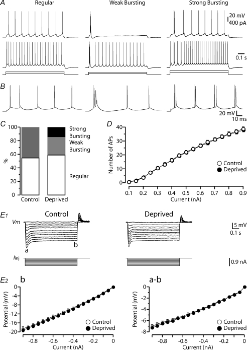
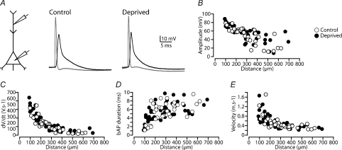
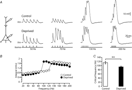
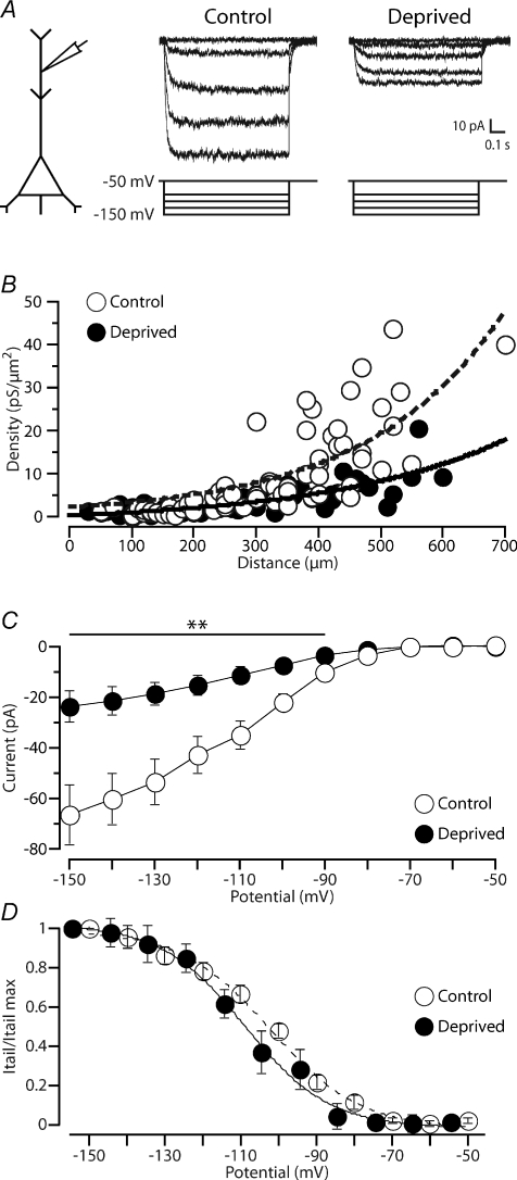
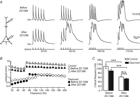
Development of the cortical map is experience dependent, with different critical periods in different cortical layers. Previous work in rodent barrel cortex indicates that sensory deprivation leads to changes in synaptic transmission and plasticity in layer 2/3 and 4. Here, we studied the impact of sensory deprivation on the intrinsic properties of layer 5 pyramidal neurons located in rat barrel cortex using simultaneous somatic and dendritic recording. Sensory deprivation was achieved by clipping all the whiskers on one side of the snout. Loss of sensory input did not change somatic active and resting membrane properties, and did not influence dendritic action potential (AP) backpropagation. In contrast, sensory deprivation led to an increase in the percentage of layer 5 pyramidal neurons showing burst firing. This was associated with a reduction in the threshold for generation of dendritic calcium spikes during high-frequency AP trains. Cell-attached recordings were used to assess changes in the properties and expression of dendritic HCN channels. These experiments indicated that sensory deprivation caused a decrease in HCN channel density in distal regions of the apical dendrite. To assess the contribution of HCN down-regulation on the observed increase in dendritic excitability following sensory deprivation, we investigated the impact of blocking HCN channels. Block of HCN channels removed differences in dendritic calcium electrogenesis between control and deprived neurons. In conclusion, these observations indicate that sensory loss leads to increased dendritic excitability of cortical layer 5 pyramidal neurons. Furthermore, they suggest that increased dendritic calcium electrogenesis following sensory deprivation is mediated in part via down-regulation of dendritic HCN channels.


Figures

Figure 1
Figure 1. Impact of sensory deprivation on somatic active and passive properties
A, traces represent three typical firing patterns recorded from layer 5 pyramidal neurons in barrel cortex, referred to as ‘regular’, ‘weak bursting’ and ‘strong bursting’. B, the first 150 ms of the top traces in A. C, bar graph of the proportion of layer 5 neurons with the different firing patterns for the indicated sensory conditions (control: n= 51; deprived: n= 88; P > 0.05). Note the regular firing pattern remains constant for each experimental condition, with strong bursting encountered solely after sensory deprivation. D, graph showing the input–output properties of recorded neurons after different sensory experiences (number of APs elicited by 900 ms somatic current injections of the indicated amplitudes). The input–output properties were unchanged after sensory deprivation (control: n= 51, open symbols; deprived: n= 88, filled symbols; P > 0.05). E1, voltage response (top) to somatic hyperpolarizing current steps (bottom) for the different conditions. The membrane potential hyperpolarization exhibited a peak (a) follow by a time-dependent ‘sag’ until reaching steady-state (b). E2, graphs of the steady-state (b, left) and peak to steady-state difference (ab, right) versus current step amplitude for the different conditions (control: n= 51, open symbols; deprived: n= 88, filled symbols; P > 0.05). Sensory deprivation did not change the IV curves of the recorded neurons. Data are shown as means ±s.e.m.
Figure 2
Figure 2. Sensory deprivation doesn't change action potential backpropagation
A, left, schematic of the experimental configuration. Right, example of somatic (grey) and dendritic bAPs (black) recorded in control and deprived neurons. APs evoked by somatic current injection and bAPs recorded ∼380 μm from the soma. B–E, graphs showing the dependence of backpropagating AP amplitude (B), rate-of-rise (C), duration (D) and velocity (E) on dendritic location for the different experimental conditions (control: open symbols and deprived: filled symbols).
Figure 3
Figure 3. Sensory deprivation increases dendritic excitability
A, left, schematic of the experimental configuration. Right, dendritic response (control: 460 μm and deprived: 470 μm from the soma) to trains of five somatic APs at the indicated frequencies (bottom) in recordings from control (top) and deprived (middle) cortex. B, plot of the integral of dendritic membrane potential versus the frequency of AP trains for the experimental conditions. Note the non-linear increase in dendritic integral, indicative of the critical frequency, is lower in deprived neurons (closed symbols) compared to control (open symbols). C, bar graph of the average critical frequency in control (n= 27) and deprived animals (n= 43). Data shown as means ±s.e.m. **P < 0.01.
Figure 4
Figure 4. Sensory deprivation leads to a decrease in HCN channel expression at distal dendritic locations
A, left, schematic diagram of the experimental configuration. Right, examples of HCN current recorded in dendritic cell-attached patches from control (550 μm from the soma) and deprived (530 μm from the soma) neurons. B, plot of dendritic HCN channel density during voltage steps to an estimated membrane potential of −150 mV versus distance of the recording location from the soma in recordings from control (open symbols, n= 68) and deprived (filled symbols, n= 54) neurons. The curves represent exponential fits to the control (dotted curve, distance constant (λ): 219 μm) and deprived (continuous curve, λ: 209 μm) data sets, respectively. C, IV curve of average peak HCN current at distal dendritic locations (∼470–480 μm from the soma) in recordings from neurons in control conditions (open symbols; n= 13) and following sensory deprivation (filled symbols; n= 10). D, HCN tail current normalized to maximum for control (open symbols, n= 13) and deprived (filled symbols, n= 10) neurons indicating no obvious impact of sensory deprivation on the voltage dependence of steady-state activation of HCN channels. Holding and test potentials were corrected for the measured −4.5 mV shift in resting membrane potential in deprived neurons (see Results). Data shown as means ±s.e.m. **P < 0.01.
Figure 5
Figure 5. Decreased dendritic Ih contributes to increased dendritic excitability following sensory deprivation
A, left, schematic diagram of the experimental configuration. Right, dendritic response to trains of five somatic APs at the indicated frequencies (bottom) in a recording from a control neuron before (top) and after (middle) bath application of ZD 7288 (50 μm). B, plot of the dendritic voltage integral versus AP frequency for control (open symbols) and deprived (filled symbols) neurons before (circles) and after (triangles) bath application of ZD 7288 (50 μm). C, block of HCN channels by bath application of ZD 7288 (50 μm) reduced the critical frequency of both control (n= 9) and deprived (n= 10) neurons to a similar extent. Data shown as means ±s.e.m. ***P < 0.001; n.s., non-significant.

References

    1. Aizenman CD, Akerman CJ, Jensen KR, Cline HT. Visually driven regulation of intrinsic neuronal excitability improves stimulus detection in vivo. Neuron. 2003;39:831–842. - PubMed
    1. Allen CB, Celikel T, Feldman DE. Long-term depression induced by sensory deprivation during cortical map plasticity in vivo. Nat Neurosci. 2003;6:291–299. - PubMed
    1. Altomare C, Terragni B, Brioschi C, Milanesi R, Pagliuca C, Viscomi C, Moroni A, Baruscotti M, DiFrancesco D. Heteromeric HCN1–HCN4 channels: a comparison with native pacemaker channels from the rabbit sinoatrial node. J Physiol. 2003;549:347–359. - PMC - PubMed
    1. Armstrong-James M, Callahan CA, Friedman MA. Thalamo-cortical processing of vibrissal information in the rat. I. Intracortical origins of surround but not centre-receptive fields of layer IV neurones in the rat S1 barrel field cortex. J Comp Neurol. 1991;303:193–210. - PubMed
    1. Armstrong-James M, Fox K. Spatiotemporal convergence and divergence in the rat S1 ‘barrel’ cortex. J Comp Neurol. 1987;263:265–281. - PubMed

Publication types

LinkOut - more resources