. 2010 Mar;31(3):359-66.
doi: 10.1093/carcin/bgp217. Epub 2009 Sep 7.

Endoglin phosphorylation by ALK2 contributes to the regulation of prostate cancer cell migration

Affiliations

Endoglin phosphorylation by ALK2 contributes to the regulation of prostate cancer cell migration

Diana Romero et al. Carcinogenesis. 2010 Mar.

Abstract

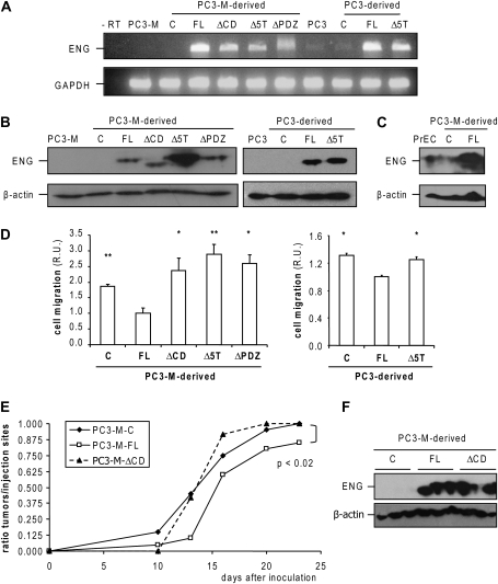
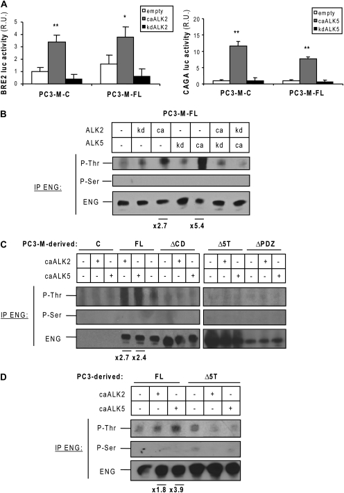
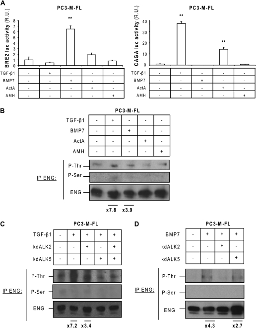
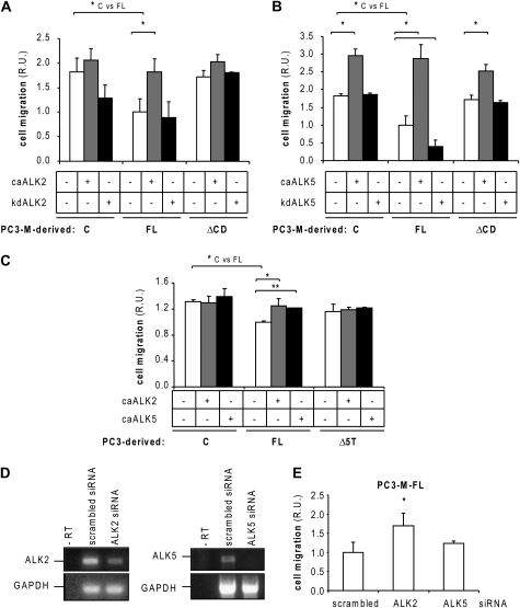
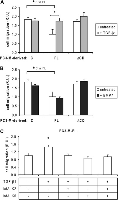
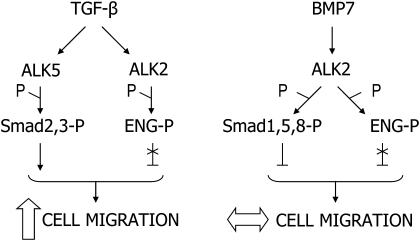
Endoglin, a transmembrane glycoprotein that acts as a transforming growth factor-beta (TGF-beta) coreceptor, is downregulated in PC3-M metastatic prostate cancer cells. When restored, endoglin expression in PC3-M cells inhibits cell migration in vitro and attenuates the tumorigenicity of PC3-M cells in SCID mice, though the mechanism of endoglin regulation of migration in prostate cancer cells is not known. The current study indicates that endoglin is phosphorylated on cytosolic domain threonine residues by the TGF-beta type I receptors ALK2 and ALK5 in prostate cancer cells. Importantly, in the presence of constitutively active ALK2, endoglin did not inhibit cell migration, suggesting that endoglin phosphorylation regulated PC3-M cell migration. Therefore, our results suggest that endoglin phosphorylation is a mechanism with relevant functional consequences in prostate cancer cells. These data demonstrate for the first time that TGF-beta receptor-mediated phosphorylation of endoglin is a Smad-independent mechanism involved in the regulation of prostate cancer cell migration.


Figures

Fig. 1.
Fig. 1.
Endoglin expression affects cell migration and tumorigenicity in PC3-M cells. (A) Reverse transcription–polymerase chain reaction and (B) western blot analysis of endoglin expression in PC3-M and PC3 cells. Glyceraldehyde-3-phosphate dehydrogenase (GAPDH) (A) and β-actin (B): controls. (C): PC3-M- or PC3-derived control cells. (C) Western blot analysis of endoglin expression in prostate epithelial cell (PrEC), PC3-M-C and FL cells. β-Actin: protein loading control. In order to obtain a detectable endoglin signal in prostate epithelial cell, the film exposure was longer than in the films shown in panel (B). (D) The effect of endoglin in the basal migration of PC3-M- and PC3-derived cells was determined as described in Material and Methods. Cells were allowed to migrate for 18 h. Five different fields per sample (n = 6) were quantified by microscopy. R.U., relative units. *P < 0.05; **P < 0.005 (Student's t-test), expressed relative to PC3-M-FL or PC3-FL. (E) In total, 1 × 106 PC3-M-control, FL- or ΔCD-endoglin-expressing cells were injected subcutaneously in both flanks of SCID mice (C and FL, n = 10; ΔCD, n = 6). The data sets were compared using the chi-square test. (F) Western blot analysis of endoglin expression in tumors harvested 19–22 days after inoculation. β-Actin: protein loading control. A minimum of three tumors per group were analyzed.
Fig. 2.
Fig. 2.
Endoglin phosphorylation by TGF-β type I receptors in prostate cancer cells. (A) PC3-M-control and FL cells were transfected with the BRE2 and CAGA luciferase reporter constructs, which were specifically activated by Smad1, 5 and 8 or Smad2 and 3, respectively, and the different receptor constructs for 48 h. The luciferase activities measured in duplicate samples were normalized and plotted (n = 6). R.U., relative units. *P < 0.05 and **P < 0.005 (Student's t-test). (B) PC3-M-FL cells were transfected with different combinations of caALK2 or caALK5, kdALK2 and kdALK5 or with an empty vector. Endoglin was immunoprecipitated and western blot was performed with P-Thr, P-Ser or total endoglin-specific antibodies. Values indicated at bottom of panel: P-Thr relative signal intensity increase compared with control. The band intensity values were quantified using ImageJ software (NIH Image), and P-Thr levels were normalized to total endoglin levels. These measurements are representative of a minimum of three independent experiments. (C) Endoglin phosphorylation by caALK2 and caALK5 in PC3-M-control, FL-, ΔCD-, Δ5T- and ΔPDZ-endoglin-expressing cells. (D) Endoglin phosphorylation by caALK2 and caALK5 in PC3 control, FL- and Δ5T-endoglin-expressing cells.
Fig. 3.
Fig. 3.
Endoglin phosphorylation by TGF-β family ligands in PC3-M cells. (A) PC3-M-FL cells were transfected with the BRE2 and CAGA luciferase reporters, and 24 h later they were treated for an additional 24 h with 5 ng/ml TGF-β, 50 ng/ml BMP7, 20 ng/ml ActA or 500 ng/ml AMH. The luciferase activities were measured as described for Figure 2A (n = 6). R.U., relative units. **P < 0.005 (Student's t-test). (B) Serum-starved PC3-M-FL cells were treated for 30 min with 5 ng/ml TGF-β1, 50 ng/ml BMP7, 20 ng/ml ActA or 500 ng/ml AMH. Endoglin phosphorylation was analyzed as described for Figure 2B. (C and D) PC3-M-FL cells were transfected with kdALK2 and/or kdALK5 or with an empty vector. Twenty-four hours later, they were serum-starved overnight. In total, 5 ng/ml TGF-β1 (C) or 50 ng/ml BMP7 (D) was added for 30 min, and endoglin phosphorylation was analyzed. Fold increases for P-Thr were determined as described above.
Fig. 4.
Fig. 4.
Effect of endoglin phosphorylation in prostate cancer cell migration. (A) Effect of caALK2 and kdALK2 or (B) caALK5 and kdALK5 in cell migration of PC3-M-control, FL and ΔCD cells. Migration assays were performed as described previously. Five different fields per sample (n = 6) were quantified by microscopy. R.U., relative units. *P < 0.05 (Student's t-test). (C) Effect of caALK2 and caALK5 in cell migration of PC3 control, FL and Δ5T cells. Migration assays were performed as described previously. Five different fields per sample (n = 6) were quantified by microscopy. R.U., relative units. *P < 0.05 (Student's t-test). (D) PC3-M-FL cells were transfected with small interfering ribonucleic acids (siRNAs) directed against ALK2, ALK5 or a scrambled sequence (control) for 48 h. ALK2 and ALK5 expression was analyzed by reverse transcription–polymerase chain reaction. Glyceraldehyde-3-phosphate dehydrogenase (GAPDH): control. (E) PC3-M-FL cells were transfected with siRNAs directed against ALK2, ALK5 or a control scrambled sequence. Twenty-four hours after transfection, they were allowed to migrate for 18 h. Cell migration assays were performed as described previously (n = 6). R.U., relative units. *P < 0.05 (Student's t-test).
Fig. 5.
Fig. 5.
Effect of TGF-β1 and BMP7 in PC3-M cell migration. (A and B) PC3-M-FL cells in suspension in serum-free media were supplemented with 5 ng/ml TGF-β1 (A) or 50 ng/ml BMP7 (B). Cell migration assay was performed as described previously (n = 6). R.U., relative units. *P < 0.05 (Student's t-test). (C) PC3-M-FL cells were transfected with kdALK2 and/or kdALK5 or with an empty vector. Twenty-four hours later, they were prepared as a cell suspension in serum-free media and supplemented with 5 ng/ml TGF-β1. Cell migration assay was performed as described previously (n = 6). R.U., relative units. *P < 0.05 (Student's t-test).
Fig. 6.
Fig. 6.
Proposed model for endoglin phosphorylation by TGF-β1 and BMP7 in prostate cancer cells and its effect in cell migration.

Similar articles

Cited by

References

    1. Ao M, et al. Transforming growth factor-beta promotes invasion in tumorigenic but not in nontumorigenic human prostatic epithelial cells. Cancer Res. 2006;66:8007–8016. - PMC - PubMed
    1. Craft CS, et al. Endoglin inhibits prostate cancer motility via activation of the ALK2-Smad1 pathway. Oncogene. 2007;26:7240–7250. - PMC - PubMed
    1. Massague J, et al. TGFbeta signaling in growth control, cancer, and heritable disorders. Cell. 2000;103:295–309. - PubMed
    1. Derynck R, et al. Smad-dependent and Smad-independent pathways in TGF-beta family signalling. Nature. 2003;425:577–584. - PubMed
    1. Bernabeu C, et al. Novel biochemical pathways of endoglin in vascular cell physiology. J. Cell. Biochem. 2007;102:1375–1388. - PMC - PubMed

Publication types

MeSH terms