. 2009 Oct;136(19):3279-88.
doi: 10.1242/dev.040071.

Disruption of the pollen-expressed FERONIA homologs ANXUR1 and ANXUR2 triggers pollen tube discharge

Affiliations

Disruption of the pollen-expressed FERONIA homologs ANXUR1 and ANXUR2 triggers pollen tube discharge

Aurélien Boisson-Dernier et al. Development. 2009 Oct.

Abstract

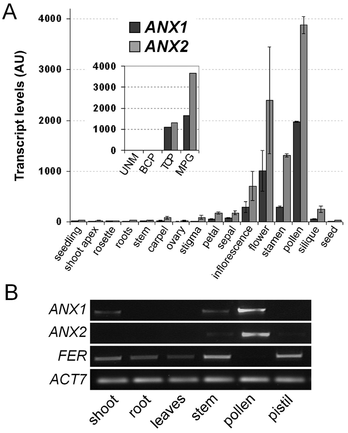
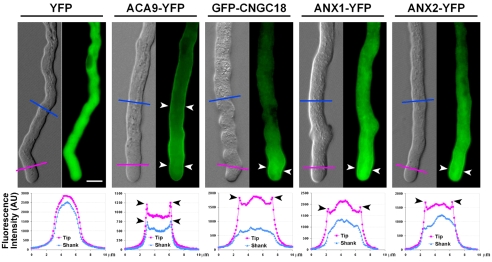
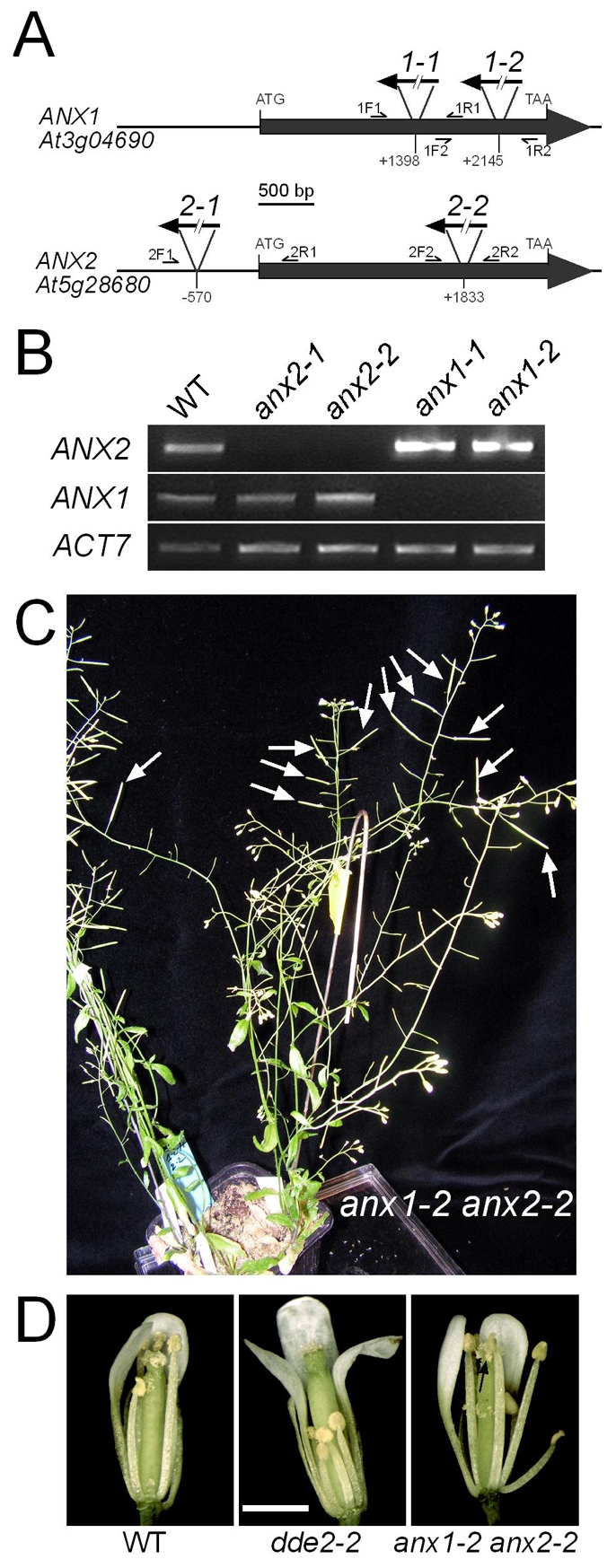
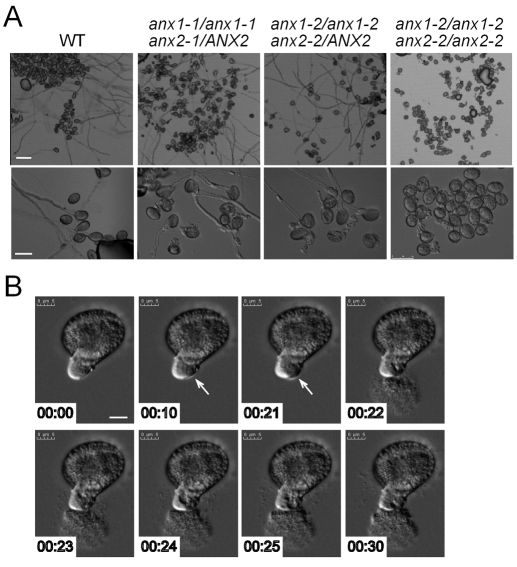
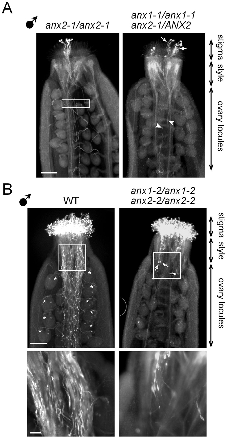
The precise delivery of male to female gametes during reproduction in eukaryotes requires complex signal exchanges and a flawless communication between male and female tissues. In angiosperms, molecular mechanisms have recently been revealed that are crucial for the dialog between male (pollen tube) and female gametophytes required for successful sperm delivery. When pollen tubes reach the female gametophyte, they arrest growth, burst and discharge their sperm cells. These processes are under the control of the female gametophyte via the receptor-like serine-threonine kinase (RLK) FERONIA (FER). However, the male signaling components that control the sperm delivery remain elusive. Here, we show that ANXUR1 and ANXUR2 (ANX1, ANX2), which encode the closest homologs of the FER-RLK in Arabidopsis, are preferentially expressed in pollen. Moreover, ANX1-YFP and ANX2-YFP fusion proteins display polar localization to the plasma membrane at the tip of the pollen tube. Finally, genetic analyses demonstrate that ANX1 and ANX2 function redundantly to control the timing of pollen tube discharge as anx1 anx2 double-mutant pollen tubes cease their growth and burst in vitro and fail to reach the female gametophytes in vivo. We propose that ANX-RLKs constitutively inhibit pollen tube rupture and sperm discharge at the tip of growing pollen tubes to sustain their growth within maternal tissues until they reach the female gametophytes. Upon arrival, the female FER-dependent signaling cascade is activated to mediate pollen tube reception and fertilization, while male ANX-dependent signaling is deactivated, enabling the pollen tube to rupture and deliver its sperm cells to effect fertilization.


Figures

Fig. 1.
Fig. 1.
ANXUR1 and ANXUR2 are preferentially expressed in Arabidopsis pollen. (A) Microarray data for ANXUR1 (At3g04690, ANX1) and ANXUR2 (At5g28680, ANX2) in different organs as retrieved from GENEVESTIGATOR (Zimmermann et al., 2004) (downloaded May 2009). ANX1 and ANX2 transcripts were detected in every tissue that contained pollen. The inset shows microarray data retrieved from Honys and Twell (Honys and Twell, 2004) that illustrates a steady increase in ANX1 and ANX2 transcript levels during the course of pollen development. UNM, uninucleate microspores; BCP, bicellular pollen; TCP, immature tricellular pollen; MPG, mature pollen grain; AU, arbitrary units. (B) RT-PCR analysis of ANX1, ANX2 and FER transcripts from cDNAs of different Arabidopsis tissues indicates that ANX1 and ANX2 are strongly and preferentially expressed in the male gametophyte, whereas FER exhibits the opposite expression pattern. ACTIN7 (ACT7) was used as a control. Amplification was performed for 26 cycles for both ACT7 and FER and for 32 cycles for ANX1 and ANX2.
Fig. 2.
Fig. 2.
ANX1-YFP and ANX2-YFP fusion proteins localize polarly to the plasma membrane of growing pollen tube tips. Fluorescence micrographs of actively growing pollen tubes (PTs) of Arabidopsis transgenic lines expressing YFP, ACA9-YFP, GFP-CNGC18, ANX1-YFP and ANX2-YFP constructs under the control of the pollen-specific ACA9 promoter. Unlike the uniform plasma membrane localization of ACA9-YFP along the PT, ANX1-YFP and ANX2-YFP fusions display polar plasma membrane localization at the tip of growing PTs, similar to GFP-CNGC18. Arrowheads point to fluorescence enrichment at the plasma membrane. Fluorescence intensity in arbitrary units (AU) across the PT shank (10 μm-long blue lines) or across the PT tip (10 μm-long purple lines), as provided below each PT, shows a similar fluorescence distribution for GFP-CNGC18, ANX1-YFP and ANX2-YFP. Scale bar: 5 μm.
Fig. 3.
Fig. 3.
Double-homozygous anx1-2 anx2-2 mutant plants are male sterile. (A) The genomic organization of the intron-less ANX1 and ANX2 genes and positions of the anx1-1, anx1-2, anx2-1 and anx2-2 T-DNA insertions. The orientation of the left border sequence of the respective T-DNAs is represented by black arrows. The positions of the primers used to genotype the mutants are indicated. (B) RT-PCR analysis from open-flower cDNAs shows no ANX1 transcripts in the ANX1 T-DNA disruption lines anx1-1 and anx1-2 and no ANX2 transcripts in the ANX2 T-DNA disruption lines anx2-1 and anx2-2. ACT7 was used as a control. Amplification was performed for 28 cycles for ACT7 and for 34 cycles for ANX1 and ANX2. (C) The double-homozygous anx1-2 anx2-2 mutant plant shows normal vegetative development, but only short pistils were observed that never developed further into siliques. This phenotype was completely reversible when wild-type pollen was used to pollinate anx1-2 anx2-2 pistils (white arrows). Conversely, when pollen from anx1-2 anx2-2 plants was deposited on dde2-2 pistils, no siliques or seeds developed (see Fig. S3 in the supplementary material). (D) Unlike male-sterile dde2-2 mutant flowers, the filaments and anthers of which fail to elongate and release pollen, anx1-2 anx2-2 flowers were normal, anthers dehisced, and pistils were efficiently self-pollinated (black arrow). Scale bar: 1 mm.
Fig. 4.
Fig. 4.
In vitro pollen growth assays reveal that anx1 anx2 mutant pollen tubes discharge spontaneously. (A) (Top row) Five hours after incubation in vitro, wild-type pollen germinated very well and produced actively growing PTs. By contrast, anx1-2/anx1-2 anx2-2/anx2-2 mutant pollen grew very poorly, and segregating anx1-1/anx1-1 anx2-1/ANX2 and anx1-2/anx1-2 anx2-2/ANX2 mutant pollen exhibited an intermediate pollen growth phenotype. (Bottom row) Higher magnification reveals that all the germinated anx1-2/anx1-2 anx2-2/anx2-2 mutant pollen had burst. Germinated pollen from independent anx1-1/anx1-1 anx2-1/ANX2 and anx1-2/anx1-2 anx2-2/ANX2 mutant alleles either produced actively growing PTs or burst, phenotypes that were observed in a 1:1 ratio as expected for segregation of anx1 anx2 pollen grains. Pollen donor genotype is indicated at the top. (B) Time-lapse imaging of anx1-2 anx2-2 mutant pollen in vitro showed that pollen discharge is an explosive phenomenon that occurs in less than 1 second after formation of either bulges or an emerging tip, or more rarely a real PT (see also Fig. S4 and Movie 3 in the supplementary material). Note that the PT stopped elongating. White arrows indicate the location of the discharge at the subapical region of the PT tip, rather than at the tip itself. Time is indicated in minutes:seconds. Scale bars: 50 μm in top row in A; 25 μm in bottom row in A; 5 μm in B.
Fig. 5.
Fig. 5.
anx1 anx2 pollen germinate normally on stigma but produce pollen tubes that fail to reach the female gametophytes. (A) Aniline Blue staining of wild-type pistils pollinated with a few anx2-1 single-mutant (left) or anx1-1/anx1-1 anx2-1/ANX2 double-mutant (right) pollen grains. Eighteen hours after pollination, most of the tips of anx2-1 PTs (left) were observed in the ovary locules (rectangle). By contrast, only about half of the tips of germinated (arrows) anx1-1/anx1-1 anx2-1/ANX2 PTs (arrowheads) were found in the ovary locules. (B) Aniline Blue staining of wild-type pistils pollinated with numerous wild-type (left) or anx1-2 anx2-2 double-homozygous mutant (right) pollen grains. Eighteen hours after manual pollination, wild-type PTs (left) had grown through the entire pistil to reach the micropyles of the ovules (asterisks). Although they germinated on the stigma, most of the anx1-2 anx2-2 mutant PTs (right) were arrested in the style, with only very few having reached the top end of the ovary (arrows). The boxed regions are shown at high magnification below and reveal the very low density of anx1-2 anx2-2 PTs (right) in the transition zone between the style and the ovary locules in comparison to wild-type PTs (left). Scale bars: 100 μm in A and in top row in B; 25 μm in bottom row in B.

References

    1. Alonso, J. M., Stepanova, A. N., Leisse, T. J., Kim, C. J., Chen, H., Shinn, P., Stevenson, D. K., Zimmerman, J., Barajas, P., Cheuk, R. et al. (2003). Genome-wide insertional mutagenesis of Arabidopsis thaliana. Science 301, 653-657. - PubMed
    1. Becker, J. D., Boavida, L. C., Carneiro, J., Haury, M. and Feijo, J. A. (2003). Transcriptional profiling of Arabidopsis tissues reveals the unique characteristics of the pollen transcriptome. Plant Physiol. 133, 713-725. - PMC - PubMed
    1. Berger, F., Hamamura, Y., Ingouff, M. and Higashiyama, T. (2008). Double fertilization-caught in the act. Trends Plant Sci. 13, 437-443. - PubMed
    1. Boavida, L. C. and McCormick, S. (2007). Temperature as a determinant factor for increased and reproducible in vitro pollen germination in Arabidopsis thaliana. Plant J. 52, 570-582. - PubMed
    1. Boavida, L. C., Vieira, A. M., Becker, J. D. and Feijo, J. A. (2005). Gametophyte interaction and sexual reproduction: how plants make a zygote. Int. J. Dev. Biol. 49, 615-632. - PubMed

Publication types

MeSH terms