. 2009 Oct;136(19):3289-99.
doi: 10.1242/dev.040451.

A directional Wnt/beta-catenin-Sox2-proneural pathway regulates the transition from proliferation to differentiation in the Xenopus retina

Affiliations

A directional Wnt/beta-catenin-Sox2-proneural pathway regulates the transition from proliferation to differentiation in the Xenopus retina

Michalis Agathocleous et al. Development. 2009 Oct.

Abstract

Progenitor cells in the central nervous system must leave the cell cycle to become neurons and glia, but the signals that coordinate this transition remain largely unknown. We previously found that Wnt signaling, acting through Sox2, promotes neural competence in the Xenopus retina by activating proneural gene expression. We now report that Wnt and Sox2 inhibit neural differentiation through Notch activation. Independently of Sox2, Wnt stimulates retinal progenitor proliferation and this, when combined with the block on differentiation, maintains retinal progenitor fates. Feedback inhibition by Sox2 on Wnt signaling and by the proneural transcription factors on Sox2 mean that each element of the core pathway activates the next element and inhibits the previous one, providing a directional network that ensures retinal cells make the transition from progenitors to neurons and glia.


Figures

Fig. 1.
Fig. 1.
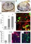
Canonical Wnt signaling promotes progenitor cell maintenance. (A) Lipofection of TCF3-VP16 or activated β-catenin increases the proportion of neuroepithelial (NE) cells as compared with GFP controls. The effect was blocked by co-expression of dominant-negative (dn) LEF1, whereas dnLEF1 alone had no significant effect on the proportion of NE cells (GFP, n=6 retinas, 391 cells; TCF3-VP16, n=7, 519 cells; activated β-catenin, n=5, 211 cells; dnLEF1+TCF3-VP16, n=6, 297 cells; dnLEF1, n=3, 321 cells). (B,B′) Cells transfected with TCF3-VP16 show typical neuroepithelial morphology and incorporate BrdU (B), unlike control cells (B′). (C-G″) TCF3-VP16-expressing cells with long processes do not stain for the Müller glial cell marker CRALPB (C), they incorporate BrdU more frequently than controls (D), and are positive for Cyclin A2 (E-F) and PCNA (G-G″). Error bars indicate s.e.m. *P<0.001 by Student's t-test. RPE, retinal pigment epithelium; ONL, outer nuclear layer; INL, inner nuclear layer; GCL, ganglion cell layer.
Fig. 2.
Fig. 2.
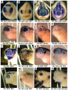
Canonical Wnt signaling does not affect retinal identity but blocks neuronal differentiation downstream of the proneural genes. (A,B) Rx:Act-β-catenin (Act β-cat) transgenic embryos (stage 23) show normal expression of Rx and En (A) and of the retinal progenitor marker Vsx1 (B). (C,D) Act β-cat transgenic embryos show normal expression of Sox2 (C), a gene required for neural competence, and of the neurogenic gene Notch (D). (E,F) Act β-cat transgenic embryos (stage 34) show normal expression of the proneural gene Xath5. (G-P) Act β-cat transgenic embryos show loss of retinal expression of the proneural target genes Brn3D (G,H), Ebf3 (I,J), ElrC (K,L) and Sbt1 (M,N), as well as of Hermes (O,P), a marker of differentiated ganglion cells. Brackets indicate the retina.
Fig. 3.
Fig. 3.
Sustained Sox2 expression promotes Müller glial cell formation. (A,B) Sox2 expression is expanded in Act β-cat transgenic embryos (A) as compared with control (B; Rx:GFP) transgenic embryos. (C-E) Lipofection of Sox2 causes a large increase in the representation of CRALBP- or R5-positive Müller cells [D, GFP, n=8, 942 cells; E, Sox2 (CRALBP), n=7, 485 cells; Sox2 (R5), n=9, 616 cells]. *P<0.001. (F-H) Sox2 causes an increase in the proportion of Müller cells born before stage 32, which are BrdU negative (G, GFP, n=5, 30 cells; H, Sox2, n=3, 31 cells). Error bars indicate s.e.m. *P<0.001.
Fig. 4.
Fig. 4.
Sox2 blocks neuronal differentiation downstream of the proneural genes. (A,B) Overexpression of Sox2 in retinal progenitors by mRNA injection at the 8-cell stage does not decrease the expression of the proneural gene Xath5 on the injected side (B), as compared with the control side (A), in stage 34 embryos. (C-L) Retinal expression of the proneural targets Brn3d (C,D), Ebf3 (E,F), ElrC (G,H) and Sbt1 (I, J) is lost, as compared with the control (uninjected) side of the embryos. Sox2-injected embryos also lose retinal expression of Hermes (K,L), a marker of differentiated ganglion cells. Brackets indicate the retina.
Fig. 5.
Fig. 5.
Blocking Notch signaling suppresses the ability of Wnt/β-catenin signaling or Sox2 to inhibit neuronal differentiation. (A) Co-expression of TCF3-VP16 and a dominant-negative form of Delta (DeltaSTU) that blocks Notch signaling in retinal progenitors, inhibits the TCF3-VP16-mediated increase in neuroepithelial cells (TCF3-VP16, n=7, 405 cells; TCF3-VP16+DeltaSTU, n=5, 234 cells). Error bars indicate s.e.m. *P<0.001. (B-E) Act β-cat transgenic embryos (stage 34) injected with DeltaSTU mRNA show restored retinal expression of the proneural target genes Brn3D (B), ElrC (C) and Sbt1 (D), and of Hermes (E), a marker of differentiated ganglion cells. (F-I) Expression of Sox2 and DeltaSTU in retinal progenitors by mRNA injection at the 8-cell stage also restored retinal expression of Brn3D (F), ElrC (G), Sbt1 (H) and Hermes (I). (J-L) Lipofection of Sox2 promotes CRALBP+/EdU- Müller cells (J arrows, L), whereas co-transfection with Su(H)DBM produces CRALBP- cells (K,L) (GFP, n=4, 192 cells; Su(H)DBM, n=6, 342 cells; Sox2, n=5, 868 cells; Sox2+Su(H)DBM, n=9, 892 cells). Error bars indicate 95% confidence interval (C.I.). *P<0.001.
Fig. 6.
Fig. 6.
Wnt/β-catenin activation, but not Sox2, promotes cell proliferation. (A-D) Electroporation with TCF3-VP16 (n=5, 493 cells), but not Sox2 (n=3, 1719 cells), significantly increases the proportion of BrdU+ cells (arrowheads) at stage 37, as compared with control GFP (n=4, 653 cells). (E) GFP, Sox2 and TCF3-VP16 were electroporated and proliferation assessed by flow cytometry at stage 37. Transfected cells display a GFP intensity above background, whereas non-electroporated retinas do not have any GFP+ cells (inset). (F) TCF3-VP16 transfection causes a 2-fold increase in the proportion of cells in the S/G2/M phases, as compared with Sox2 or GFP transfection, indicating a larger proliferating cell fraction (see inset). (G) Co-lipofection of the dominant-negative Sox2BD(-) does not inhibit BrdU incorporation caused by TCF3-VP16 at stage 41 (GFP, n=12, 2615 cells; Sox2BD(-), n=9, 670 cells; TCF3-VP16, n=11, 1539 cells; TCF3-VP16+Sox2BD(-), n=10, 1242 cells. Error bars indicate 95% C.I. *P<0.001 compared with GFP control.
Fig. 7.
Fig. 7.
Sox2 and Cyclin E1 can recapitulate the effects of Wnt/β-catenin signaling on the proliferation and maintenance of neuroepithelial morphology. (A) GFP-lipofected retinas show a typical cell type distribution at stage 41. (B-C′) Lipofection of Sox2+Cyclin E1 results in BrdU-positive cells that do not stain for the Müller cell marker CRALBP (C′). (D) The increase in BrdU-positive, neuroepithelial-like cells is significant in the double transfection versus the Cyclin E1-only control (**P<0.001). Cyclin E1 lipofection causes a slight increase in BrdU-positive central retina cells (GFP, n=10, 2642 cells; Sox2, n=11, 2043 cells; Cyclin E1, n=13, 2388 cells; Sox2+Cyclin E1, n=10, 1688 cells. Error bars indicate 95% C.I. *P<0.001. (E-I) Co-expression of Sox2 and Cyclin E1 promotes mitosis, as seen with anti-phospho-histone H3 staining (H,I), in contrast to overexpression of GFP (E), Sox2 (F) or Cyclin E1 (G) alone (GFP, n=10, 1308 cells; Sox2, n=6, 474 cells; Cyclin E1, n=6, 605 cells; Sox2+Cyclin E1, n=11, 1697 cells). Error bars indicate 95% C.I. **P<0.005.
Fig. 8.
Fig. 8.
Sox2 inhibits canonical Wnt/β-catenin signaling. (A,B) mRFP-only-injected cells co-express the TOP-dGFP reporter (A, arrowheads), unlike Sox2-injected cells (B). (C) The proportion of TOP-dGFP+ cells out of the total injected (RFP+) cells is reduced in the presence of Sox2 (mRFP, n=13, 3471 cells; Sox2, n=10, 3420 cells). (D) mRNAs for mRFP (tracer) alone, mRFP+Sox2 or mRFP+Sox2BD(-) (dominant-negative Sox2) were injected into dorsal animal cells of cleavage-stage embryos. TOP-dGFP DNA was subsequently lipofected into stage 17 optic vesicles. (E-G) In a Sox2-injected embryo, the cells that express GFP (E) are those that do not express Sox2 (gaps indicated by arrowheads in F). (H) In retinal cells expressing Sox2 or Sox2BD(-), there is a 2-fold reduction in the proportion of TOP-dGFP+ cells that express mRFP (mRFP, 682 cells; Sox2, 228 cells; Sox2BD(-), 437 cells). Error bars indicate 95% C.I. *P<0.01.
Fig. 9.
Fig. 9.
Xath5 injection suppresses Sox2 protein levels. (A-F) Control embryos injected with GFP alone and dexamethasone (DEX) treated (A,B), or co-injected with Xath5GR and GFP without DEX (C,D), did not show downregulation of Sox2 protein in the injected areas. By contrast, embryos co-injected with Xath5GR and GFP and DEX treated (E,F) displayed a pronounced Sox2 downregulation in Xath5-expressing cells. (G-H‴) Staining 6 hours after the start of DEX treatment shows a downregulation of Sox2 (G″) and EdU (G′) in areas of the retina that overexpress Xath5GR (G). Areas overexpressing Xath5GR are outlined in a magnified view in H-H‴. Occasionally, a reduction in Sox2 is seen in cells that are still cycling (arrowheads, H‴).
Fig. 10.
Fig. 10.
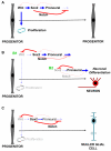
A model for the role of the Wnt-Sox2 pathway in the transition from a progenitor to a differentiated cell. (A) Wnt signaling activation in a neuroepithelial cell activates Sox2 and the proneural genes, but through Sox2 and Notch it blocks proneural activity and neuronal differentiation. Wnt also independently maintains proliferation, and this leads to progenitor maintenance and expansion. (B) The build-up of Sox2 switches off Wnt, inhibiting proliferation (B1), and then the accumulation of proneural activity switches off Sox2, relieving the inhibition of neuronal differentiation and leading to neurogenesis (B2). (C) Alternatively, if Sox2 levels remain high it will limit proneural activity and neuronal differentiation will be blocked, but Wnt signaling will also be inhibited, leading to cell cycle exit and glial differentiation.

Similar articles

Cited by

References

    1. Baker, J. C., Beddington, R. S. and Harland, R. M. (1999). Wnt signaling in Xenopus embryos inhibits bmp4 expression and activates neural development. Genes Dev. 13, 3149-3159. - PMC - PubMed
    1. Bani-Yaghoub, M., Tremblay, R. G., Lei, J. X., Zhang, D., Zurakowski, B., Sandhu, J. K., Smith, B., Ribecco-Lutkiewicz, M., Kennedy, J., Walker, P. R. et al. (2006). Role of Sox2 in the development of the mouse neocortex. Dev. Biol. 295, 52-66. - PubMed
    1. Blackshaw, S., Harpavat, S., Trimarchi, J., Cai, L., Huang, H., Kuo, W. P., Weber, G., Lee, K., Fraioli, R. E., Cho, S. H. et al. (2004). Genomic analysis of mouse retinal development. PLoS Biol. 2, E247. - PMC - PubMed
    1. Bunt-Milam, A. H. and Saari, J. C. (1983). Immunocytochemical localization of two retinoid-binding proteins in vertebrate retina. J. Cell Biol. 97, 703-712. - PMC - PubMed
    1. Burns, C. J., Zhang, J., Brown, E. C., Van Bibber, A. M., Van Es, J., Clevers, H., Ishikawa, T. O., Taketo, M. M., Vetter, M. L. and Fuhrmann, S. (2008). Investigation of Frizzled-5 during embryonic neural development in mouse. Dev. Dyn. 237, 1614-1626. - PMC - PubMed

Publication types

MeSH terms