. 2009 Oct;136(19):3323-33.
doi: 10.1242/dev.038422.

The cell adhesion molecules Echinoid and Friend of Echinoid coordinate cell adhesion and cell signaling to regulate the fidelity of ommatidial rotation in the Drosophila eye

Affiliations

The cell adhesion molecules Echinoid and Friend of Echinoid coordinate cell adhesion and cell signaling to regulate the fidelity of ommatidial rotation in the Drosophila eye

Jennifer L Fetting et al. Development. 2009 Oct.

Abstract

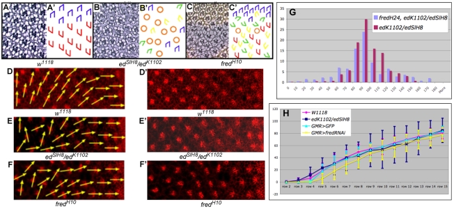
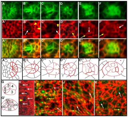
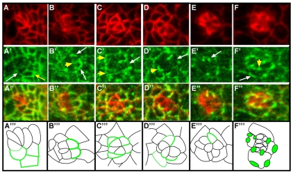
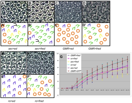
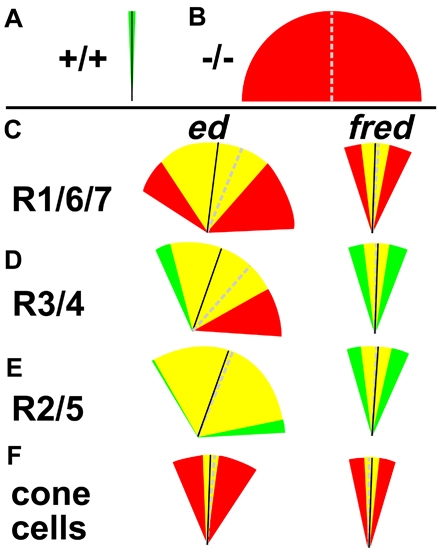
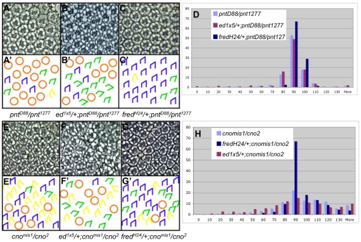
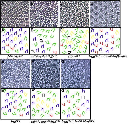
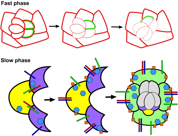
Directed cellular movements are a universal feature of morphogenesis in multicellular organisms. Differential adhesion between the stationary and motile cells promotes these cellular movements to effect spatial patterning of cells. A prominent feature of Drosophila eye development is the 90 degrees rotational movement of the multicellular ommatidial precursors within a matrix of stationary cells. We demonstrate that the cell adhesion molecules Echinoid (Ed) and Friend of Echinoid (Fred) act throughout ommatidial rotation to modulate the degree of ommatidial precursor movement. We propose that differential levels of Ed and Fred between stationary and rotating cells at the initiation of rotation create a permissive environment for cell movement, and that uniform levels in these two populations later contribute to stopping the movement. Based on genetic data, we propose that ed and fred impart a second, independent, ;brake-like' contribution to this process via Egfr signaling. Ed and Fred are localized in largely distinct and dynamic patterns throughout rotation. However, ed and fred are required in only a subset of cells - photoreceptors R1, R7 and R6 - for normal rotation, cells that have only recently been linked to a role in planar cell polarity (PCP). This work also provides the first demonstration of a requirement for cone cells in the ommatidial rotation aspect of PCP. ed and fred also genetically interact with the PCP genes, but affect only the degree-of-rotation aspect of the PCP phenotype. Significantly, we demonstrate that at least one PCP protein, Stbm, is required in R7 to control the degree of ommatidial rotation.


Figures

Fig. 1.
Fig. 1.
ed and fred mutant ommatidia misrotate. (A-C′) Tangential sections through adult Drosophila eyes (A-C) and corresponding schematics (A′-C′). (A) Wild type (w1118). Ommatidia come in two chiral forms, depicted in blue in the dorsal half and red in the ventral half of the eye. (B) Some edSlH8/edK1102 ommatidia under- or over-rotate (green and yellow trapezoids, respectively), and some contain an incorrect number of photoreceptors (orange circles). (C) Some ommatidia in fredH10 clones rotate correctly whereas others under- or over-rotate. (D-F′) The ed and fred orientation phenotypes result from aberrant ommatidial rotation. Anti-Arm staining (red) highlights the apical surface and outlines cell boundaries. Yellow vectors bisect R8 and run through the R3/R4 interface, highlighting the angle of orientation of each ommatidium. (D) Wild-type ommatidia follow a smooth progression of rotation. Ommatidial precursors in both edK1102/edSlH8 (E) and GMR>fredRNAi (F) knockdown eye discs misrotate. D′-F′ show D-F without arrows. (G) Reduction of fred activity enhances the ed mutant phenotype. Bar chart illustrating the percentage of ommatidia (y-axis) that are oriented at the angles indicated (x-axis) in edk1102/edSlH8 and edK1102, fredH24/edSlH8 eyes. (H) Graphical representation of data from D-F plotted as the mean angle of orientation (MAO) of ommatidia in each of four genotypes in rows 2-15. Error bars indicate the variance (s.d.). w1118 is the control for edK1102/edSlH8; GMR>GFP is the control for GMR>fredRNAi. The s.d. of ed and fred ommatidia is significantly different from that of the controls between rows 7-15. Trapezoid color for all schematics: blue and red, wild-type; green, under-rotated; yellow, over-rotated; black, fail to rotate; orange circles, incorrect number of photoreceptors.
Fig. 2.
Fig. 2.
Ed localization is dynamic throughout rotation. (A-F‴) Anti-Arm (A-F, green) and anti-Ed (A′-F′, red) staining, and merge (A″-F″), in sequentially older ommatidial precursors in Drosophila third instar eye disc. Corresponding schematics (A‴-F‴) are shown with Ed localization in ommatidial precursors represented by solid red lines, Ed localization in cells outside the ommatidial precursors shown in black, and dashed red lines indicating cell boundaries where Ed is below detectable levels. The intensity of Ed staining correlates with the line weight. (A-A‴) In row 1, Ed is localized in all cells. (B-B‴) By row 3, Ed levels have diminished in R8, R2 and R5 (white arrow). Ed puncta are visible (yellow arrowheads). (C-C‴) Just prior to the start of rotation, Ed levels drop in the photoreceptor cells (see also H); Ed is visible at the R3/R4 (white arrow), R2/R3 and R4/R5 interfaces and in puncta (yellow arrowhead). (D-D‴) Ed levels increase in the photoreceptors as rotation progresses (yellow arrow). (E-E‴) In row 8, Ed remains high in the photoreceptor and cone cells (white arrow), and levels equalize between rotating and non-rotating cells (yellow arrow). (F-F‴) At the completion of rotation, Ed is enriched at the cone cell/interommatidial cell (IOC) (yellow arrow) and the cone cell/photoreceptor cell (white arrow) boundaries. (G,G′) Key to the schematics in A‴-F‴. cc, cone cell. (H) Low-magnification image of an eye disc stained with anti-Ed antibody. Just before rotation begins, ommatidia with low levels of Ed appear as `holes' in the staining pattern (white arrows). Mitotic cells, which also resemble `holes' (yellow arrowhead), are distinct. (I,J) Ed (red) vesicles colocalize with (I) Rab5-GFP (green) and (J) Rab7-GFP (green) positive puncta in both IOCs (puncta shown by yellow arrows) and photoreceptor cells (puncta shown by white arrows). (K) Vesicular Ed (red) does not colocalize with anti-Rab11 staining (green) in recycling endosomes in either IOCs (yellow arrows) or photoreceptors (white arrows).
Fig. 3.
Fig. 3.
Fred localization is dynamic throughout rotation. (A-F‴) Anti-Arm (A-F, red) and anti-Fred (A′-F′, green) staining, and (A″-F″) merge, in increasingly older ommatidial precursors in Drosophila third instar eye disc. (A‴-F‴) Corresponding schematics, in which Fred localization is represented by green lines and line weight correlates with the intensity of Fred staining. (A-A‴) In row 3, Fred levels are enriched in R3 (white arrow), R4 (not evident in this image), and in the mystery cells (1-2 cells that lie between R3 and R4 in the developing ommatidial cluster but are ultimately expelled from the maturing ommatidial precursor; yellow arrow). (B-B‴) Just prior to the initiation of rotation, Fred localizes to the lateral edges of R3 and R4 (white arrows) and the R3/R4 boundary (yellow arrowhead). (C-C‴) In row 6, Fred begins to disappear from R3 (white arrow), but remains high in R4 and at the R3/R4 boundary. The newly added R1 and R6 contain high levels of Fred (yellow arrowheads). (D-D‴) In row 7, Fred disappears from R3 but is still high in R4 and at the R3/R4 boundary (white arrow). A bright band of Fred highlights the interface between R7 and R8 (yellow arrowhead), and Fred can still be seen faintly in R1 and R6. (E-E‴) By row 9, Fred is no longer present at the R3/R4 boundary, outlining only the periphery of R4 (white arrow). (F-F‴) At the end of rotation, Fred is enriched at the interfaces between the cone cells and the IOCs (white arrow) and also at the boundaries between the photoreceptors and the cone cells (yellow arrowhead).
Fig. 4.
Fig. 4.
Misexpression of ed or fred results in under-rotation. (A-F′) Sections through adult Drosophila eyes (A-F) and corresponding schematics (A′-F′) of ed and fred misexpression lines. (A,B) sev>ed and sev>fred ommatidia frequently under-rotate; very few ommatidia are missing photoreceptors. (C) GMR>ed tissue is severely disrupted, precluding analysis of angles of orientation. (D) Most ommatidia in GMR>fred adult eyes under-rotate. Additional defects (spacing, morphology) might be due to disrupted Egfr signaling. (E) Most ommatidia rotate 90° in ro>ed eyes; some ommatidia are missing photoreceptors. (F) By contrast, many ommatidia under-rotate in ro>fred eyes. (G) Graph of larval rotation, or MAO, for ommatidia in rows 2-15. y-axis, degree of rotation. Ommatidia in misexpression lines are under-rotated in rows 4-15 compared with controls (GMR>GFP). Error bars indicate s.d.
Fig. 5.
Fig. 5.
ed and fred are required in R1, R6, R7 and the cone cells for correct ommatidial rotation. (A) Schematic representation of wild-type MAO (black line; 90.6°) and wild-type variance (s.d., green wedge; 1.7). (B) Schematic representation of hypothetical mutant MAO (dashed gray line) and s.d. (red wedges). (C,F) ed and fred are required in photoreceptors R1, R6, R7 and the cone cells for rotation. (D,E) ed and fred are not required in R3/R4 or R2/R5 for rotation. The difference between the s.d. of the genetically wild-type and the genetically mutant populations is not significant. In C-F: black line, MAO when designated cells are genetically wild-type for ed or fred; dashed gray line, MAO when designated cells are genetically mutant for ed or fred; green wedges, genetically wild-type s.d; red wedges, genetically mutant s.d.; yellow wedges, overlap.
Fig. 6.
Fig. 6.
ed and fred interact genetically with pnt and cno. (A-C′) Sections through adult Drosophila eyes (A-C) and corresponding schematics (A′-C′). (A) pnt1277/pntΔ88 mutant eyes exhibit both over- and under-rotated ommatidia. (B) ed1x5/+, pntΔ88/pnt1277. Reducing ed activity enhances the pnt phenotype (i.e. the s.d. increases). (C) fredH24/+, pntΔ88/pnt1277. Reducing fred activity suppresses the pnt phenotype virtually to wild type. (D) Bar chart of angles of ommatidial orientation for pntΔ88/pnt1277, ed1x5/+, pntΔ88/pnt1277 and fredH24/+, pntΔ88/pnt1277. x-axis, MAO; y-axis, percentage. (E-G′) Sections (E-G) and corresponding schematics (E′-G′) for adult eyes of the indicated genotypes. (E) In cnomis1/cno2 mutant eyes, most ommatidia over-rotate. (F) ed1x5/+, cnomis1/cno2. Reducing ed activity enhances the cno phenotype. (G) fredH24/+; cnomis1/cno2. Reducing fred activity suppresses the cno phenotype. (H) Bar chart of angles of ommatidial orientation in cnomis1/cno2, ed1x5/+, cnomis1/cno2 and fredH24/+; cnomis1/cno2 adult eyes. x-axis, MAO; y-axis, percentage.
Fig. 7.
Fig. 7.
ed and fred interact genetically with different subsets of the PCP genes. (A-G′) Sections through adult Drosophila eyes (A-G) and corresponding schematics (A′-G′). Red trapezoids (this figure only) indicate dorsoventral inversions. (A) fzN21/fzJ22 mutant eyes exhibit both over- and under-rotated ommatidia. ed interacts specifically with the subset of PCP genes required in R3: fz, dgo and fmi. (B) ed1x5/+; fzN21/fzJ22. Reducing ed activity enhances the fz rotation phenotype without affecting the chirality phenotype. fred interacts with two PCP genes that are required in R4 for correct polarity: stbm and fmi. (C) stbm153 mutant eyes exhibit both over- and under-rotated ommatidia. (D) fredH24/+, stbm153/stbm153. Reducing fred activity strongly suppresses the stbm rotation phenotype. The fmifrz3 phenotype (E) is enhanced by both loss of ed function in ed1x5/+, fmifrz3/fmifrz3 (F) and loss of fred function in fredH24/+, fmifrz3/fmifrz (G).
Fig. 8.
Fig. 8.
Ed and Fred contribute to both phases of rotation. (Above) Differential levels or expression domains of Ed and Fred in rotating and non-rotating cells create a permissive environment for the faster phase of rotation. Levels of Ed are equivalent in cells within nascent ommatidial preclusters and IOCs (solid red lines of equal weight in the left panel). Immediately before rotation, cells that will rotate actively reduce their levels/distribution of Ed and Fred (middle; reduced Ed levels, thin red line; reduced number of cells expressing Fred, green). A decrease in adhesion (dashed red line, right) between rotating and stationary cells enables rotation to proceed. (Below) Ed and Fred regulate Egfr signaling during the slow phase of ommatidial rotation. When photoreceptors R1, R6 (purple) and R7 (yellow) join the cluster, they contain high levels of both Ed (red bars) and Fred (green bars). During the fast phase, Ed/Fred binding reduces inhibition by Ed of the Egf receptor (blue bars, left). Robust Egfr signaling inhibits Cno (blue hexagons) activity and, consequently, few stable adherens junctions (AJs) form (orange squares). Concurrent with the slower phase of rotation, Ed levels increase in R1, R6 and R7. Ed associates with the Egf receptor, inhibiting Egfr signaling (middle). As a result, Cno activity increases and stable AJs form between moving and stationary cells, effectively applying a brake at the rotation interface (middle). Rotation-specific signaling events shift to a new rotation interface upon recruitment of the cone cells (light green) into the cluster. At the completion of rotation, levels of AJ proteins (Ed, Fred, Cno and Arm) are high, an indication that these two subsets of cells adhere strongly to one another.

References

    1. Bai, J., Chiu, W., Wang, J., Tzeng, T., Perrimon, N. and Hsu, J. (2001). The cell adhesion molecule Echinoid defines a new pathway that antagonizes the Drosophila EGF receptor signaling pathway. Development 128, 591-601. - PubMed
    1. Bastock, R., Strutt, H. and Strutt, D. (2003). Strabismus is asymmetrically localised and binds to Prickle and Dishevelled during Drosophila planar polarity patterning. Development 130, 3007-3014. - PubMed
    1. Boettner, B., Harjes, P., Ishimaru, S., Heke, M., Fan, H. Q., Qin, Y., Van Aelst, L. and Gaul, U. (2003). The AF-6 homolog canoe acts as a Rap1 effector during dorsal closure of the Drosophila embryo. Genetics 165, 159-169. - PMC - PubMed
    1. Brown, K. E. and Freeman, M. (2003). Egfr signalling defines a protective function for ommatidial orientation in the Drosophila eye. Development 130, 5401-5412. - PubMed
    1. Chae, J., Kim, M. J., Goo, J. H., Collier, S., Gubb, D., Charlton, J., Adler, P. N. and Park, W. J. (1999). The Drosophila tissue polarity gene starry night encodes a member of the protocadherin family. Development 126, 5421-5429. - PubMed

Publication types

MeSH terms