Comparative Study
. 2009 Sep;66(9):938-46.
doi: 10.1001/archgenpsychiatry.2009.115.

Differential targeting of the CA1 subfield of the hippocampal formation by schizophrenia and related psychotic disorders

Affiliations
Comparative Study

Differential targeting of the CA1 subfield of the hippocampal formation by schizophrenia and related psychotic disorders

Scott A Schobel et al. Arch Gen Psychiatry. 2009 Sep.

Abstract

Context: Because schizophrenia and related disorders have a chronic time course and subtle histopathology, it is difficult to identify which brain regions are differentially targeted.

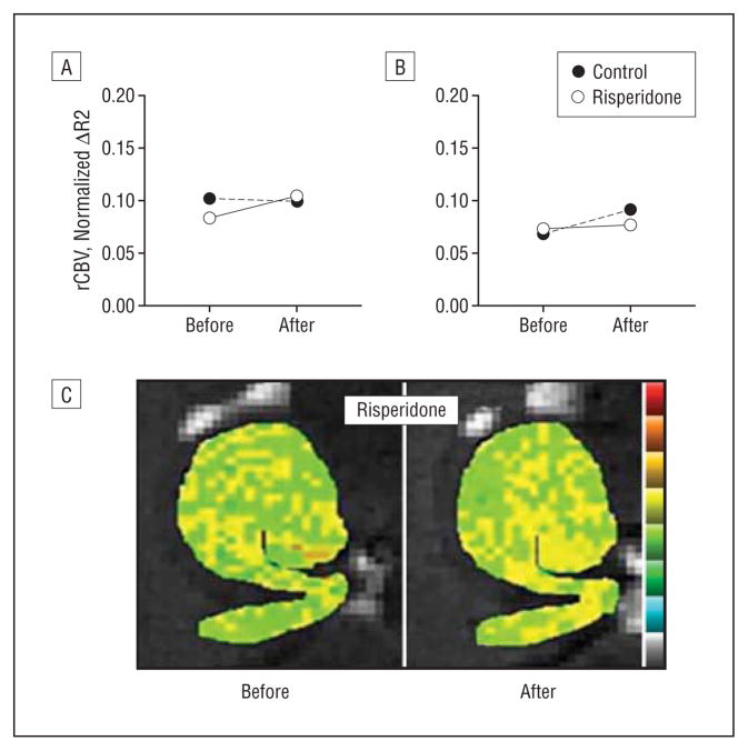
Objective: To identify brain sites differentially targeted by schizophrenia, we applied a high-resolution variant of functional magnetic resonance imaging to clinically characterized patients and matched healthy controls and to a cohort of prodromal subjects who were prospectively followed up. Additionally, to explore the potential confound of medication use, the fMRI variant was applied to rodents receiving an antipsychotic agent.

Design: Cross-sectional and prospective cohort designs.

Setting: Hospital clinic and magnetic resonance imaging laboratory.

Participants: Eighteen patients with schizophrenia, 18 controls comparable in age and sex, and 18 prodromal patients followed up prospectively for 2 years. Ten C57-B mice received an antipsychotic agent or vehicle control.

Main outcome measures: Regional cerebral blood volume (CBV), as measured with magnetic resonance imaging, and symptom severity, as measured with clinical rating scales.

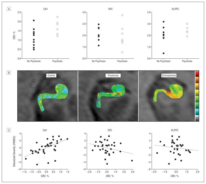
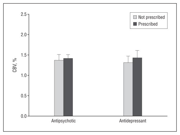
Results: In a first between-group analysis that compared patients with schizophrenia with controls, results revealed abnormal CBV increases in the CA1 subfield and the orbitofrontal cortex and abnormal CBV decreases in the dorsolateral prefrontal cortex. In a second longitudinal analysis, baseline CBV abnormalities in the CA1 subfield differentially predicted clinical progression to psychosis from a prodromal state. In a third correlational analysis, CBV levels in the CA1 subfield differentially correlated with clinical symptoms of psychosis. Finally, additional analyses of the human data set and imaging studies in mice suggested that antipsychotic agents were not confounding the primary findings.

Conclusions: Taken as a whole, the results suggest that the CA1 subfield of the hippocampal subregion is differentially targeted by schizophrenia and related psychotic disorders. Interpreted in the context of previous studies, these findings inform underlying mechanisms of illness progression.


Figures

Figure 1
Figure 1
Dysfunction in multiple regions in brain areas implicated in schizophrenia. A, Using high-resolution T1-weighted images, resting-state cerebral blood volume (CBV) mapping is a variant of functional magnetic resonance imaging that, as shown, can visualize the anterior and posterior hippocampus (a superior oblique view of the hippocampus is shown on the top left); subregions within the anterior hippocampus (green indicates entorhinal cortex; light blue, dentate gyrus; dark blue, CA3; red, CA1; yellow, subiculum); the posterior hippocampus (white); the frontal lobe in coronal section (inferior regions indicate orbitofrontal cortex and gyrus rectus; superior-lateral regions, dorsolateral cortex); the basal ganglia (medial regions indicate ventral caudate and nucleus accumbens; lateral regions, ventral putamen); and the amygdala (superior region indicates dorsal amygdala; inferior, basolateral amygdala). B, An increase in CBV between the 2 groups (control, schizophrenia) was observed selectively for the CA1 subfield of the hippocampal formation and the orbitofrontal cortex (OFC), while a CBV decrease was observed in the dorsolateral prefrontal cortex (DLPFC).
Figure 2
Figure 2
Resting-state cerebral blood volume (CBV) mapping in mice. A, A horizontal high-resolution T2-weighted magnetic resonance image is generated with a high-field 9.4-T scanner and used to obtain resting-state CBV maps of the mouse hippocampal formation. Green indicates entorhinal cortex; white, dentate gyrus; yellow, CA3; red, CA1; dark blue, subiculum. B, A horizontal brain section with Nissl staining shows the correspondence between the magnetic resonance imaging scan and the histological anatomy of the individual hippocampal subregions. ERC indicates entorhinal cortex; SUB, subiculum; CA1, CA1 subfield; CA3, CA3 subfield; DG, dentate gyrus.
Figure 3
Figure 3
The CA1 subfield is a site of dysfunction selectively associated with clinical features. A, Cerebral blood volume (CBV) measured in the CA1 subfield, but not the orbitofrontal cortex (OFC) or dorsolateral prefrontal cortex (DLPFC), was significantly elevated at baseline, comparing the prodromal subjects who clinically progressed to psychosis with those who did not. B, Individual CBV maps of the hippocampal formation are shown for a healthy control, a prodromal subject, and a patient with schizophrenia. The CBV maps are color coded such that warmer colors reflect higher CBV values. Higher CBV was observed in the CA1 subfield of the prodromal subject, and higher CBV was observed in the CA1 and subiculum in the patient with schizophrenia. C, CA1 CBV, but not OFC or DLPFC CBV correlated with positive symptoms, in particular, delusional severity. PANSS indicates Positive and Negative Symptom Scale.
Figure 4
Figure 4
Cerebral blood volume (CBV) measured in the CA1 subfield compares the prodromal subjects who were prescribed antidepressant or antipsychotic drugs with those who were not.
Figure 5
Figure 5
Relative cerebral blood volume (rCBV) maps were generated in mice before and after long-term administration of the dopamine blocking agent risperidone. Risperidone had no effect on hippocampal CBV, as shown for averaged group data measured in the CA1 subfield (A) and entorhinal cortex (B) as well as a hippocampal CBV map from a single mouse before and after receiving risperidone (C).

References

    1. Shenton ME, Dickey CC, Frumin M, McCarley RW. A review of MRI findings in schizophrenia. Schizophr Res. 2001;49(1–2):1–52. - PMC - PubMed
    1. Zhao X, Lein ES, He A, Smith SC, Aston C, Gage FH. Transcriptional profiling reveals strict boundaries between hippocampal subregions. J Comp Neurol. 2001;441(3):187–196. - PubMed
    1. Small SA, Nava AS, Perera GM, Delapaz R, Stern Y. Evaluating the function of hippocampal subregions with high-resolution MRI in Alzheimer’s disease and aging. Microsc Res Tech. 2000;51(1):101–108. - PubMed
    1. Harrison PJ. The neuropathology of schizophrenia: a critical review of the data and their interpretation. Brain. 1999;122(pt 4):593–624. - PubMed
    1. Yung AR, McGorry PD. The prodromal phase of first-episode psychosis: past and current conceptualizations. Schizophr Bull. 1996;22(2):353–370. - PubMed

Publication types

MeSH terms

Substances