. 2010 Jul;19(7):1095-107.
doi: 10.1089/scd.2009.0217.

Reconstitution of marrow-derived extracellular matrix ex vivo: a robust culture system for expanding large-scale highly functional human mesenchymal stem cells

Affiliations

Reconstitution of marrow-derived extracellular matrix ex vivo: a robust culture system for expanding large-scale highly functional human mesenchymal stem cells

Yanlai Lai et al. Stem Cells Dev. 2010 Jul.

Abstract

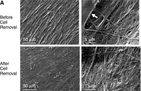
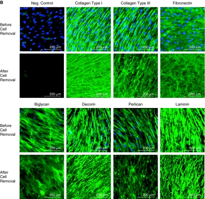
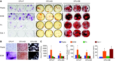
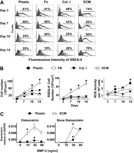
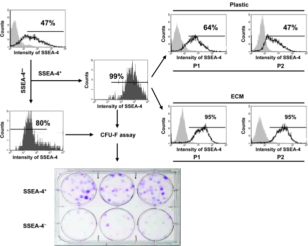
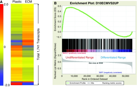
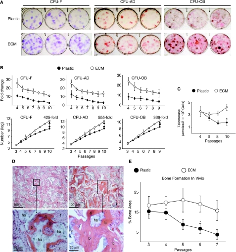
The difficulty in long-term expansion of mesenchymal stem cells (MSCs) using standard culture systems without the loss of their stem cell properties suggests that a critical feature of their microenvironment necessary for retention of stem cell properties is absent in these culture systems. We report here the reconstitution of a native extracellular matrix (ECM) made by human marrow cells ex vivo, which consists of at least collagen types I and III, fibronectin, small leucine-rich proteoglycans such as biglycan and decorin, and major components of basement membrane such as the large molecular weight proteoglycan perlecan and laminin. Expansion of human MSCs on this ECM strongly promoted their proliferation, retained their stem cell properties with a low level of reactive oxygen species (ROS), and substantially increased their response to BMP-2. The quality of the expanded cells following each passage was further tested by an in vivo transplantation assay. The results showed that MSCs expanded on the ECM for multiple passages still retained the same capacity for skeletogenesis. In contrast, the bone formation capacity of cells expanded on plastic was dramatically diminished after 6-7 passages. These findings suggest that the marrow stromal cell-derived ECM is a promising matrix for expanding largescale highly functional MSCs for eventualuse in stem cell-based therapy. Moreover, this system should also be invaluable for establishment of a unique tissue-specific ECM, which will facilitate control of the fate of MSCs for therapeutic applications.


Figures

FIG. 1.
FIG. 1.
Characteristics of human marrow stromal cell-derived extracellular matrix (ECM). (A) Scanning electron microscopy (SEM) images of stromal cell-derived ECM before and after cell removal. Left panels: low magnification; and right panels: high magnification. The structure of the ECM appeared to be similar before and after cell removal. The arrow denotes a cell. (B) Confocal fluorescence images showing localization of collagen types I and III, fibronectin, biglycan, decorin, perlecan, and laminin in the ECM elaborated by human bone marrow stromal cells before and after cell removal. The distribution of cells was visualized with DAPI staining (blue), and matrix proteins by immunofluorescence (green).
FIG. 1.
FIG. 1.
Characteristics of human marrow stromal cell-derived extracellular matrix (ECM). (A) Scanning electron microscopy (SEM) images of stromal cell-derived ECM before and after cell removal. Left panels: low magnification; and right panels: high magnification. The structure of the ECM appeared to be similar before and after cell removal. The arrow denotes a cell. (B) Confocal fluorescence images showing localization of collagen types I and III, fibronectin, biglycan, decorin, perlecan, and laminin in the ECM elaborated by human bone marrow stromal cells before and after cell removal. The distribution of cells was visualized with DAPI staining (blue), and matrix proteins by immunofluorescence (green).
FIG. 2.
FIG. 2.
Stromal cell-derived extracellular matrix (ECM) enhances human mesenchymal stem cells (MSCs) in colony formation. (A) The appearance of CFU-F, CFU-AD, and CFU-OB colonies generated on the various substrata. Freshly isolated human bone marrow mononuclear cells were placed into uncoated plastic (Plastic), or plastic coated with a cell-free ECM (ECM), fibronectin (Fn), or collagen type I (Col. I) at 3 × 105 cells per 10 cm2 area. After 24 h of incubation, non-adherent cells were removed and cultures maintained in α-MEM containing 15% FBS. After 14 days of culture, CFU-F colonies were visualized with crystal violet staining. CFU-OB colonies were generated by cells cultured in osteoblast differentiation medium. After 25 days of culture, CFU-OB colonies were visualized with von Kossa staining. CFU-adipocytes (CFU-AD) colonies were formed by cells cultured in adipogenic medium. After 10 days of culture, CFU-AD colonies were visualized with Oil Red O staining. (B) Microscopic views of CFU-F, CFU-AD, and CFU-OB colonies formed on plastic or on the ECM. Original magnification: 100×. (C) Quantification of average size and intensity per colony formed on the various substrata using the ImageJ program. Osteocalcin secretion in supernatant collected from CFU-OB was measured using a Metra Osteocalcin EIA kit (QUIDEL Corporation, San Diego, CA). *P < 0.05, n = 3 vs. plastic or plastic coated with fibronectin (Fn) or collagen type I (Col. I). P < 0.05, n = 3 vs. plastic or plastic coated with Fn.
FIG. 3.
FIG. 3.
Stromal cell-derived extracellular matrix (ECM) promotes human mesenchymal stem cell (MSC) proliferation and suppresses reactive oxygen species (ROS) formation. (A) Flow cytometric analysis of SSEA-4 expression by human MSCs from passage 2. Single-cell suspensions derived from cultures on uncoated plastic (Plastic), a cell-free ECM, or fibronectin (Fn), or collagen type I (Col. I) for various days were analyzed by fluorescence-activated cell sorting (FACS). Cells stained with primary nonspecific antibody (isotype, IgG) served as negative controls (gray peaks). (B) SSEA-4 and ROS analysis. Other cell aliquots were used to determine cell number (left panel), the number of SSEA-4+ cells (middle panel), and ROS content (right panel) expressed as arbitrary units (AU) of DCF fluorescence per 105 cells. *P < 0.05, n = 3 vs. plastic, plastic coated with fibronectin (Fn) or collagen type I (Col. I) at the same time point. (C) Enhanced BMP-2 responsiveness of MSCs cultured on ECM. Cells were cultured on ECM or uncoated plastic (Plastic) or plastic coated with fibronectin (Fn) or collagen type I (Col. I) in the expansion medium for 10 days, and then cultured in osteoblast differentiation medium with 2% fetal bovine serum (FBS) overnight and then treated for 3 days with varying doses of BMP-2, as indicated. Gene expression of osteocalcin and bone sialoprotein was determined by quantitative RT-PCR (TaqMan). n = 3; *P < 0.05, value at the lowest dose needed for the stimulation vs. vehicle control.
FIG. 4.
FIG. 4.
Marrow stromal cell-derived extracellular matrix (ECM) retains SSEA-4+ cells and enriches colony-forming cells. Freshly isolated human bone marrow mononuclear cells were cultured on tissue culture plastic at an initial seeding 3 × 105 cells/cm2 until 70% confluence (2–3 weeks) in the expansion medium. After removal of non-adherent cells, the cultured bone marrow adherent cells were detached and stained with a specific antibody against SSEA-4. SSEA-4+ cells and SSEA-4 cells were sorted using fluorescence-activated cell sorting (FACS). CFU-F assay was performed to determine the frequency of CFU-F in the sorted SSEA-4+ and SSEA-4 cell populations. In addition, SSEA-4+ cells were subcultured on either ECM or tissue culture plastic (Plastic) for 2 passages (P1 and P2). SSEA-4 expression was analyzed by FACS following each passage. For a negative control (gray peak), cells were stained with primary non-specific antibody (isotype). Simultaneously, the sorted SSEA-4+ or SSEA-4 cells were placed onto tissue culture plastic at 300 cells per well (∼10 cm2 area) in triplicate and cultured for 14 days in 3 mL α-MEM containing 15% FBS. CFU-F colonies were then visualized with crystal violet staining.
FIG. 5.
FIG. 5.
Global gene expression patterns for human mesenchymal stem cells (MSCs) cultured on extracellular matrix (ECM) compared to cells cultured on plastic. (A) Gene expression signatures of human MSCs maintained on plastic versus ECM for 12 days. They are presented by hierarchical clustering of 1,741 transcripts that were significantly up- or down-regulated by the ECM as compared to plastic. Color bar represents the range of expression levels indicated by log2 scale. (B) Enrichment plot of the 721 up-regulated transcripts on the ECM. The majority of this gene set was overrepresented within a ranked list of genes expressed by undifferentiated bone marrow stem cell (BMSC), shown in red. Normalized enrichment score (NES) was 1.76 [Actual ES divided by Mean (ESs against all permutations of the dataset)]; and a Family Wise-error Rate (FWER) P value was 0.016, which estimates the probability that the normalized enrichment score represents a false positive finding.
FIG. 6.
FIG. 6.
Stromal cell-derived extracellular matrix (ECM) promotes replication of CFUs, and retains the ability of mesenchymal stem cells (MSCs) to form skeletal tissue in vivo. (A) Appearance of CFU-F, CFU-AD, and CFU-OB assayed after 7 passages of expansion on plastic or ECM. (B) Cell replication. Upper panels: replication of colony-forming cells expanded on the ECM versus plastic, expressed as fold changes in number of colonies with increasing passage number. The replicative activity of MSCs maintained on the ECM was significantly higher (P < 0.05) than those of MSCs maintained on plastic at all time points. Lower panels: growth kinetics of colony-forming cells (log scale) expanded on ECM versus plastic with increasing passage number. *P < 0.05, value at the earliest passage when cells expanded on ECM showed increased colony-forming activity versus plastic. (C) Telomerase activity in cells expanded on ECM versus plastic with increasing passage number. *P < 0.05 (by ANOVA), ECM versus plastic (before P8). (D) Histology of ossicle produced by implantation of P7 human bone marrow cells. While bone was formed by cells expanded on plastic (left panels), as well as by cells expanded on ECM (right panels); high magnification (lower panels) of areas selected in upper panels clearly showed more robust bone formation in the latter. b, bone; f, fibrous tissue; and ha, HA/TCP. (E) Following each passage, the cells (1 × 106) were loaded into HA/TCP ceramic powder and transplanted subcutaneously into the dorsal surface of 10-week-old immunodeficient mice. Three implants for each group were harvested at 8 weeks post-implantation. The extent of new bone formed in the implants was histomorphometrically determined as areas measured by using the ImageJ analysis software. n = 3; *P < 0.05, value at the earliest passage versus that at the passage 3 or 4. Color images available online at www.liebertonline.com/scd.

Similar articles

Cited by

References

    1. Prockop DJ. Marrow stromal cells as stem cells for non-hematopoietic tissues. Science. 1997;276:71–74. - PubMed
    1. Dennis JE. Merriam A. Awadallah A. Yoo JU. Johnstone B. Caplan AI. A quadripotential mesenchymal progenitor cell isolated from the marrow of an adult mouse. J Bone Miner Res. 1999;14:700–709. - PubMed
    1. Ferrari G. Cusella-De AG. Coletta M. Paolucci E. Stornaiuolo A. Cossu G. Mavilio F. Muscle regeneration by bone marrow derived myogenic progenitors. Science. 1998;279:1528–1530. - PubMed
    1. Kassem M. Stem cells: potential therapy for age-related diseases. Ann N Y Acad Sci. 2006;1067:436–442. - PubMed
    1. Banerjee M. Bhonde RR. Autologous bone marrow transplantation/mobilization: a potential regenerative medicine for systemic degenerative disorders and healthy living. Med Hypotheses. 2007;68:1247–1251. - PubMed

Publication types

MeSH terms

LinkOut - more resources