. 2010 Feb;16(2):605-15.
doi: 10.1089/ten.TEA.2009.0518.

Stem/progenitor cell-mediated de novo regeneration of dental pulp with newly deposited continuous layer of dentin in an in vivo model

Affiliations

Stem/progenitor cell-mediated de novo regeneration of dental pulp with newly deposited continuous layer of dentin in an in vivo model

George T-J Huang et al. Tissue Eng Part A. 2010 Feb.

Abstract

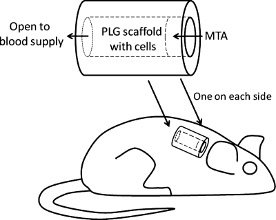
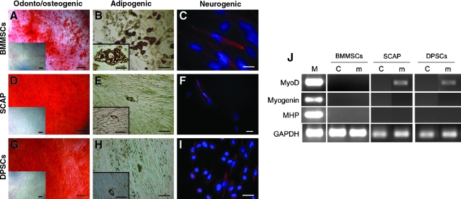
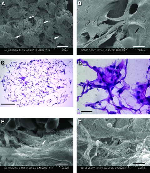
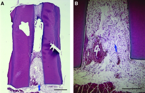
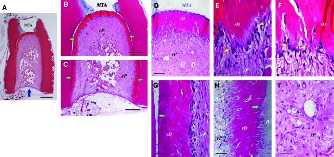
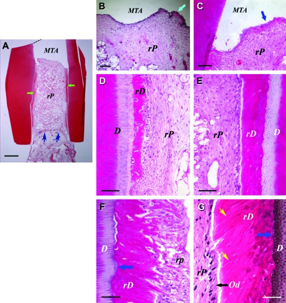
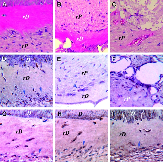
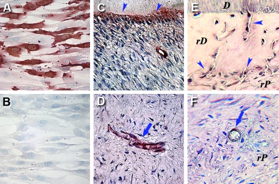
The ultimate goal of this study is to regenerate lost dental pulp and dentin via stem/progenitor cell-based approaches and tissue engineering technologies. In this study, we tested the possibility of regenerating vascularized human dental pulp in emptied root canal space and producing new dentin on existing dentinal walls using a stem/progenitor cell-mediated approach with a human root fragment and an immunocompromised mouse model. Stem/progenitor cells from apical papilla and dental pulp stem cells were isolated, characterized, seeded onto synthetic scaffolds consisting of poly-D,L-lactide/glycolide, inserted into the tooth fragments, and transplanted into mice. Our results showed that the root canal space was filled entirely by a pulp-like tissue with well-established vascularity. In addition, a continuous layer of dentin-like tissue was deposited onto the canal dentinal wall. This dentin-like structure appeared to be produced by a layer of newly formed odontoblast-like cells expressing dentin sialophosphoprotein, bone sialoprotein, alkaline phosphatase, and CD105. The cells in regenerated pulp-like tissue reacted positively to anti-human mitochondria antibodies, indicating their human origin. This study provides the first evidence showing that pulp-like tissue can be regenerated de novo in emptied root canal space by stem cells from apical papilla and dental pulp stem cells that give rise to odontoblast-like cells producing dentin-like tissue on existing dentinal walls.


Figures

FIG. 1.
FIG. 1.
SCID mouse subcutaneous study model for pulp/dentin regeneration. The canal space of human tooth root fragments (∼6–7 mm long) was enlarged to ∼1–2.5 mm in diameter. One end of the canal opening was sealed with MTA cement. SCID, severe combined immunodeficient; MTA, mineral trioxide aggregate.
FIG. 2.
FIG. 2.
Multiple lineage differentiation properties of SCAP and DPSCs compared with BMMSCs. (A, D, G) Odonto/osteogenic induction and Alizarin Red staining of matrix mineralization. Insets: uninduced control. All scale bars including insets: 200 μm. (B, E, H) Adipogenic induction and Oil Red O staining of the accumulated lipid droplets in cells. Inset images: higher magnification views of the adipocyte-like cells from the same culture shown in the main images. Scale bars: main images, 100 μm; insets, 50 μm. (C, F, I) Neurogenic induction. βIII-tubulin: red fluorescence; 4',6-diamidino-2-phenylindole dihydrochloride (DAPI): blue fluorescence. Scale bars: (C) 20 μm; (F, I) 30 μm. (J) Myogenic induction and RT-polymerase chain reaction analysis of gene expression. M, RNA from cultured human myoblasts (obtained from Dr. Wesley M. Jackson, NIH/NIAMS, Bethesda, MD); C, uninduced control; m, induction in myogenic medium. SCAP, stem cells from apical papilla; DPSC, dental pulp stem cells; BMMSCs, bone marrow mesenchymal stem cells.
FIG. 3.
FIG. 3.
Cell attachment and growth in the PLG scaffold in vitro. Scanning electronic microscopy of DPSCs seeded onto PLG after (A) 10 days and (B)14 days in culture. (C, D) H&E staining of DPSCs seeded onto PLG after 7 weeks in culture. (E, F) Scanning electronic microscopy of SCAP seeded onto PLG after 8 weeks in culture. Scale bars: (A) 200 μm; (B, D, E) 20 μm; (C) 250 μm; and (F) 1 μm. Arrows in (A) indicate attached cells, in (B) scaffold surface, in (E and F) fiber-like matrix. PLG, poly-D,L-lactide and glycolide; H&E, hemotoxylin and eosin. Color images available online at www.liebertonline.com/ten.
FIG. 4.
FIG. 4.
Histological analysis of control group in the mouse subcutaneous model. (A) A root fragment with empty canal space was transplanted into a SCID mouse for 3 months before being removed and processed for H&E staining. Mouse subcutaneous tissue ingrown from the canal opening (arrow). (B) Higher magnification view showing the ingrown fatty tissue (arrow). Scale bars: (A) 1 mm; (B) 0.5 mm.
FIG. 5.
FIG. 5.
Histological analysis of in vivo pulp/dentin regeneration using SCAP. A root fragment was prepared and the canal space inserted with SCAP/PLG and transplanted into a SCID mouse for 3 months. The sample was harvested and processed for H&E staining. D, original dentin; rD, regenerated dentin-like tissue; rP, regenerated pulp-like tissue. Blue arrow in (A) indicates the blood supply entrance; green arrows in (B) and (C) indicate continuous layer of uniformed thickness of rD; yellow arrows in (E) and (F) indicate the region of well-aligned odontoblast-like cells with polarized cell bodies; green arrows in (G and H) indicate junctions between D and rD. Scale bars: (A) 1 mm; (B and C) 500 μm; (D) 100 μm; (E and F) 20 μm; and (G–I) 50 μm.
FIG. 6.
FIG. 6.
Histological analysis of in vivo pulp/dentin regeneration using DPSCs. Samples were prepared using the same procedures as described in Figure 5, except that the sample was harvested from the SCID mouse 4 months after implantation. D, original dentin; rD, regenerated dentin-like tissue; rP, regenerated pulp-like tissue. Od, odontoblast-like cells. Green arrows in (A) indicate rD; blue arrows in (A) indicate the entrance of blood supply; blue arrows in (B and C) indicate the thin layer of rD under MTA cement; blue arrows in (F and G) indicate the junction of D and rD; black arrow in (G) indicates well-aligned odontoblast-like cells. Yellow arrows in (G) indicate dentinal tubule-like structures. Scale bars: (A) 1 mm; (B) 200 μm; (C–E) 100 μm; and (F and G) 50 μm. Color images available online at www.liebertonline.com/ten.
FIG. 7.
FIG. 7.
Immunohistochemical analysis of pulp tissue regenerated in vivo using SCAP. (A–C) H&E staining; immunostaining for (D–F) human mitochondria; (G) dentin sialoprotein; (H) alkaline phosphatase and (I) bone sialoprotein. D, original dentin; rD, regenerated dentin-like tissue; rP, regenerated pulp-like tissue. Arrows indicate cells with positive staining. Color images available online at www.liebertonline.com/ten.
FIG. 8.
FIG. 8.
Immunocyto/histochemical staining of CD105. Cultured SCAP immunostained with (A) anti-CD105 antibody or (B) nonimmune IgG as negative control. (C) Positive staining of odontoblasts (arrowheads) in a sample of natural human dental pulp/dentin tissue. (D) A blood vessel in the natural pulp is positive for CD105 (arrow). (E) Positive immunostaining is seen in odontoblast-like cells lining against rD or inside rD (arrowheads). (F) CD105-positive cells are seen in rP and around a blood vessel (arrow). rD, regenerated dentin-like tissue; rP, regenerated pulp-like tissue. Color images available online at www.liebertonline.com/ten.

References

    1. Mooney D.J. Powell C. Piana J. Rutherford B. Engineering dental pulp-like tissue in vitro. Biotechnol Prog. 1996;12:865. - PubMed
    1. Bohl K.S. Shon J. Rutherford B. Mooney D.J. Role of synthetic extracelluar matrix in development of engineered dental pulp. J Biomaterials Science. (Polymer Edition) 1998;9:749. - PubMed
    1. Buurma B. Gu K. Rutherford R.B. Transplantation of human pulpal and gingival fibroblasts attached to synthetic scaffolds. Eur J Oral Sci. 1999;107:282. - PubMed
    1. Huang G.T.J. Sonoyama W. Liu Y. Liu H. Wang S. Shi S. The hidden treasure in apical papilla: the potential role in pulp/dentin regeneration and bioroot engineering. J Endod. 2008;34:645. - PMC - PubMed
    1. Huang G.T.-J. Apexification: the beginning of its end. Int Endod J. 2009;42:855. - PubMed

Publication types

MeSH terms