. 2009 Oct;8(7):699-713.
doi: 10.1111/j.1601-183X.2009.00514.x. Epub 2009 Jun 22.

Analysis of the mouse mutant Cloth-ears shows a role for the voltage-gated sodium channel Scn8a in peripheral neural hearing loss

Affiliations
Free PMC article

Analysis of the mouse mutant Cloth-ears shows a role for the voltage-gated sodium channel Scn8a in peripheral neural hearing loss

F E Mackenzie et al. Genes Brain Behav. 2009 Oct.
Free PMC article

Abstract

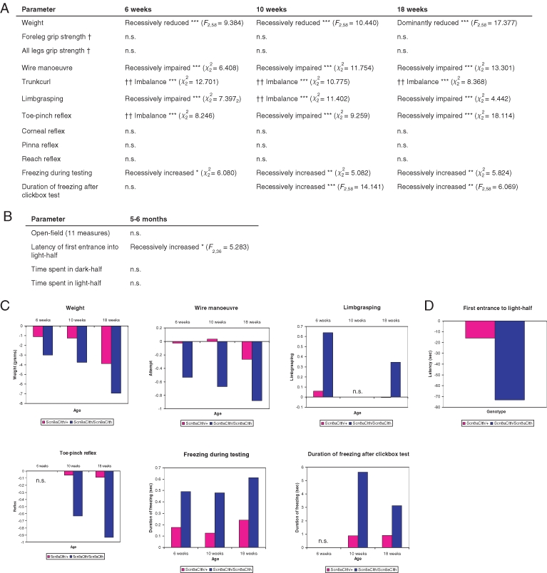
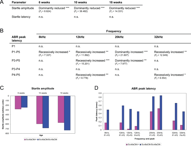
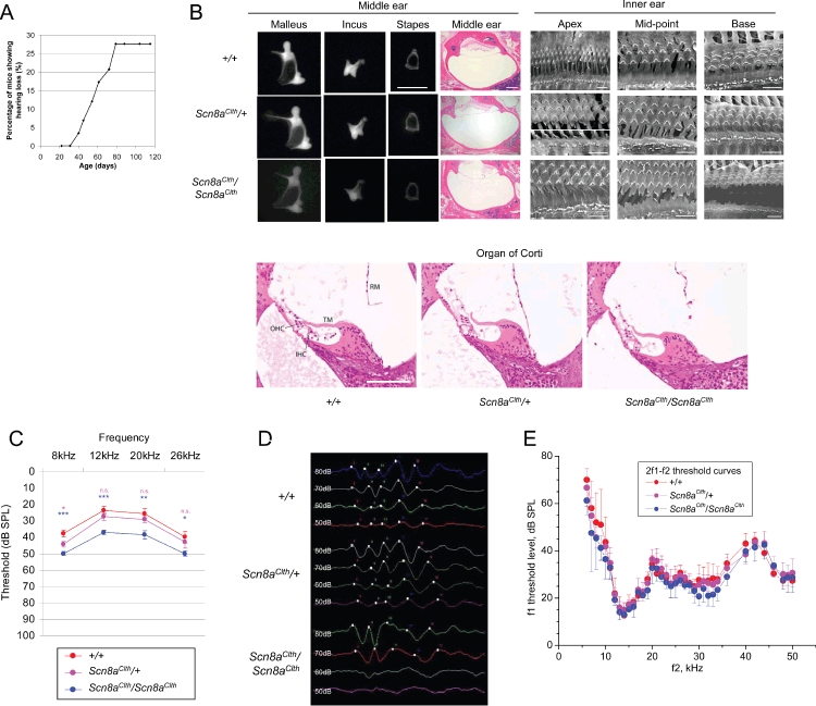
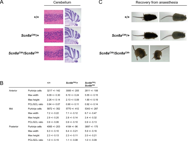
Deafness is the most common sensory disorder in humans and the aetiology of genetic deafness is complex. Mouse mutants have been crucial in identifying genes involved in hearing. However, many deafness genes remain unidentified. Using N-ethyl N-nitrosourea (ENU) mutagenesis to generate new mouse models of deafness, we identified a novel semi-dominant mouse mutant, Cloth-ears (Clth). Cloth-ears mice show reduced acoustic startle response and mild hearing loss from approximately 30 days old. Auditory-evoked brainstem response (ABR) and distortion product otoacoustic emission (DPOAE) analyses indicate that the peripheral neural auditory pathway is impaired in Cloth-ears mice, but that cochlear function is normal. In addition, both Clth/Clth and Clth/+ mice display paroxysmal tremor episodes with behavioural arrest. Clth/Clth mice also show a milder continuous tremor during movement and rest. Longitudinal phenotypic analysis showed that Clth/+ and Clth/Clth mice also have complex defects in behaviour, growth, neurological and motor function. Positional cloning of Cloth-ears identified a point mutation in the neuronal voltage-gated sodium channel alpha-subunit gene, Scn8a, causing an aspartic acid to valine (D981V) change six amino acids downstream of the sixth transmembrane segment of the second domain (D2S6). Complementation testing with a known Scn8a mouse mutant confirmed that this mutation is responsible for the Cloth-ears phenotype. Our findings suggest a novel role for Scn8a in peripheral neural hearing loss and paroxysmal motor dysfunction.


Figures

Figure 5:
Figure 5:
(a) Statistical analysis for genotypic differences of weight, neurological, neuromuscular and behavioural phenotypes of Scn8aClth mice at 6, 10 and 18 weeks (n = 73), where Ft,u denotes the F-statistic on t degrees of freedom. Continuous numeric data were analysed under linear models, binary data using generalized linear models and ordinal data using proportion odds logistic regression. Phenotypes significantly different to wild types are given with the best fit of inheritance. n.s., not significant; *P < 0.05; **P < 0.01; ***P < 0.001 (unadjusted P values). † = Only +/+ and Scn8aClth/Scn8aClth were tested. †† = Where an imbalance of design confounded statistical analysis, manual analysis of individual data points showed that Scn8aClth/Scn8aClth displayed increased trunk curling and limb grasping and impaired toe-pinch reflex compared with other genotypes. (b) Statistical analysis for genotypic differences of anxiety and locomotion behaviour of Scn8aClth mice at 5–6 months old (n = 39), where Ft,u denotes the F-statistic on t degrees of freedom. Tests were performed using the open-field and light-dark box behavioural paradigms. Analysis was performed under a linear model. Phenotypes significantly different to wild types are given with the best fit of inheritance. n.s., not significant; *P < 0.05; **P < 0.01; ***P < 0.001 (unadjusted P values). (c) Graphical representations of significantly different weight, neurological, motor and behavioural phenotypes in Scn8aClth mice, showing data for adjusted P values < 0.05. Data shown are estimate values for Scn8aClth/+ and Scn8aClth/Scn8aClth mice, having adjusted for sex and mating identifier, where +/+ is normalized to = 0. n.s., not significant. (d) Graphical representation of significantly different anxiety/locomotor-related phenotypes in Scn8aClth mice compared with wild types, showing data for adjusted P values < 0.05.. Data shown are estimate values for Scn8aClth/+ and Scn8aClth/Scn8aClth mice, having adjusted for sex and mating identifier, where +/+ is normalized to = 0. n.s., not significant
Figure 1:
Figure 1:
(a) Haplotypes of backcross and intercross progeny segregating the Cloth-ears mutation. The Cloth-ears (Clth) mutation arose on a mutagenized (BALB/cAnN × C3H/HeH)F1 individual. Sixty-four backcross progeny with a reduced startle response and/or an intermittent tremor and 26 intercross progeny with or without a continuous tremor were mapped across chromosome 8 and genotyped at recombinant markers. The genetic data indicates that the mutagenized chromosome was from the BALB/cAnN strain. The Cloth-ears mutation is non-recombinant with the SNP markers Scn8a_SNP and rs4231023 and maps to the interval D15Mit97.2–rs8266857. (b) Chromatograms of the Cloth-ears mutation from wild-type (+/+) and Scn8aClth/Scn8aClth DNA. Arrow: base 2942 of the Scn8a coding sequence, showing an adenine (A) in wild-type mice and a thymine (T) in Scn8aClth/Scn8aClth mice (arrow). The predicted amino acid sequence from wild-type and Scn8aClth/Scn8aClth mice is also indicated, highlighting the D981V change. (c) Position of the Cloth-ears mutation (D981V) in the SCN8A amino acid sequence. D981V resides within the predicted domain 2 (D2) of the SCN8A peptide, six amino acids downstream of the S6 transmembrane segment. The amino acid sequence of domain 2 (D2) is shown in black; sequence of domain 2 segment 6 (D2S6) is shown in pink and the position of the Cloth-ears mutation (D981V) is shown in bold red. (d) Schematic of the voltage-gated sodium channel α- and β-subunit proteins (adapted from Meisler & Kearney 2005). The α-subunit comprises four homologous domains (D1–D4 or DI–DIV), which are shown in different colours, each containing six transmembrane segments (S1–S6). S4 segments function as voltage sensors and the S5–S6 loops form the outer pore. A pore structure is formed by the transmembrane segments in the membrane (inset). The location of the Cloth-ears mutation is shown (red star). (e) Alignment of peptide sequences of SCN8A orthologues and other mouse SCN α-subunits (Ensembl) in the region of amino acid 981 (arrow). Alignments were made using clustal-W web-based software.
Figure 3:
Figure 3:
(a) Statistical analysis for genotypic differences of hearing phenotypes of Scn8aClth mice at 6, 10 and 18 weeks (n = 73), where Ft,u denotes the F-statistic on t degrees of freedom. Analysis was performed under a linear model (see Methods). Startle amplitudes and maximal latencies were measured in response to 40 ms soundbursts of white noise at 110 dB SPL using the startlebox paradigm. Parameters significantly different to wild types are given with the best fit of inheritance. n.s., not significant; *P < 0.05; **P < 0.01; ***P < 0.001 (unadjusted P values). (b) Statistical analysis for genotypic differences of ABR peak latencies of Scn8aClth mice at 5–7 months old, where Ft,u denotes the F-statistic on t degrees of freedom. Analysis was performed under a linear model (see Methods). Parameters significantly different to wild types are given with the best fit of inheritance. n.s., not significant; *P < 0.05; **P < 0.01; ***P < 0.001 (unadjusted P values). (c and d) Graphical representation of significantly different startle response and ABR peak latency parameters in Scn8aClth mice compared with wild types, showing data for adjusted P values < 0.05. Startle response was measured by placing mice in a soundproof chamber containing a loudspeaker to generate sound. Startle amplitude was calculated in arbitary units using an accelerometer connected to the chamber floor to measure floor displacement upon sound presentation. Data shown are estimate values for Scn8aClth/+ and Scn8aClth/Scn8aClth mice, having adjusted for sex and mating identifier, where +/+ is normalized to = 0.
Figure 2:
Figure 2:
(a) Age of onset and penetrance of reduced startle response in Scn8aClth mice. Startle response analysis of 58 age-matched, N4 mice from a (Clth/+× +/+) cross, comprising ungenotyped Scn8aClth/+ and +/+ progeny, showed that 27.6% of the cohort display a reduced startle response to a 90 dB SPL, 20 kHz single tone burst, indicating reduced penetrance. As mice are ungenotyped, these data include both +/+ and Clth/+ mice. The onset of reduced startle response was between 31 and 79 days. (b) Representative histology of middle and inner ears of Scn8aClth mice. No morphological differences in structure, size or bone density of middle ear ossicles from 6–10-month-old Scn8aClth/+ and Scn8aClth/Scn8aClth mice were seen (scale bar, 1 mm). The small protusion on the stapes seen in the +/+ sample has detached with the incus during dissection in the Scn8aClth/+ sample. H&E-stained 8 μm dorsal sections of the middle ear of 8-month-old Scn8aClth/+ and Scn8aClth/Scn8aClth mice shows that the middle ear cavity is free from effusive matter and the epithelium is smooth, indicating the absence of otitis media, and the tympanic membrane is intact (scale bar, 500 μm). H&E-stained 5 μm cochlear sections from 6-month-old Scn8aClth/+ and Scn8aClth/Scn8aClth mice shows a normal organ of Corti (OHC, outer hair cell; IHC, inner hair cell; TM, tectorial membrane; RM, Reissner's membrane; scale bar, 100 μm). In some samples, Reissner's membrane has detached during processing. Scanning electron microscopy of the exposed organ of Corti from 6- to 7-month-old Scn8aClth/+ and Scn8aClth/Scn8aClth mice shows three rows of OHCs and IHCs in all samples (scale bar, 8 μm). No patterning abnormalities or degeneration of stereocilia or hair cells was seen. Some artefacts of sample preparation can be seen (spherical blebs) in +/+ and mutant samples. (c) Mean and SEM of ABR hearing thresholds of 3-month-old sex-matched +/+ (n = 7), Scn8aClth/+ (n = 7) and Scn8aClth/Scn8aClth (n = 8) mice at 8, 12, 20 and 26 kHz. Thresholds of Scn8aClth/+ mice were significantly different from +/+ mice at 8 kHz (Scn8aClth/+ vs. +/+: 8 kHz, P = 0.0363; 12 kHz, P = 0.2570; 20 kHz, P = 0.3288 and 26 kHz, P = 0.5602). Thresholds of Scn8aClth/Scn8aClth mice were significantly different from +/+ mice across all four frequencies tested (Scn8aClth/Scn8aClth vs. +/+: 8 kHz, P = 0.0007; 12 kHz, P = 0.0006; 20 kHz, P = 0.0071 and 26 kHz, P = 0.0342). Scn8aClth/Scn8aClth mice showed an average threshold increase of 10–14 dB SPL. Significance levels are indicated in pink for Scn8aClth/+ and in blue for Scn8aClth/Scn8aClth. n.s., not significant; *P < 0.05; **P < 0.01; ***P < 0.001. (d) Representative ABR traces from 5–7-month-old sex-matched +/+ (n = 5), Scn8aClth/+ (n = 5) and Scn8aClth/Scn8aClth (n = 5) mice at 80, 70, 60 and 50 dB SPL to 8 kHz tones. Peaks 1–5 are labelled I–V. ABR of Scn8aClth/Scn8aClth mice shows a merged morphology of peaks 3 and 4, and peak 4 shows a smaller amplitude than peak 3. Peaks 3–4 and 4–5 interpeak latencies appear prolonged. At 60 and 50 dB SPL, a response is seen from Scn8aClth/Scn8aClth mice, but peaks are much less defined than from +/+ and Scn8aClth/+ mice. (e) Threshold curves (mean ± SD ) for DPOAE at frequency 2f1f2. The horizontal axis is f2 frequency. The vertical axis is the level of f1 to generate emission of 0 dB SPL. Level of f2 was 10 dB below the f1 level. Ratio f2/f1 was 1.23. Red symbols represent mean values for +/+ (n = 3), magenta symbols represent mean values for Scn8aClth/+ (n = 6) and blue symbols represent mean values for Scn8aClth/Scn8aClth (n = 4) mice. There was no significant difference between the three genotypes at any frequency tested
Figure 4:
Figure 4:
(a) H&E-stained coronal sections of cerebellum from 8-month-old mice show no Purkinje cell loss in any region (scale bar, 50 μm). Histopathology of the cerebellum of 8.5-month-old mice using cresyl violet on coronal sections shows no neuronal mislocalization (scale bar, 1 mm). (b) Area and layer measurements and Purkinje cell counts of Scn8aClth mice. Although Scn8aClth/Scn8aClth cerebellums are slightly smaller and show slightly reduced Purkinje cell counts compared with +/+ and Scn8aClth/+, the Purkinje cell layer (PCL) to granule cell layer (GCL) ratio is normal, indicating normal overall cerebellar structure. Area measurements are in millimetres. (c) Abnormal dystonic postures displayed by Scn8aClth/Scn8aClth mice during recovery from anaesthesia were not seen in +/+ or Scn8aClth/+ mice. Postures were maintained for up to 1 min.

Similar articles

Cited by

References

    1. Atar O, Avraham KB. Therapeutics of hearing loss: expectations vs reality. Drug Discov Today. 2005;10:1323–1330. - PubMed
    1. Brown SD, Hardisty-Hughes RE, Mburu P. Quiet as a mouse: dissecting the molecular and genetic basis of hearing. Nat Rev Genet. 2008;9:277–290. - PubMed
    1. Buchner DA, Seburn KL, Frankel WN, Meisler MH. Three ENU-induced neurological mutations in the pore loop of sodium channel Scn8a (Na(v)1.6) and a genetically linked retinal mutation, rd13. Mamm Genome. 2004;15:344–351. - PubMed
    1. Burgess DL, Kohrman DC, Galt J, Plummer NW, Jones JM, Spear B, Meisler MH. Mutation of a new sodium channel gene, Scn8a, in the mouse mutant ’motor endplate disease’. Nat Genet. 1995;10:461–465. - PubMed
    1. Caldwell JH, Schaller KL, Lasher RS, Peles E, Levinson SR. Sodium channel Na(v)1.6 is localized at nodes of ranvier, dendrites, and synapses. Proc Natl Acad Sci U S A. 2000;97:5616–5620. - PMC - PubMed

MeSH terms