. 2009 Dec;100(12):2275-82.
doi: 10.1111/j.1349-7006.2009.01318.x. Epub 2009 Aug 19.

Maintenance of HCT116 colon cancer cell line conforms to a stochastic model but not a cancer stem cell model

Affiliations

Maintenance of HCT116 colon cancer cell line conforms to a stochastic model but not a cancer stem cell model

Kazuharu Kai et al. Cancer Sci. 2009 Dec.

Abstract

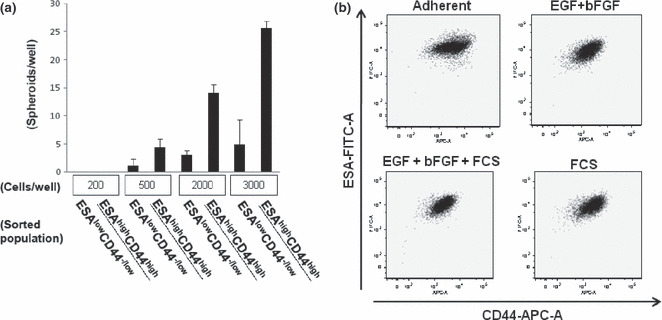
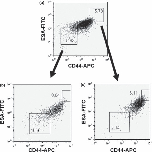
The cancer stem cell (CSC) model, in which a small population of cells within a tumor possesses the ability to self-renew and reconstitute the phenotype of primary tumor, has gained wide acceptance based on evidence over the past decade. It has also been reported that cancer cell lines contain a CSC subpopulation. However, phenotypic differences between CSCs and non-CSCs in cancer cell lines are not better defined than in primary tumors. Furthermore, some cell lines do not have a CSC population, revealed as a side population and expression of CD133. Thus, the identification of CSCs in cancer cell lines remains elusive. Here, we investigated the CSC hierarchy within HCT116 colon cancer cells, which do not have a CD133-positive subpopulation. We examined the expression of alternative CSC markers epithelial specific antigen (ESA) and CD44 in floating-sphere-derived cells, which are known to be the cells of enriching CSCs. Sphere-derived HCT116 cells exhibited heterogeneous expression of ESA and CD44. The two major subpopulations of HCT116 sphere cells (ESA(low)CD44(-/low) and ESA(high)CD44(high)) exhibited a biological/proliferative hierarchy of sphere-forming and soft agar colony-forming activity. However, there was no difference between the two subpopulations in the incidence of xenograft tumors. When ESA(low)CD44(-/low) cells were allowed to aggregate and re-form floating-spheres, the biological/proliferative hierarchy of parental HCT116 spheres was reconstituted, in terms of ESA and CD44 expression. Thus, HCT116 cells have plasticity when they are set in floating-spheres, suggesting that maintenance of the HCT116 cell line conforms to a stochastic model, not a CSC model.


Figures

Figure 1
Figure 1
CD133 is not an appropriate cancer stem cell (CSC) marker for HCT116 cells. (a) Expression of CD133 in the human colon cancer cell lines WiDr, HT29, DLD‐1, and HCT116. Dashed line, isotype control Ab. PE, phycoerythrin. (b) HCT116 cell spheres in floating cultures. (c) FACS analysis of CD133 expression in sphere‐derived HCT116 cells. SCC‐A, side scatter‐area.
Figure 2
Figure 2
Floating culture sphere‐formation induces a biological/proliferative, but not cancer stem cell (CSC) hierarchy, in HCT116 cells. (a) Representative FACS dot plots showing the expression of epithelial specific antigen (ESA) and CD44 in adherent and sphere‐derived HCT116 cells. APC, allophycocyanin. (b) Analysis of sphere‐formation (left) and colony‐formation in agar (right) by FACS‐sorted subpopulations of sphere‐derived HCT116 cells. (c) Representative subcutaneous tumors were derived from 5.0 × 103 ESAlowCD44−/low (left, arrow) and ESAhighCD44high (right, arrow) HCT116 sphere cells. Corresponding sections were stained with H&E (upper) and subjected to immunohistochemical analysis with anti‐CD44 (middle) and anti‐ESA (lower) Abs (magnification, ×40). (d) Volume of tumors derived from 5.0 × 103 cells from each subpopulation, as described for (c), 8 weeks after inoculation. Data represents the means ± SEM of four mice inoculated as one series of inoculations.
Figure 3
Figure 3
HCT116 sphere‐formation is due to cellular aggregation and exhibits a stable phenotype, independent of extrinsic growth factors. (a) Sphere‐formation assay of fractionated sphere‐derived HCT116 cells plated at the indicated cell densities. Data represents the means ± SEM of at least three wells for each density. ESAhighCD44high is underlined to distinguish it clearly from ESAlowCD44−/low. (b) FACS analysis of sphere‐derived HCT116 cells cultured under floating culture conditions in the presence of epidermal growth factor (EGF) and basic‐fibroblast growth factor (b‐FGF) (top right), EGF, bFGF, and FCS (bottom left), or FCS (bottom right). The phenotype of adherent HCT116 cells cultured in the presence of 10% FCS was analyzed as a control (top left).
Figure 4
Figure 4
Structural and biological characterization of HCT116 spheres. (a) Expressions of epithelial specific antigen (ESA) and CD44 (top), and E‐cadherin and β‐catenin (bottom) in HCT116 spheres were analyzed by fluorescence microscopy. Nuclei were counterstained with DAPI (blue staining). (b) Immunohistochemical analysis of frozen sections of HCT116 spheres using anti‐phospho‐p38 (top left), anti‐pan‐p38 (top right), anti‐APG8b (bottom left), and anti‐BrdU (bottom right) Abs. (c) Cell‐cycle analysis of HCT116 sphere‐derived cells. Representative staining pattern for ESA and CD44 in sphere‐derived HCT116 cells (top left) is shown. Hoechst 33342 and Pyronin Y staining pattern of intact sphere‐derived HCT116 cells (top right), and fractionated ESAlowCD44−/low (bottom left, arrow) and ESAhighCD44high (bottom right, arrow) cells are also shown.
Figure 5
Figure 5
Reconstitution of the phenotype of sphere‐derived HCT116 cells following aggregation of ESAlowCD44−/low and ESAhighCD44high subpopulations. Representative FACS dot plots of (a) intact sphere‐derived HCT116 cells at the time of sorting and fractionated (b) ESAlowCD44−/low and (c) ESAhighCD44high subpopulations after culturing at an aggregative density of 5000 cells/mL. APC, allophycocyanin.

References

    1. Bonnet D, Dick JE. Human acute myeloid leukemia is organized as a hierarchy that originates from a primitive hematopoietic cell. Nat Med 1997; 3: 730–7. - PubMed
    1. Visvader JE, Lindeman GJ. Cancer stem cells in solid tumours: accumulating evidence and unresolved questions. Nat Rev Cancer 2008; 8: 755–68. - PubMed
    1. Dick JE. Stem cell concepts renew cancer research. Blood 2008; 112: 4793–807. - PubMed
    1. Kondo T. Stem cell‐like cancer cells in cancer cell lines. Cancer Biomark 2007; 3: 245–50. - PubMed
    1. Kondo T, Setoguchi T, Taga T. Persistence of a small subpopulation of cancer stem‐like cells in the C6 glioma cell line. Proc Natl Acad Sci U S A 2004; 101: 781–6. - PMC - PubMed

Publication types