. 2009 Sep 8:6:19.
doi: 10.1186/1742-9994-6-19.

Sponge budding is a spatiotemporal morphological patterning process: Insights from synchrotron radiation-based x-ray microtomography into the asexual reproduction of Tethya wilhelma

Affiliations

Sponge budding is a spatiotemporal morphological patterning process: Insights from synchrotron radiation-based x-ray microtomography into the asexual reproduction of Tethya wilhelma

Jörg U Hammel et al. Front Zool. .

Abstract

Background: Primary agametic-asexual reproduction mechanisms such as budding and fission are present in all non-bilaterian and many bilaterian animal taxa and are likely to be metazoan ground pattern characters. Cnidarians display highly organized and regulated budding processes. In contrast, budding in poriferans was thought to be less specific and related to the general ability of this group to reorganize their tissues. Here we test the hypothesis of morphological pattern formation during sponge budding.

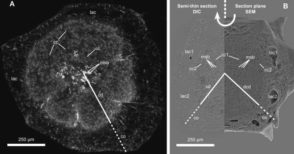
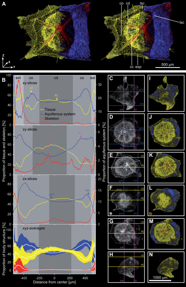
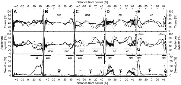
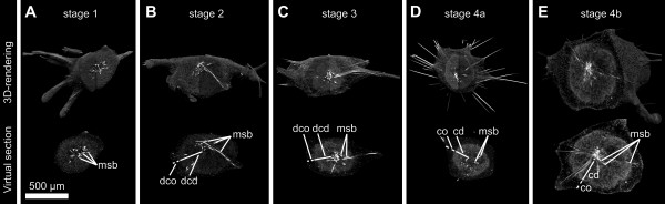
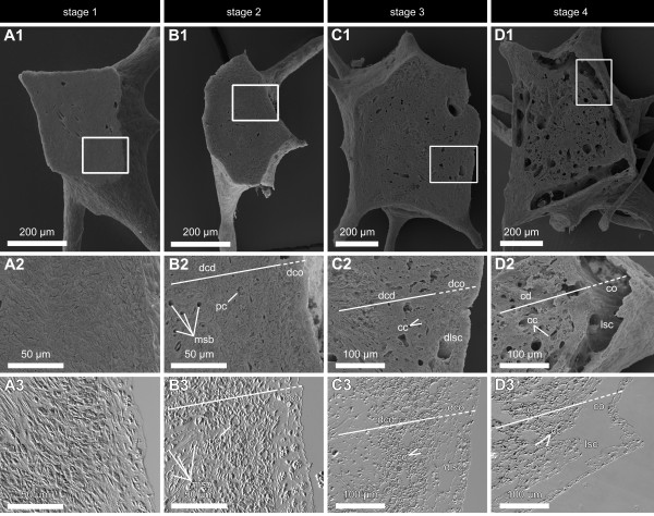
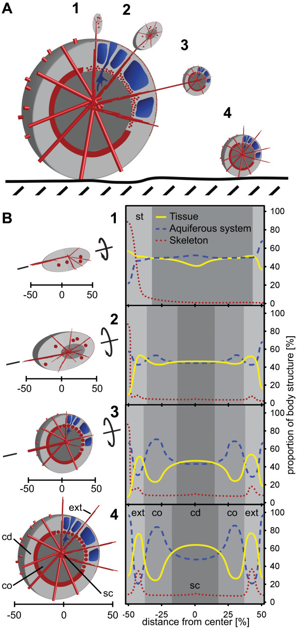
Results: We investigated the budding process in Tethya wilhelma (Demospongiae) by applying 3D morphometrics to high resolution synchrotron radiation-based x-ray microtomography (SR-muCT) image data. We followed the morphogenesis of characteristic body structures and identified distinct morphological states which indeed reveal characteristic spatiotemporal morphological patterns in sponge bud development. We discovered the distribution of skeletal elements, canal system and sponge tissue to be based on a sequential series of distinct morphological states. Based on morphometric data we defined four typical bud stages. Once they have reached the final stage buds are released as fully functional juvenile sponges which are morphologically and functionally equivalent to adult specimens.

Conclusion: Our results demonstrate that budding in demosponges is considerably more highly organized and regulated than previously assumed. Morphological pattern formation in asexual reproduction with underlying genetic regulation seems to have evolved early in metazoans and was likely part of the developmental program of the last common ancestor of all Metazoa (LCAM).


Figures

Figure 1
Figure 1
Morphology of a detached T. wilhelma bud (stage 4). Comparison between (A) a virtual section from an SR-μCT data set and (B) 'combined scanning electron histology' data consisting of a semi-thin section imaged using DIC-microscopy and SEM of the corresponding surface after sectioning (cc - choanocyte chamber, cd - choanoderm, co - cortex, dcd - developing choanoderm, lac - lacunae, msb - megasclere bundle).
Figure 2
Figure 2
Detailed visualization and analysis of the overall morphology of a stage 4 T. wilhelma bud (data set E, see Additional file 2) based on SR μCT x-ray absorption and volumetric measurements. (A) Stereo pair rendering with segmentation of morphological structures: sponge tissue (yellow) separated into cortex (co) and choanoderm (cd) with developed choanocyte chambers (cc), exopinacoderm (exp), skeleton (red) and aquiferous system (blue) with lacunar system cavities (lsc). (B) Related volumetric measurements. Proportions [%] of sponge tissue, skeleton and aquiferous system measured on 1.4 μm slices. Proportional volume is given for all three spatial directions (x, y and z axes) and as xyz averages with standard deviations in relation to the sponge centre (x, y, z = 0,0,0 μm); arrows and lower case letters refer to C - N (slice images). Main body structures and body extensions (ext) are marked in grayscale. (C - N) examples of 1.4 μm slices in grayscale (left column) and colored x-ray absorption-based segmentation of morphological elements (right column). Two slices are presented per dataset direction: xy slices (C, D, I & J), zx slices (E, F, K & L), zy slices (G, H, M & N); lower case letters and lines in grayscale slice images mark the corresponding positions of the orthogonal planes shown as examples in C - N.
Figure 3
Figure 3
Volume analysis of body structures in T. wilhelma buds based on SR-μCT. (A) stage 1 bud without choanoderm/cortex differentiation; (B) stage 2 bud without a separated choanoderm but displaying the first differentiated aquiferous system canals; (C) stage 3 bud with an early developing choanoderm (dcd) and developing cortex (dco), (D - E) stage 4 buds with differentiated choanoderm (cd) and cortex (co) regions. Graphs represent relative volumetric proportions of all main morphological sponge structures: tissue (top row), aquiferous system (middle row) and skeleton (bottom row). Graph patterns typical for distinctly developed sponge regions are marked (sc - skeleton centre, ext - body extension (filaments), st - stalk). Volumetric results are given for the three main axes of the 3D data sets: x-axis (dashed), y-axis (dotted) and z-axis (solid).
Figure 4
Figure 4
3D volume renderings of stage 1 to 4 buds of T. wilhelma and corresponding virtual sections from SR-μCT data elucidating the development of distinct morphological structures. (A) stage 1 bud without choanoderm/cortex differentiation (msb - megasclere bundle); (B) stage 2 bud without a separated choanoderm (dcd - developing choanoderm, dco - developing cortex) but displaying the first differentiated aquiferous system canals; (C) stage 3 bud with an early developing choanoderm (dcd) and developing cortex (dco), (D - E) stage 4 buds with differentiated choanoderm (cd) and cortex (co) regions, the latter displaying lacunar cavities.
Figure 5
Figure 5
Corresponding SEM and DIC images of stage 1 to stage 4 buds of T. wilhelma A1 - D1. SEM images of median section planes of the four bud stages of T. wilhelma, Framed areas are represented in A2 - D2 (SEM) and the DIC microimages of corresponding semi-thin sections in A3 - D3. In stage 1 buds (A) cells are densely packed and evenly distributed. Stage 2 buds (B) display early developing cortex (dco) and choanoderm (dcd) regions as well as few numbers of primordial choanocytes (pc) and megasclere bundles (msb). Developing lacunar system cavities (dlsc) are found in stage 3 (C) as separated developing choanoderm and cortex regions. In stage 4 (D) clearly developed lacunar system cavities (lsc), choanocyte chambers (cc) and distinguished cortex (co) and choanoderm (cd) are present.
Figure 6
Figure 6
Scheme of bud development in T. wilhelma. (A) Four bud stages are characteristic, with the first three connected to the mother sponge by a stalk: Skeletal elements in red (megasclere bundles and aster spheres); megasclere bundles partly simplified as cylinders; Tissue in grey, separated into cortex (light grey) and choanoderm (dark grey). (B) Details of bud stages (left) and schematic graphs of morphological functional unit distribution. There are indications of rotational symmetry along the initial connecting stalk (st) in stages one to three (compare Additional file 4). Stage 4 buds display an adult-like body morphology with point symmetry to the skeleton centre (sc; see Additional file 5). Choanoderm development starts in stage 2, accompanied by the development of the megaster spheres in stage 3. Differentiation into a cortex (co) and choanoderm (cd) is characterized by the development of the aquiferous system (larger canals in stage 2; lacunae in stage 3). Body extensions (ext) (filaments) are found in stage 4 buds. For further details see text.

References

    1. Fell PE. Porifera. In: Adivodi KG, Adivodi RG, editor. Reproductive biology of invertebrates: Asexual propagation and reproductive strategies. Chichester; New York: John Wiley & Sons; 1993. pp. 1–44.
    1. Schmidt-Rhaesa A. The evolution of organ systems. Oxford: Oxford University Press; 2007.
    1. Schulze FE. Über Trichoplax adhaerens. Abhdl Königl Preuss Akad Wiss. 1891. pp. 1–23.
    1. Campbell RD. Cnidaria. In: Giese AC, Pearse JS, editor. Reproduction of marine invertebrates Acoelomate and Pseudocoelomate Metazoans. I. New York: Academic Press; 1974. pp. 133–200.
    1. Pianka HD. Ctenophora. In: Giese AC, Pearse JS, editor. Reproduction of marine invertebrates Acoelomate and Pseudocoelomate Metazoans. I. New York: Academic Press; 1974. pp. 201–266.