. 2009 Sep 8:6:80.
doi: 10.1186/1742-4690-6-80.

The formation of cysteine-linked dimers of BST-2/tetherin is important for inhibition of HIV-1 virus release but not for sensitivity to Vpu

Affiliations

The formation of cysteine-linked dimers of BST-2/tetherin is important for inhibition of HIV-1 virus release but not for sensitivity to Vpu

Amy J Andrew et al. Retrovirology. .

Abstract

Background: The Human Immunodeficiency virus type 1 (HIV-1) Vpu protein enhances virus release from infected cells and induces proteasomal degradation of CD4. Recent work identified BST-2/CD317 as a host factor that inhibits HIV-1 virus release in a Vpu sensitive manner. A current working model proposes that BST-2 inhibits virus release by tethering viral particles to the cell surface thereby triggering their subsequent endocytosis.

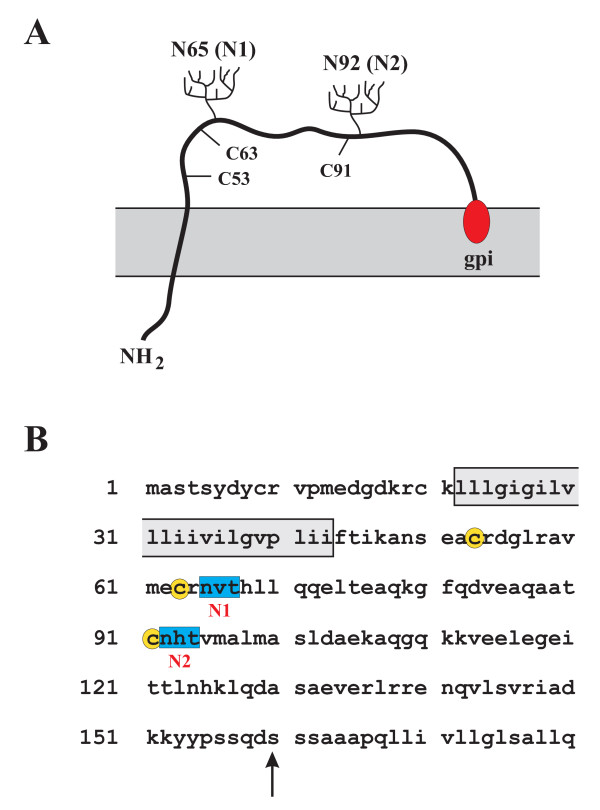
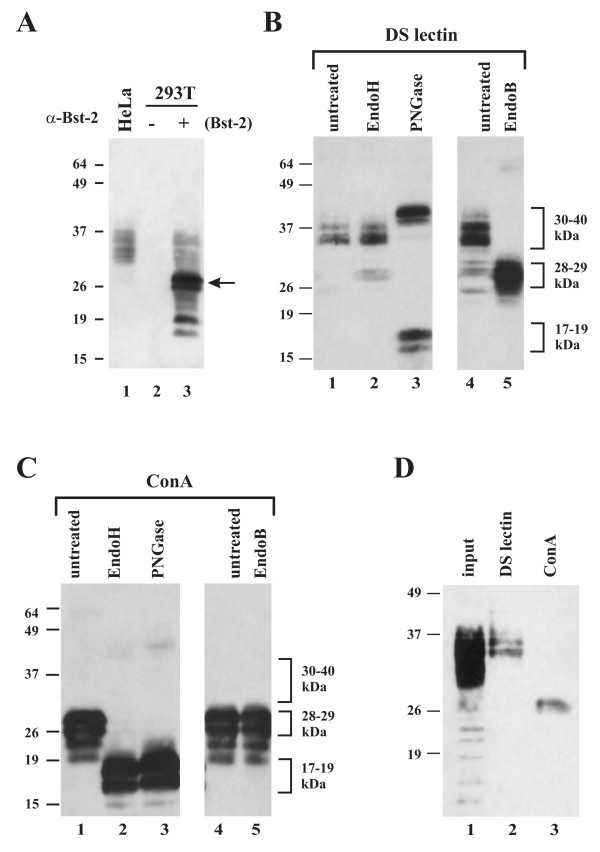
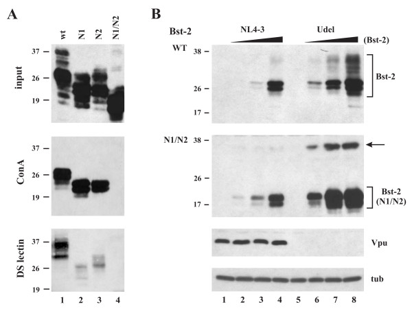
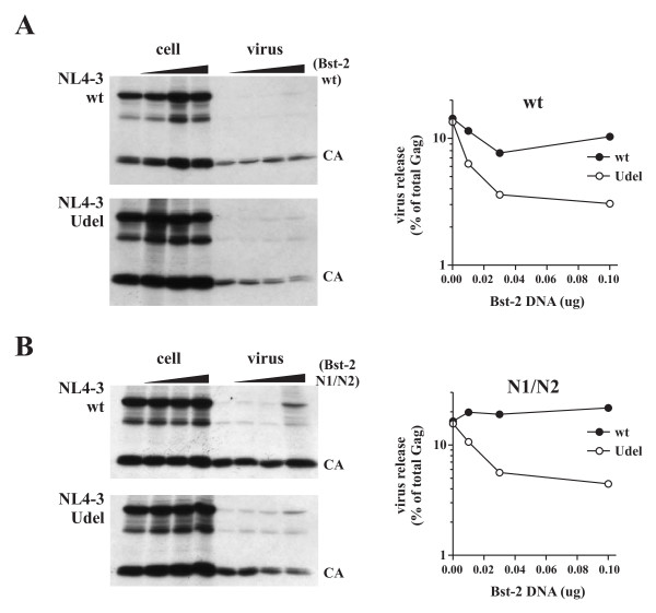
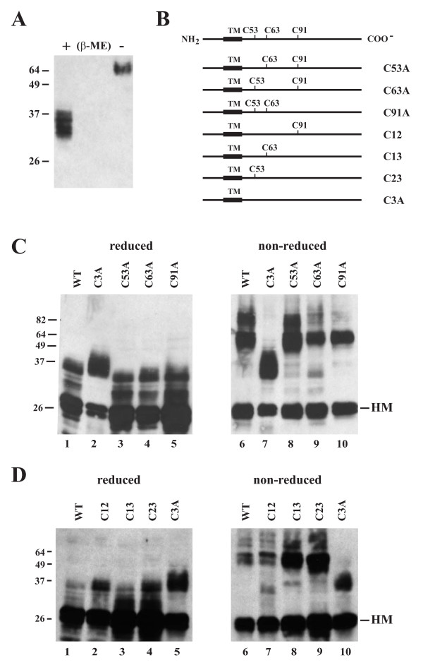
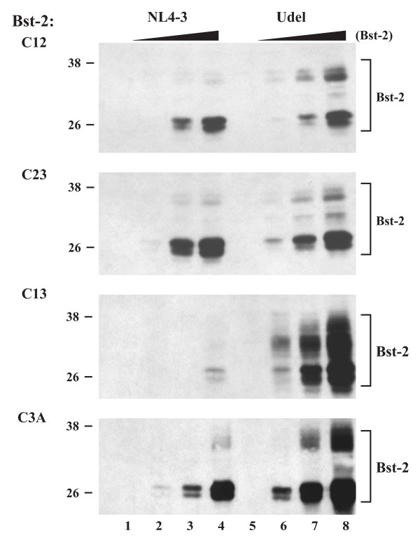
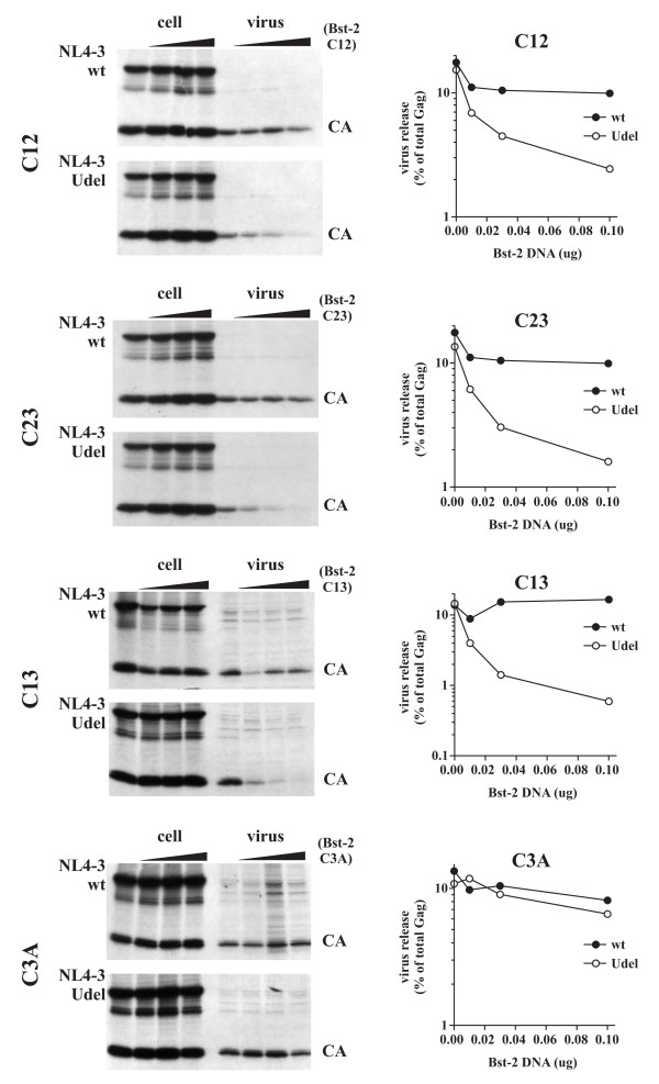
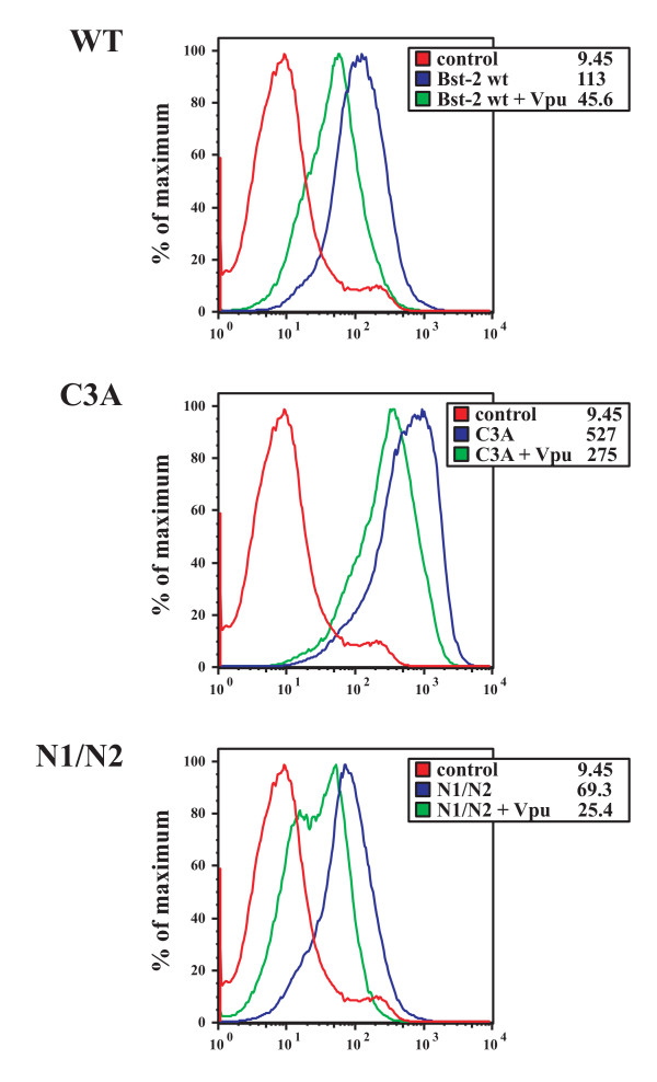
Results: Here we defined structural properties of BST-2 required for inhibition of virus release and for sensitivity to Vpu. We found that BST-2 is modified by N-linked glycosylation at two sites in the extracellular domain. However, N-linked glycosylation was not important for inhibition of HIV-1 virus release nor did it affect surface expression or sensitivity to Vpu. Rodent BST-2 was previously found to form cysteine-linked dimers. Analysis of single, double, or triple cysteine mutants revealed that any one of three cysteine residues present in the BST-2 extracellular domain was sufficient for BST-2 dimerization, for inhibition of virus release, and sensitivity to Vpu. In contrast, BST-2 lacking all three cysteines in its ectodomain was unable to inhibit release of wild type or Vpu-deficient HIV-1 virions. This defect was not caused by a gross defect in BST-2 trafficking as the mutant protein was expressed at the cell surface of transfected 293T cells and was down-modulated by Vpu similar to wild type BST-2.

Conclusion: While BST-2 glycosylation was functionally irrelevant, formation of cysteine-linked dimers appeared to be important for inhibition of virus release. However lack of dimerization did not prevent surface expression or Vpu sensitivity of BST-2, suggesting Vpu sensitivity and inhibition of virus release are separable properties of BST-2.


Figures

Figure 1
Figure 1
Schematic of the BST-2 structure. (A) BST-2 is a type 2 integral membrane protein. The N-terminus localizes to the cytoplasm. The BST-2 ectodomain contains three cysteine residues (C53, C63, C91) and two potential sites for N-linked glycosylation (N65, N92). The C-terminus of BST-2 is modified by the addition of a glycosyl-phosphatidylinositol (gpi) anchor. (B) Predicted amino acid sequence of human BST-2. The predicted transmembrane region is indicated by a box. Signals for N-linked glycosylation are marked in blue; cysteine residues in the BST-2 extracellular domain are highlighted in yellow. The arrow points to the predicted site of cleavage for the addition of the gpi anchor [31].
Figure 2
Figure 2
Comparison of endogenous BST-2 in HeLa cells to BST-2 expressed in transiently transfected 293T cells. (A) 293T cells were transfected with wt BST-2 (lane 3). A mock transfected culture from HeLa (lane 1) and 293T cells (lane 2) was analyzed in parallel. Whole cell lysates were processed for immunoblotting as described in Methods. The arrow identifies a BST-2 species in transfected 293T cells not seen in HeLa cells. (B & C) Endoglycosidase analysis of transiently expressed BST-2. 293T cells were transfected with pcDNA-BST-2. BST-2 was enriched by adsorption to either datura stramonium lectin resin (DS lectin) (B) or Concanavalin A resin (ConA) (C) as described in Methods. DS lectin or ConA bound proteins were either left untreated (lanes 1 & 4) or treated with endoglycosidase H (EndoH) (lanes 2), Peptide: N-Glycosidase F (PNGase) (lanes 3), or endo-β-galactosidase (EndoB) (lanes 5) as described in Methods. Proteins were visualized by immunoblot analysis using a BST-2 specific antibody. (D) HeLa extracts were adsorbed to DS lectin (lane 2) and ConA resin (lane 3) as described for panels B & C. Total input lysate is shown in lane 1. A high mannose form of endogenous BST-2 was enriched on the ConA resin.
Figure 3
Figure 3
Immunoblot analysis of BST-2 glycosylation mutants. (A) 293T cells were transfected with wt BST-2, single glycosylation site mutants N1 & N2, or the double glycosylation site mutant N1/N2. BST-2 specific proteins were identified by immunoblotting using a BST-2-specific polyclonal antibody (top panel). Aliquots of the same samples were adsorbed to either ConA (middle panel) or DS lectin (lower panel) as described in Methods. Eluates were analyzed by immunoblotting using a BST-2-specific polyclonal antibody. (B) 293T cells were transfected with 5 μg each of NL4-3 wt (lanes 1-4) or NL4-3/Udel (lanes 5-8) either in the absence of BST-2 (lanes 1 & 5) or in the presence of 0.01 μg (lanes 2 & 6), 0.03 μg (lanes 3 & 7), or 0.1 μg (lanes 4 & 8) BST-2 DNA. Cells were harvested 20 h post transfection. A fraction of the cells was used for immunoblot analysis; the other part was used for metabolic labelling (Fig. 4). Whole cell lysates were prepared and used for immunoblot analysis using a BST-2-specific polyclonal antibody (top two panels). The blots were then sequentially reprobed with antibodies to Vpu (third panel) or tubulin (lower panel). Representative samples shown in the lower panels were from the N1/N2 blot. Proteins are identified on the right. The arrow points to a form of BST-2 N1/N2 whose migration in the gel is consistent with a dimer.
Figure 4
Figure 4
Glycosylation of BST-2 is not required for inhibition of virus release and for sensitivity to Vpu. (A) Analysis of wt BST-2. (B) Analysis of BST-2 N1/N2. (A & B) Cells were metabolically labeled for 90 min with [35S]-methionine as described in Methods and cell lysates and cell-free supernatants were subjected to immunoprecipitation by an HIV-positive patient serum. Immunoprecipitates were subjected to SDS-PAGE and proteins were visualized by fluorography (left panels). Virus release was quantified by phosphoimage analysis using a Fujifilm FLA7000. Virus release was calculated independently for each sample by determining the percentage of cell-free CA protein relative to the total intra- and extra-cellular Gag protein. Solid circles represent wt NL4-3. Open circles represent NL4-3/Udel. Data are presented as a function of BST-2 concentration on a semi-log plot.
Figure 5
Figure 5
BST-2 forms cysteine-linked dimers. (A) HeLa cells were lysed in reducing (+β-ME) or non-reducing (-β-ME) sample buffer as described in Methods. Lysates were separated by SDS-PAGE and subjected to immunoblot analysis using a BST-2-specific polyclonal antibody. (B) Schematic representation of mutants analyzed in this experiment. In all cases, cysteine residues were mutated to alanine by site-directed mutagenesis. Remaining cysteine residues are shown for each mutant. (C) 293T cells were transfected with wt BST-2 (lanes 1 & 6), the triple cysteine mutant C3A (lanes 2 & 7), or individual cysteine mutants (lanes 3-5 & 8-10). Cells were harvested 24 h post transfection, washed in PBS, and split into two equal samples. One set of samples was mixed with an equal volume of reducing sample buffer (lanes 1-5); the second set was mixed with sample buffer lacking β-ME (lanes 6-10). Cell lysates were separated by SDS-PAGE and subjected to immunoblotting using a BST-2 specific antibody. (D) 293T cells were transfected with wt BST-2 (lanes 1 & 6), the triple cysteine mutant C3A (lanes 5 & 10), or double-cysteine mutants (lanes 2-4 & 7-9). Samples were analyzed under reducing and non-reducing conditions as in panel C. (HM) marks the position of the high-mannose forms of BST-2 in the gels.
Figure 6
Figure 6
Dimerization of BST-2 is not a prerequisite for sensitivity to Vpu. 293T cells were transfected with 5 μg of wt pNL4-3 (lanes 1-4) or pNL4-3/Udel (lanes 5-8) in the absence of BST-2 (lanes 1 & 5) or together with 0.01 (lanes 2 & 6), 0.03 (lanes 3 & 7), and 0.1 μg (lanes 4 & 8) of vectors encoding cysteine mutants of BST-2 containing only one remaining cysteine (C12, C23, C13) or no cysteine at all (C3A). Twenty-four hours later, cell lysates were prepared from a fraction of the cells as described in the Methods and subjected to immunoblot analysis using a BST-2-specific antibody.
Figure 7
Figure 7
Dimerization of BST-2 is important for inhibition of virus release. Transfected 293T cells from figure 6 were metabolically labeled for 90 minutes and analyzed as described for figure 4. Proteins were identified by fluorography (left panels). Quantification of virus release was performed as described for figure 4 and is shown on the right.
Figure 8
Figure 8
Monomeric BST-2 and non-glycosylated BST-2 are expressed at the cell surface and are sensitive to Vpu-induced down modulation. 293T cells were transfected with 0.1 μg each of wt BST-2, BST-2 C3A, or BST-2 N1/N2 together with 1 μg each of pEGFP-N1 in the presence or absence of 1 μg pcDNA-Vphu. All samples were adjusted to 5 μg total DNA with empty vector DNA. Cells were harvested 24 h after transfection and stained with an antibody to BST-2. As a control, 293T cells were transfected with pEGFP-N1 and empty vector in the absence of BST-2 and stained with BST-2 antibody (Ctrl). Samples were subjected to FACS analysis as described in Methods and gated for GFP-positive cells. The red line represents the control of GFP+/BST-2- cells. Blue lines represent BST-2 staining in the absence of Vpu and green lines indicate BST-2 staining in the presence of Vpu. Numbers in the boxed legends represent mean fluorescence intensities for each sample.

Similar articles

Cited by

References

    1. Strebel K, Klimkait T, Martin MA. A novel gene of HIV-1, vpu, and its 16-kilodalton product. Science. 1988;241:1221–1223. doi: 10.1126/science.3261888. - DOI - PubMed
    1. Cohen EA, Terwilliger EF, Sodroski JG, Haseltine WA. Identification of a protein encoded by the vpu gene of HIV-1. Nature. 1988;334:532–534. doi: 10.1038/334532a0. - DOI - PubMed
    1. Willey RL, Maldarelli F, Martin MA, Strebel K. Human immunodeficiency virus type 1 Vpu protein induces rapid degradation of CD4. J Virol. 1992;66:7193–7200. - PMC - PubMed
    1. Willey RL, Maldarelli F, Martin MA, Strebel K. Human immunodeficiency virus type 1 Vpu protein regulates the formation of intracellular gp160-CD4 complexes. J Virol. 1992;66:226–234. - PMC - PubMed
    1. Klimkait T, Strebel K, Hoggan MD, Martin MA, Orenstein JM. The human immunodeficiency virus type 1-specific protein vpu is required for efficient virus maturation and release. J Virol. 1990;64:621–629. - PMC - PubMed

Publication types

MeSH terms