

Myelin sheaths are formed with proteins that originated in vertebrate lineages
- PMID: 19737434
- PMCID: PMC3781599
- DOI: 10.1017/S1740925X09990238
Myelin sheaths are formed with proteins that originated in vertebrate lineages
Abstract
All vertebrate nervous systems, except those of agnathans, make extensive use of the myelinated fiber, a structure formed by coordinated interplay between neuronal axons and glial cells. Myelinated fibers, by enhancing the speed and efficiency of nerve cell communication allowed gnathostomes to evolve extensively, forming a broad range of diverse lifestyles in most habitable environments. The axon-covering myelin sheaths are structurally and biochemically novel as they contain high portions of lipid and a few prominent low molecular weight proteins often considered unique to myelin. Here we searched genome and EST databases to identify orthologs and paralogs of the following myelin-related proteins: (1) myelin basic protein (MBP), (2) myelin protein zero (MPZ, formerly P0), (3) proteolipid protein (PLP1, formerly PLP), (4) peripheral myelin protein-2 (PMP2, formerly P2), (5) peripheral myelin protein-22 (PMP22) and (6) stathmin-1 (STMN1). Although widely distributed in gnathostome/vertebrate genomes, neither MBP nor MPZ are present in any of nine invertebrate genomes examined. PLP1, which replaced MPZ in tetrapod CNS myelin sheaths, includes a novel 'tetrapod-specific' exon (see also Möbius et al., 2009). Like PLP1, PMP2 first appears in tetrapods and like PLP1 its origins can be traced to invertebrate paralogs. PMP22, with origins in agnathans, and STMN1 with origins in protostomes, existed well before the evolution of gnathostomes. The coordinated appearance of MBP and MPZ with myelin sheaths and of PLP1 with tetrapod CNS myelin suggests interdependence - new proteins giving rise to novel vertebrate structures.
Figures
 ).
).
 ) for complete identity, white letters on gray background for blocks of identity (>2) and conserved substitutions (
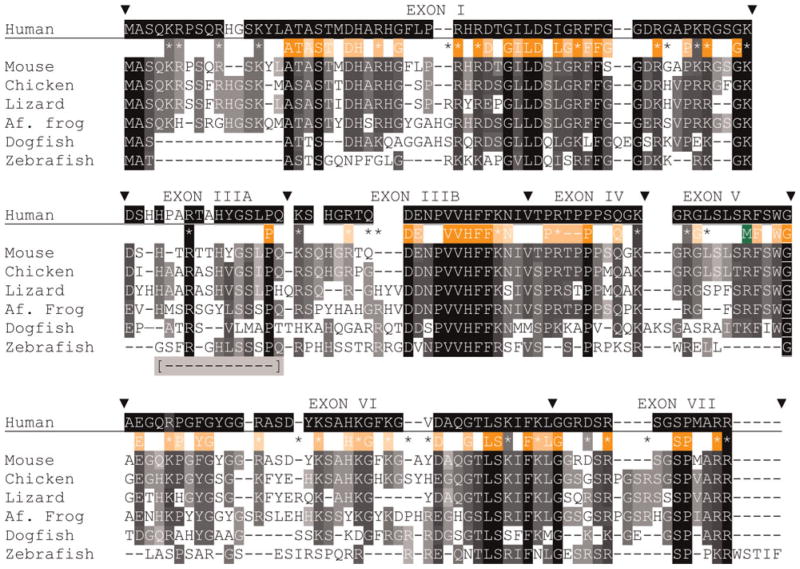
 ); the background shading is set to the number of sequences with the same amino acid – darker background are for greater number of sequences. For simplicity, conserved substitutions are considered the same as identical amino acids. Exon boundaries are marked (θ) with exon numbers above the human sequence. Positions of all basic (K, R) amino acids are shown with coloring indicating complete identity (
 ) and (
 ) conservation and presence in sequences of one or two species (*). Sixty-nine HMM logo (PFAM) amino acids were selected based on individual or conserved substitution contributions >1.0 and shown with white lettering on orange backgrounds. As above, darker background shading indicates greater sequence identity. Letters and basic residue designations are placed between human and murine sequences. A single arginine methylation site is shown below the human sequence (M). A portion of exon III, absent in the common zebrafish 88 amino acid isoform is indicated [
 ] and assigned IIIA (see supplementary Table 3 online).
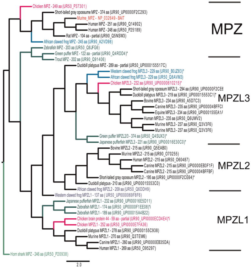
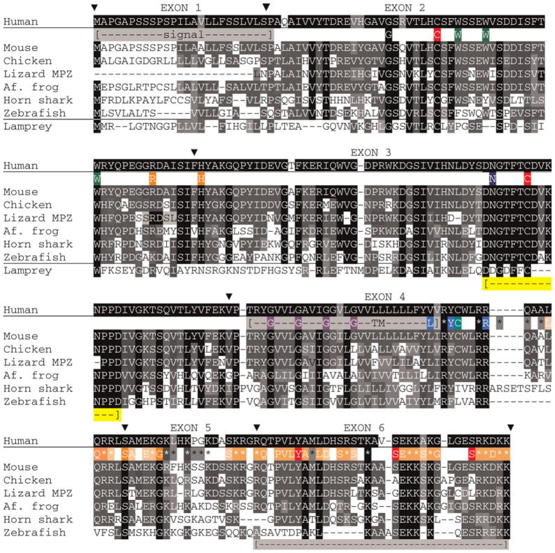 ]). Included are the single glycosylation site (N), two cysteine residues (C) that form the disulfide bond, a third (C) that is acylated in mammals. Two tryptophan residues (W) suggested from the X-ray diffraction data (Shapiro et al., 1996) to intercalate the apposing lipid bilayers, a glycine zipper (G) in the transmembrane domain, a tyrosine (Y) and two serine (S) represent phosphorylation sites. The (L), (Y) and (R) are cholesterol recognition motifs are highlighted. Amino acids that form the myelin-PO_C PFAM HMM logo are depicted as in Fig. 4, i.e. they are shaded in orange. A region of high conservation in exon 3 is also marked ([
]).
]). Included are the single glycosylation site (N), two cysteine residues (C) that form the disulfide bond, a third (C) that is acylated in mammals. Two tryptophan residues (W) suggested from the X-ray diffraction data (Shapiro et al., 1996) to intercalate the apposing lipid bilayers, a glycine zipper (G) in the transmembrane domain, a tyrosine (Y) and two serine (S) represent phosphorylation sites. The (L), (Y) and (R) are cholesterol recognition motifs are highlighted. Amino acids that form the myelin-PO_C PFAM HMM logo are depicted as in Fig. 4, i.e. they are shaded in orange. A region of high conservation in exon 3 is also marked ([
]).Similar articles
-
Evolution of myelin ultrastructure and the major structural myelin proteins.Brain Res. 2016 Jun 15;1641(Pt A):43-63. doi: 10.1016/j.brainres.2015.10.037. Epub 2015 Oct 28. Brain Res. 2016. PMID: 26519753 Review.
-
Adhesive properties of proteolipid protein are responsible for the compaction of CNS myelin sheaths.J Neurosci. 1995 Aug;15(8):5502-13. doi: 10.1523/JNEUROSCI.15-08-05502.1995. J Neurosci. 1995. PMID: 7543946 Free PMC article.
-
Myelin tetraspan family proteins but no non-tetraspan family proteins are present in the ascidian (Ciona intestinalis) genome.Biol Bull. 2005 Aug;209(1):49-66. doi: 10.2307/3593141. Biol Bull. 2005. PMID: 16110093
-
The evolution of lipophilin genes from invertebrates to tetrapods: DM-20 cannot replace proteolipid protein in CNS myelin.J Neurosci. 2000 Jun 1;20(11):4002-10. doi: 10.1523/JNEUROSCI.20-11-04002.2000. J Neurosci. 2000. PMID: 10818135 Free PMC article.
-
The myelin proteolipid DMα in fishes.Neuron Glia Biol. 2010 May;6(2):109-12. doi: 10.1017/S1740925X09000131. Epub 2009 Jun 10. Neuron Glia Biol. 2010. PMID: 19508742 Review.
Cited by
-
Motor Exit Point (MEP) Glia: Novel Myelinating Glia That Bridge CNS and PNS Myelin.Front Cell Neurosci. 2018 Oct 2;12:333. doi: 10.3389/fncel.2018.00333. eCollection 2018. Front Cell Neurosci. 2018. PMID: 30356886 Free PMC article. Review.
-
Single-cell sequencing reveals glial cell involvement in development of neuropathic pain via myelin sheath lesion formation in the spinal cord.J Neuroinflammation. 2024 Aug 31;21(1):213. doi: 10.1186/s12974-024-03207-3. J Neuroinflammation. 2024. PMID: 39217340 Free PMC article.
-
Cryo-EM, X-ray diffraction, and atomistic simulations reveal determinants for the formation of a supramolecular myelin-like proteolipid lattice.J Biol Chem. 2020 Jun 26;295(26):8692-8705. doi: 10.1074/jbc.RA120.013087. Epub 2020 Apr 7. J Biol Chem. 2020. PMID: 32265298 Free PMC article.
-
Roles of Progesterone, Testosterone and Their Nuclear Receptors in Central Nervous System Myelination and Remyelination.Int J Mol Sci. 2020 Apr 30;21(9):3163. doi: 10.3390/ijms21093163. Int J Mol Sci. 2020. PMID: 32365806 Free PMC article. Review.
-
The Effects of Insulin on Immortalized Rat Schwann Cells, IFRS1.Int J Mol Sci. 2021 May 23;22(11):5505. doi: 10.3390/ijms22115505. Int J Mol Sci. 2021. PMID: 34071138 Free PMC article.
References
-
- Aricescu AR, Jones EY. Immunoglobulin superfamily cell adhesion molecules: zippers and signals. Current Opinion in Cell Biology. 2007;19:543–550. - PubMed
-
- Avila RL, Tevlin BR, Lees JP, Inouye H, Kirschner DA. Myelin structure and composition in zebrafish. Neurochemical Research. 2007;32:197–209. - PubMed
-
- Baines AJ. Comprehensive analysis of all triple helical repeats in beta-spectrins reveals patterns of selective evolutionary conservation. Cell and Molecular Biology Letters. 2003;8:195–214. - PubMed
-
- Birchmeier C, Nave KA. Neuregulin-1, a key axonal signal that drives Schwann cell growth and differentiation. Glia. 2008;56:1491–1497. - PubMed
Publication types
MeSH terms
Substances
Grants and funding
LinkOut - more resources
Full Text Sources
Research Materials
Miscellaneous
