Review
. 2009 Sep 4;138(5):838-54.
doi: 10.1016/j.cell.2009.08.021.

Caspases and kinases in a death grip

Affiliations
Review

Caspases and kinases in a death grip

Manabu Kurokawa et al. Cell. .

Abstract

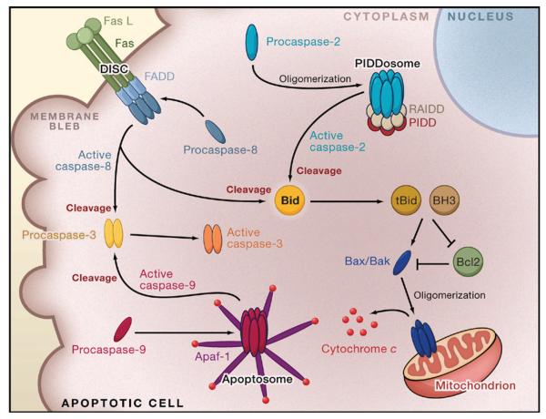
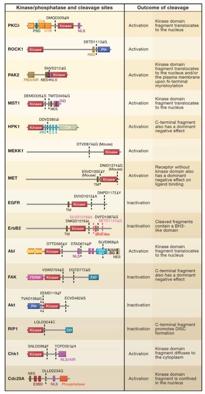
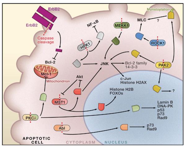
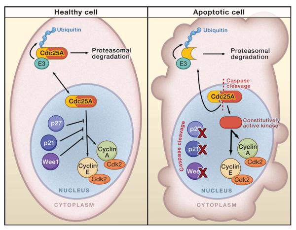
The complex process of apoptosis is orchestrated by caspases, a family of cysteine proteases with unique substrate specificities. Accumulating evidence suggests that cell death pathways are finely tuned by multiple signaling events, including direct phosphorylation of caspases, whereas kinases are often substrates of active caspases. Importantly, caspase-mediated cleavage of kinases can terminate prosurvival signaling or generate proapoptotic peptide fragments that help to execute the death program and facilitate packaging of the dying cells. Here, we review caspases as kinase substrates and kinases as caspase substrates and discuss how the balance between cell survival and cell death can be shifted through crosstalk between these two enzyme families.


Figures

Figure 1
Figure 1. Extrinsic and Intrinsic Apoptotic Pathways
Three major apoptotic pathways, initiated by the apical caspase-2, −9, and −8/10, are depicted. In the extrinsic pathway of apoptosis, caspase-8 (or caspase-10) is activated upon the binding of a cognate ligand such as Fas ligand through the formation of the death-inducing signaling complex (DISC), which includes a death receptor (for example, Fas) and a death domain-containing adaptor protein (for example, Fas-associated death domain or FADD). Once active, caspase-8 directly cleaves and activates executioner caspases (for example, caspase-3) and/or the BH3-only protein Bid, which activates the intrinsic apoptotic pathway by triggering oligomerization (activation) of the proapoptotic proteins Bax and Bak. Bax/ Bak activation is promoted by cleaved Bid (tBid), as well as other BH3-only proteins; the antiapoptotic Bcl-2 family members regulate the inhibition of Bax/Bak. The intrinsic pathway leads to the release of cytochrome c from the intermembrane space of the mitochondria to the cytoplasm. Binding of cytochrome c induces oligomerization of the adaptor protein Apaf-1, which recruits and activates caspase-9 (through formation of the apoptosome), leading to activation of executioner caspases. Caspase-2 is activated through the PIDDosome, which consists of the two adaptor proteins, PIDD (p53-induced protein with a death domain) and RAIDD (RIP-associated ICH-1/CED-3 homologous protein with a death domain). Only a few direct caspase-2 substrates have been identified, and apoptotic pathways induced by the activation of caspase-2 appear to be mediated at least in part by the cleavage of Bid.
Figure 2
Figure 2. Caspase Cleavage Sites
Many kinases and phosphatases are subject to proteolytic cleavage by caspases. Kinase/phosphatase domains and other regulatory regions of these enzymes are shown with their caspase-mediated cleavage sites (↓) and the effects of caspase cleavage on their catalytic activity (activation or inactivation). FERM, the band 4.1 homology and ERM domain; FAT, focal adhesion targeting region; PH, pleckstrin homology domain; DD, death domain; PSD, pseudosubstrate domain; NLS, nuclear localization signal; RBD, Rho-binding domain; PBD, p21-binding domain; AIR, autoinhibitory region; NES, nuclear export signal; DiD, dimerization domain; PR, proline-rich region; CH, citron homology domain; TM, transmembrane domain; SH2, src homology 2 domain; SH3, src homology 3 domain; DNA BD, DNA-binding domain; Actin BD, actin-binding domain; E3BD, E3 ligase-binding domain.
Figure 3
Figure 3. Caspase-Mediated Activation of Kinases Promotes Apoptosis
Caspase cleavage induces the constitutive activation of a kinase by removing an inhibitory domain (for example, PKCδ, ROCK1, PAK2, MST1, HPK1, and MEKK1) and/or triggering the redistribution of a cleaved kinase domain due to the loss or gain of a targeting motif in the cleaved protein (for example, PKCδ, PAK2, MST1, ErbB2, and Abl). In some cases, caspase cleavage generates a fragment that does not contain a kinase domain and acts as a dominant negative (HPK1) or gains a new function (ErbB2). Caspase cleavage products may acquire new substrates (or binding partners) or phosphorylate the same substrates with aberrantly high kinase activity, resulting in activation or inactivation of proor antiapoptotic signaling pathways.
Figure 4
Figure 4. Increased Cdk2 Activity during Apoptosis
In healthy cells (left), activity of Cdk2, which forms a complex with Cyclin A or Cyclin E, is inhibited by p21Cip1/Waf1 and/or p27Kip1. Cdk2 activity is also regulated by inhibitory phosphorylations modulated by the Wee1 kinase and Cdc25A phosphatase. In cells undergoing apoptosis (right), p21Cip1/Waf1, p27Kip1, and Wee1 are proteolytically inactivated by caspases. In contrast, caspase-mediated cleavage of Cdc25A generates a constitutively active phosphatase fragment with a nuclear localization signal that confines the phosphatase fragment in the nucleus. Caspase-mediated cleavage also separates the active Cdc25A fragment from the E3 ligase-binding domains at the N-terminal half of the phosphatase, resulting in increased stability of the active phosphatase fragment. Consequently, caspase activation can indirectly trigger an increase in Cdk2 activity.

Similar articles

Cited by

References

    1. Allan LA, Clarke PR. Phosphorylation of caspase-9 by CDK1/cyclin B1 protects mitotic cells against apoptosis. Mol. Cell. 2007;26:301–310. - PubMed
    1. Allan LA, Morrice N, Brady S, Magee G, Pathak S, Clarke PR. Inhibition of caspase-9 through phosphorylation at Thr 125 by ERK MAPK. Nat. Cell Biol. 2003;5:647–654. - PubMed
    1. Arnold R, Liou J, Drexler HC, Weiss A, Kiefer F. Caspase-mediated cleavage of hematopoietic progenitor kinase 1 (HPK1) converts an activator of NFκB into an inhibitor of NFκB. J. Biol. Chem. 2001;276:14675–14684. - PubMed
    1. Arnold R, Frey CR, Müller W, Brenner D, Krammer PH, Kiefer F. Sustained JNK signaling by proteolytically processed HPK1 mediates IL-3 independent survival during monocytic differentiation. Cell Death Differ. 2007;14:568–575. - PubMed
    1. Alvarado-Kristensson M, Andersson T. Protein phosphatase 2A regulates apoptosis in neutrophils by dephosphorylating both p38 MAPK and its substrate caspase 3. J. Biol. Chem. 2005;280:6238–6244. - PubMed

Publication types

LinkOut - more resources