. 2009 Sep 4;138(5):1005-18.
doi: 10.1016/j.cell.2009.06.023.

The DLK-1 kinase promotes mRNA stability and local translation in C. elegans synapses and axon regeneration

Affiliations

The DLK-1 kinase promotes mRNA stability and local translation in C. elegans synapses and axon regeneration

Dong Yan et al. Cell. .

Abstract

Growth cone guidance and synaptic plasticity involve dynamic local changes in proteins at axons and dendrites. The Dual-Leucine zipper Kinase MAPKKK (DLK) has been previously implicated in synaptogenesis and axon outgrowth in C. elegans and other animals. Here we show that in C. elegans DLK-1 regulates not only proper synapse formation and axon morphology but also axon regeneration by influencing mRNA stability. DLK-1 kinase signals via a MAPKAP kinase, MAK-2, to stabilize the mRNA encoding CEBP-1, a bZip protein related to CCAAT/enhancer-binding proteins, via its 3'UTR. Inappropriate upregulation of cebp-1 in adult neurons disrupts synapses and axon morphology. CEBP-1 and the DLK-1 pathway are essential for axon regeneration after laser axotomy in adult neurons, and axotomy induces translation of CEBP-1 in axons. Our findings identify the DLK-1 pathway as a regulator of mRNA stability in synapse formation and maintenance and also in adult axon regeneration.


Figures

Figure 1
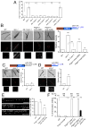
Figure 1. Loss of function in mak-2 suppresses rpm-1(lf)
(A) MAK-2 is a member of the MAPKAPK family and has two isoforms that differ at their N-termini. Mouse MK2 is NP_032577. (B) mak-2(lf) suppresses motor neuron presynaptic morphology defects of rpm-1 cell-autonomously. Images are juIs1[Punc-25-SNB-1::GFP] in the adult dorsal cord. SNB-1::GFP puncta are uniformly-shaped and evenly distributed in wild type and mak-2, but are irregular and fewer in rpm-1 mutants. Right panel, quantification of SNB-1::GFP puncta # (n ≥ 12 animals per genotype; data shown as mean ± SEM). (C) mak-2 suppresses touch neuron axon termination defects of rpm-1 cell-autonomously. Images are muIs32[Pmec-7-GFP] in adults. The ALM axon (red arrows) ends before the tip of the nose in wild type, but overextends and frequently loops in rpm-1 mutants. The PLM axon (blue arrows) terminates posterior to the ALM cell body (*) in wild type, but overextends and turns ventrally in rpm-1. The PLM synaptic branch (purple arrows) is frequently missing in rpm-1. Axon termination defects of PLM and ALM neurons are quantified as % of total # of animals. Quantification of PLM synaptic branch shows similar pattern (not shown). (D) Expression of MAK-2(EE) induces gain of function phenotypes. (E) MAK-2 rescuing activity is abolished by mutation of Thr250 and Thr262 or of Lys103 in the ATP-binding site, but is not affected by deletion of the NLS or NES. (B–E) Unless noted, alleles are rpm-1(ju44) and mak-2(ok2394). Promoters: Prgef-1 (F25B3.3) for pan-neural, Pmyo-3 for body wall muscles; Pmec-4 for touch neurons; Punc-25 for GABA motor neurons. N, total # of animals; TL, total # of transgenic lines; RL, # of transgenic lines showing rescuing activities. Statistics in this and subsequent figures, ANOVA (panel B) or Fisher exact test (C–E). *, P < 0.05; **, P < 0.01; ns, not significant. Scales, 10 μm.
Figure 2
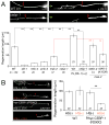
Figure 2. Loss of function in cebp-1 suppresses rpm-1(lf)
(A) CEBP-1 contains a bZip domain most similar to those of C/EBP proteins. (B) cebp-1(lf) suppresses motor neuron presynaptic morphology defects of rpm-1 cell-autonomously (juIs1 marker). Right panel, quantification of SNB-1::GFP puncta (n ≥ 10 animals per genotype; data shown as mean ± SEM). (C) cebp-1(lf) suppresses the touch axon overextension of rpm-1 cell-autonomously (muIs32 marker). (D) The bZip domain is required for cebp-1 function. (E) CEBP-1(R290C) and CEBP-1(S289L) have dominant-negative activity. (B–E) Unless noted, alleles are rpm-1(ju44) and cebp-1(ju659). Quantitation, labels, details of mutant phenotypes, promoters and statistics as in Figure 1. Scales, 10 μm.
Figure 3
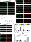
Figure 3. rpm-1 and the MAP kinases regulate levels of cebp-1 mRNA via its 3′ UTR
(A) Quantitative RT-PCR analysis shows up-regulation of cebp-1 mRNA in rpm-1(lf), which is eliminated in dlk-1, pmk-3 or mak-2 mutants (n=3). The control transcript ama-1 encodes the large subunit of RNA polymerase II (Sanford et al., 1983). (B–D) Transgenic expression of mCherry reporters in GABA motor neurons and quantification of fluorescence (n = 35 per genotype). Blue lines and letters denote inclusion of the cebp-1 3′ UTR in the transgene. (B) CEBP-1 with its own 3′ UTR is more highly expressed in rpm-1(lf) and mak-2(EE) transgenic animals than in wild type, dlk-1, and MAK-2(AA) transgenic animals. (C) A CEBP-1 transgene with the unc-54 3′UTR is expressed at similar levels in rpm-1 and wild type. (D) The cebp-1 3′UTR confers up-regulation of mCherry in rpm-1. (E) Expression of cebp-1 with the unc-54 3′UTR causes abnormal motor synapses. Data in A–E shown as mean ± SEM; n = 30 animals per genotype; statistics, Anova. (F) Overexpression of cebp-1 with its own UTR causes PLM axon overextension; transgenes with the unc-54 3′ UTR cause further defects in ALM. Overexpression of cebp-1 from muscles does not affect neuronal morphology. Quantitation, labels, details of mutant phenotypes, promoters and statistics as in Figure 1; GL, # of transgenic lines exhibiting gain-of-function abnormalities. Scales, 10 μm.
Figure 4
Figure 4. MAK-2 and CEBP-1 are present at motor neuron synapses
(A) Functional mCherry::MAK-2 in GABA motor neurons. Top panels are motor neuron soma in animals coexpressing Punc-25-mCherry::MAK-2 (red) and Histone::GFP (zuIs178). MAK-2 is present both in nucleus and cytoplasm. Bottom panels are motor neuron synapses in the dorsal cord coexpressing mCherry::MAK-2 with Punc-25-SNB-1::GFP (juIs1, left), or with RPM-1::GFP (juIs77, right). MAK-2 partially colocalizes with SNB-1 but not with RPM-1 (quantitation in Figure S7). (B) Functional mCherry::CEBP-1 in motor neuron cell bodies (top panels) and synapses (bottom panels) using the same markers as in (A). CEBP-1 shows nuclear and cytoplasmic localization in soma, and co-localizes with SNB-1 but is excluded from areas of RPM-1 at synapses (Figure S7). Synapse morphology (SNB-1::GFP) is unaltered in these transgenic animals. (C) mCherry::PHR::MAK-2 is confined to motor neuron synapses (left panels), and is excluded from the nucleus (N) in the soma (right panel). (D) Synaptic targeting of constitutively active MAK-2(EE), but not wild type MAK-2(+) or inactive MAK-2(AA), alters synapse morphology and reduces SNB-1::GFP puncta number (juIs1). (E) Quantification of the SNB-1::GFP phenotypes in D (n ≥ 30 per genotype). (F) Induction of un-regulated cebp-1 in adult neurons alters synapse morphology and number (juIs1). Quantification is on the right (n ≥ 30 for each group). 15°C–15°C indicates animals cultured continuously at 15°C; 15°C–25°C indicates animals grown at 15°C until late L4, then shifted to 25°C for 24 h. Data shown as mean ± SEM; statistics, Anova. Scales, 10 μm.
Figure 5
Figure 5. cebp-1 protein and mRNA are localized in touch neuron axon and synapses
(A) Expression of functional GFP::CEBP-1 in touch neurons. GFP is present along the axon (left panel), in the cell body (middle) and synapse (right). (B) GFP::NLS::MS2 reporter detects mRNA of cebp-1 in touch neuron axons. Illustrations of the constructs are shown above each image. Axonal fluorescence intensity as a fraction of peak cell body intensity is plotted versus distance along the axon from the cell body. GFP::NLS::MS2 alone (juEx1982) is concentrated in the cell body, and faintly seen along the axon. Coexpression of juEx1982 with cebp-1 transgenes containing six MS2 binding sites in the cebp-1 3′ UTR (juEx1984), but not in unc-54 3′ UTR (juEx2013), results in increased punctate GFP in axons. Axonal expression of GFP::NLS::MS2 + CEBP-1-cebp-1 3′ UTR is decreased in dlk-1(lf) animals, but significantly elevated in rpm-1(lf). (C) Average GFP::NLS::MS2 fluorescence intensity in axons when co-expressed with juEx1984 as % of cell body intensity (n = 15 per genotype). Data shown as mean ± SEM; statistics, Anova. Scale, 10 μm.
Figure 6
Figure 6. The DLK-1 pathway and CEBP-1 are required for touch axon regeneration in adults
(A) Regeneration of PLM axon (muIs32 marker) is reduced in loss of function mutants in each MAP kinase and cebp-1. mak-2(lf) has modest effects on regeneration compared to dlk-1(lf); the defect is rescued by wild type MAK-2 but not by MAK-2(K103R). Overexpression of DLK-1 enhances regrowth in a CEBP-1-dependent manner. Images are at 0 h and 24 h; quantitation at 24 h. Alleles are dlk-1(ju476), mkk-4(ju91), pmk-3(ok169), cebp-1(ju659), juEx1301[Prgef-1-dlk-1(+)] as dlk-1(++), juEx2111[Pmec-4-GFP::MAK-2(+)], and juEx2262[Pmec-4::MAK-2(K103R)]. n, # of animals shown below genotype or in column. (B) Heat-shock induced expression of cebp-1(R290C) transgene in adults blocks PLM regeneration. n, # of animals, in columns. Data shown as mean ± SEM; statistics, Anova. Scales, 10 μm.
Figure 7
Figure 7. Axotomy induces translation of CEBP-1 in a MAP kinase dependent manner and involves the 3′ UTR of cebp-1
Shown are images of Dendra reporter transgenes expressed in touch neurons, with illustration of constructs above. Green images are unconverted or newly synthesized Dendra; red images show photoconverted Dendra (Dendra*). (A) Under normal conditions synthesis of new Dendra::CEBP-1 from transgenes containing the cebp-1 3′ UTR (juEx1915) is slow. Quantification data are collected from soma, and axons of 60 μm away from soma (n = 10 for each time point). (B) Axotomy of PLM axons (middle), but not laser wounding of nearby epidermis (top panels), increases synthesis of Dendra::CEBP-1 (juEx1915). The increase is blocked by dlk-1(lf) or cycloheximide, and reduced by mak-2(lf). White arrows mark the laser cutting sites. (C) Axotomy of PLM axons does not induce new protein synthesis from a Dendra::CEBP-1 transgene lacking cebp-1 3′ UTR. (D) Quantitation of B and C (n = 12 for each group). (E, F) cebp-1 3′ UTR is sufficient to confer axotomy-induced protein synthesis (n = 12 for each group). Data shown as mean ± SEM. Statistics, Anova or t-test. Scales, 10 μm.

References

    1. Abrams B, Grill B, Huang X, Jin Y. Cellular and molecular determinants targeting the Caenorhabditis elegans PHR protein RPM-1 to perisynaptic regions. Dev Dyn. 2008;237:630–639. - PMC - PubMed
    1. Alberini CM, Ghirardi M, Metz R, Kandel ER. C/EBP is an immediate-early gene required for the consolidation of long-term facilitation in Aplysia. Cell. 1994;76:1099–1114. - PubMed
    1. Bernardi A, Spahr PF. Nucleotide sequence at the binding site for coat protein on RNA of bacteriophage R17. Proc Natl Acad Sci U S A. 1972;69:3033–3037. - PMC - PubMed
    1. Bertrand E, Chartrand P, Schaefer M, Shenoy SM, Singer RH, Long RM. Localization of ASH1 mRNA particles in living yeast. Mol Cell. 1998;2:437–445. - PubMed
    1. Bloom AJ, Miller BR, Sanes JR, DiAntonio A. The requirement for Phr1 in CNS axon tract formation reveals the corticostriatal boundary as a choice point for cortical axons. Genes Dev. 2007;21:2593–2606. - PMC - PubMed

Publication types

MeSH terms

Substances

LinkOut - more resources