Review
. 2009 Nov 30;169(2):183-99.
doi: 10.1016/j.resp.2009.08.015. Epub 2009 Sep 6.

Current tissue engineering and novel therapeutic approaches to axonal regeneration following spinal cord injury using polymer scaffolds

Affiliations
Review

Current tissue engineering and novel therapeutic approaches to axonal regeneration following spinal cord injury using polymer scaffolds

Nicolas N Madigan et al. Respir Physiol Neurobiol. .

Abstract

This review highlights current tissue engineering and novel therapeutic approaches to axonal regeneration following spinal cord injury. The concept of developing 3-dimensional polymer scaffolds for placement into a spinal cord transection model has recently been more extensively explored as a solution for restoring neurologic function after injury. Given the patient morbidity associated with respiratory compromise, the discrete tracts in the spinal cord conveying innervation for breathing represent an important and achievable therapeutic target. The aim is to derive new neuronal tissue from the surrounding, healthy cord that will be guided by the polymer implant through the injured area to make functional reconnections. A variety of naturally derived and synthetic biomaterial polymers have been developed for placement in the injured spinal cord. Axonal growth is supported by inherent properties of the selected polymer, the architecture of the scaffold, permissive microstructures such as pores, grooves or polymer fibres, and surface modifications to provide improved adherence and growth directionality. Structural support of axonal regeneration is combined with integrated polymeric and cellular delivery systems for therapeutic drugs and for neurotrophic molecules to regionalize growth of specific nerve populations.


Figures
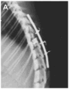
Fig. 1
Fig. 1
Polymer scaffold in situ. Lateral radiograph showing a well-aligned scaffold within the spinal canal after 4 weeks. The spine has been fixed and the scaffold contains barium contrast within the polymer. From Rooney et al. (2008) with permission.
Fig. 2
Fig. 2
The effect of spine stabilization on scaffold alignment. Three-dimensional magnetic resonance microscopy (MRM) in coronal ((A) and (B)) and axial ((C) and (D)) images 4 weeks after scaffold placement into the transected cord with ((A) and (C)) and without ((B) and (D)) spine fixation. From Rooney et al. (2008) with permission.
Fig. 3
Fig. 3
Scaffold polymer porosity. Porosity is controlled in a polyethylene glycol based hydrogel by varying the size of the porogen. Scanning electron microscopy (SEM) of lyophilized hydrogel ((a)–(c)) shows a highly porous structure with open, interconnected pores surrounded by polymer walls. Magnetic resonance microscopy (MRM) of scaffolds in their hydrated state ((d)–(f)) are compared. The distribution of sodium chloride particles was in the range of 100–500 μm. Scaffold with no porosity ((a) and (d)), porous scaffold with 75% porogen content and 300 μm particle size, ((b) and (e)), and porous scaffold with 75% porogen content and 500 μm particle size ((c) and (f)). Figure from Dadsetan et al. (2008) with permission.
Fig. 4
Fig. 4
Macroarchitectural design. Various injection-molding strategies for spinal cord scaffolds to align fascicular bundles. (A) Templated agarose cast over polystyrene fibres produces linear aligned channels of 200 μm diameter for a hemisection model (from Stokols et al. (2006) with permission. (B) Cylinder, tube, multichannel and open-path designs, from Wong et al. (2008) with permission, for casting in poly (ε-caprolactone). (C) PLGA multichannel scaffolds cast over parallel metal wiring provide dorsal and ventral channels, (left, bar 500 μm, from Moore et al. (2006) which may be elaborated into molds of complex anatomical design, (right, Friedman et al. (2002) both figures with permission).
Fig. 5
Fig. 5
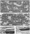
Oriented axonal growth. Microengineering strategies with oriented extension of neurite outgrowth in relation to micropatterned grooves and nanofibres. SEM imaging of PC12 cell growth on a collagen type I-coated PLGA film (A) and on a laminin peptide-coated PLGA film (B) each with a laser-etched groove size of 10 μm. Bar 40 μm, from Yao et al. (2009). Neurite extension along the length of electrospun PCL fibres of a diameter ranging from 0.8 +/− 0.7 μm (C) and 3.7 +/− 0.5 μm (D), from Yao et al. (2009) with permission.
Fig. 6
Fig. 6
Freeze dried agarose scaffolds in a complete transection model, figure from Stokols and Tuszynski (2006) with permission. Upper panel: scanning electron microscopic images of scaffolds in (A) longitudinal or (B) cross-sectional orientation shows the arrangement of channels in a honeycomb structure. Scale bar is 100 μm. Lower panel: neurofilament labeling demonstrates penetration and linear growth of axons within channels of scaffolds. (A) Scaffold lacking growth factor. (B) Scaffold loaded with 2 μg recombinant human BDNF into walls and matrix-filled lumen of individual channels. Magnitude of linear axonal growth is significantly increased. (C) Best example of linear axonal growth through complete length of channel. Scale bars = 100 μm.
Fig. 7
Fig. 7
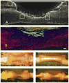
Scaffolds in situ. A dorsal hemisection injury (A) is filled with a fibrin matrix, as visualized with fibrinogen immunofluorescence (B). Diverse applications have been developed with this polymer, including affinity-based drug elution from heparin complexes and embryonic stem cell differentiation. Bar is 200 μm. Adapted from (Johnson et al., 2009) with permission. ((C)–(F)) A complete spinal cord transection is bridged with chitosan scaffolds loaded with brain-derived neural stem cells ((C) and (D)) or Schwann cells ((E) and (F)) as seen from a dorsal and lateral aspect. Neuronal tissue bridges have developed from the transected cord stumps. Figure from Zahir et al. (2008) with permission.
Fig. 8
Fig. 8
Neurofilament staining of axons in transverse sections through a PLGA scaffold after 1 month in vivo. Fluorescent microscopy image of one transverse section of a scaffold (loaded with NSCs) stained with an antibody against neurofilament with the region in which axons were counted encircled by a white line (A). Transverse sections of one channel from the NSC group (B), one from the SC group (C), and one from the control group (D) stained for neurofilament using an AEC chromogen. Magnification: 25× (A); 100× ((B)–(D)). Dot plots of axonal counts per cord in the NSC-, SC-, and control-treated groups (E). Data points represent individual animals. 95% Confidence interval was used for each group. *Medians vary significantly (p < 0.05). Figure from Olson et al. (2009) with permission.
Fig. 9
Fig. 9
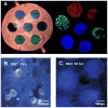
(A) Cells of different types can be placed in a scaffold. PCL-fumarate scaffold of 2-mm diameter loaded with SpL201 cells (blue), Schwann cells from a GFP rat (green) and rat Mesenchymal stem cells labeled with red quantum dots. Subsequently the scaffold can be placed in a way that ventral and dorsal orientation is retained. (B) Neurospheres of neural stem cells suspended in scaffold channels at 2 h grow to fill the channel diameter (C) within 3 days, figure from Olson et al. (2009) with permission. (For interpretation of the references to color in this figure legend, the reader is referred to the web version of the article.)
Fig. 10
Fig. 10
((A) and (B)) Light micrographs of the microsphere-loaded chitosan channels. The thickness of the channels when hydrated is approximately 200 μm, of which the secondary chitosan layer (indicated by arrows) contributes about 20 μm. (C) Scanning electron microscopy shows microspheres (arrowheads) embedded by the secondary chitosan coating. Figure from Kim et al. (2008) with permission.

References

    1. Adams DN, Kao EY, Hypolite CL, Distefano MD, Hu WS, Letourneau PC. Growth cones turn and migrate up an immobilized gradient of the laminin IKVAV peptide. J Neurobiol. 2005;62:134–147. - PubMed
    1. Balgude AP, Yu X, Szymanski A, Bellamkonda RV. Agarose gel stiffness determines rate of DRG neurite extension in 3D cultures. Biomaterials. 2001;22:1077–1084. - PubMed
    1. Bamber NI, Li H, Lu X, Oudega M, Aebischer P, Xu XM. Neurotrophins BDNF and NT-3 promote axonal re-entry into the distal host spinal cord through Schwann cell-seeded mini-channels. Eur J Neurosci. 2001;13:257–268. - PubMed
    1. Bellamkonda R. Peripheral nerve regeneration: an opinion on channels, scaffolds and anisotropy. Biomaterials. 2006;27:3515–3518. - PubMed
    1. Bellamkonda R, Ranieri JP, Bouche N, Aebischer P. Hydrogel-based three-dimensional matrix for neural cells. J Biomed Mater Res. 1995;29:663–671. - PubMed