. 2009 Sep 28;206(10):2067-77.
doi: 10.1084/jem.20090545. Epub 2009 Sep 8.

Loss of SOCS3 expression in T cells reveals a regulatory role for interleukin-17 in atherosclerosis

Affiliations

Loss of SOCS3 expression in T cells reveals a regulatory role for interleukin-17 in atherosclerosis

Soraya Taleb et al. J Exp Med. .

Abstract

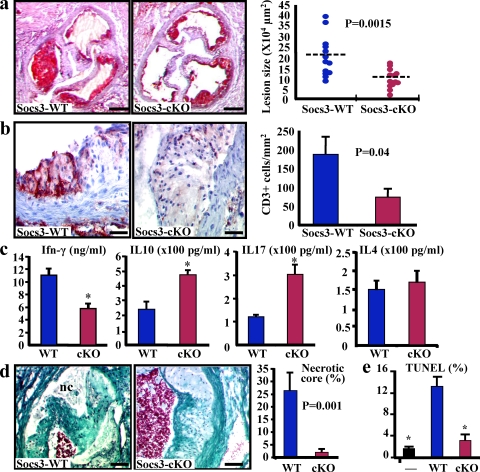
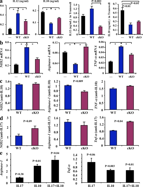
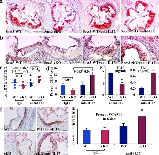
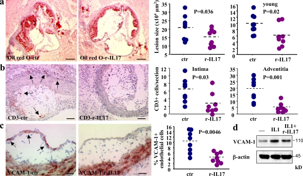
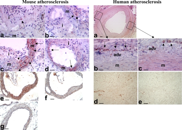
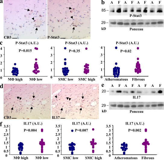
Atherosclerosis is an inflammatory vascular disease responsible for the first cause of mortality worldwide. Recent studies have clearly highlighted the critical role of the immunoinflammatory balance in the modulation of disease development and progression. However, the immunoregulatory pathways that control atherosclerosis remain largely unknown. We show that loss of suppressor of cytokine signaling (SOCS) 3 in T cells increases both interleukin (IL)-17 and IL-10 production, induces an antiinflammatory macrophage phenotype, and leads to unexpected IL-17-dependent reduction in lesion development and vascular inflammation. In vivo administration of IL-17 reduces endothelial vascular cell adhesion molecule-1 expression and vascular T cell infiltration, and significantly limits atherosclerotic lesion development. In contrast, overexpression of SOCS3 in T cells reduces IL-17 and accelerates atherosclerosis. We also show that in human lesions, increased levels of signal transducer and activator of transcription (STAT) 3 phosphorylation and IL-17 are associated with a stable plaque phenotype. These results identify novel SOCS3-controlled IL-17 regulatory pathways in atherosclerosis and may have important implications for the understanding of the increased susceptibility to vascular inflammation in patients with dominant-negative STAT3 mutations and defective Th17 cell differentiation.


Figures

Figure 1.
Figure 1.
SOCS3 deletion in T cells promotes IL-17 and IL-10 production, inhibits macrophage apoptosis, and limits atherosclerotic lesion development. (a) Atherosclerotic lesion size in the aortic root of chimeric Ldlr−/− SOCS3-WT or SOCS-cKO mice. Data are representative of four independent experiments throughout this work. Dashed lines indicate mean values. (b) Lesion T cell infiltration (red staining; mean values ± SEM). Data are representative of three experiments. (c) Reduced IFN-γ but increased IL-10 and IL-17 production in supernatants of CD3-stimulated CD4+ T cells (means ± SEM of five mice per group and two experiments; *, P < 0.05). (d) Necrotic core (nc; trichrome staining) size (means ± SEM) in the aortic root. Representative of two experiments and 13 mice per group. (e) Macrophage apoptosis (TUNEL staining) after coincubation with CD4+ cells from SOCS3-WT or SOCS-cKO mice (means ± SEM). Representative of three experiments (*, P < 0.05). Bars: (a) 250 µm; (b) 30 µm; (d) 60 µm.
Figure 2.
Figure 2.
SOCS3 deletion in T cells promotes an antiinflammatory macrophage phenotype. (a) IL-12 and IL-10 production by macrophages after coincubation with CD4+ cells from SOCS3-WT or SOCS-cKO mice in the presence or absence of Transwells. Mean values ± SEM of three to four mice per group are shown (*, P < 0.05). (b) TNF-α, NOS2, and arginase-1 mRNA expression (arbitrary units relative to GAPDH mRNA expression) by macrophages after coincubation with CD4+ cells. Mean values ± SEM of three to four mice per group and three independent experiments are shown (*, P < 0.05). (c and d) Quantitative analysis (relative to incubation without neutralizing antibodies) of NOS2, arginase-1, and TNF-α mRNA expression in macrophages after coincubation with CD4+ cells in the presence of either anti–IL-10 (c) or anti–IL-17 (d) neutralizing antibodies. (e) Quantitative analysis (relative to control without incubation with recombinant cytokines) of arginase-1 and TNF-α mRNA expression in macrophages after incubation with IL-10, IL-17 or IL-10, and IL-17. Values in c–e represent mean values ± SEM of three mice per group and three different experiments.
Figure 3.
Figure 3.
SOCS3-controlled IL-17 production protects against vascular inflammation and atherosclerotic lesion development. Representative photomicrographs (a and b) and quantitative analysis (c and d) of atherosclerotic lesion size (a and c) and lesion T cell infiltration (b and d) in the aortic root of chimeric Ldlr−/− SOCS3-WT or SOCS-cKO mice treated either with a neutralizing anti–IL-17A antibody (anti–IL-17) or an isotype-matched control (IgG; n = 7–8 mice per group). The anti–IL-17 in vivo experiment was repeated twice. p-values are shown in c and d. Dashed lines indicate mean values. (e) Increased IL-4 but reduced IL-10 production in supernatants of CD3-stimulated CD4+ T cells recovered at the time of sacrifice from chimeric Ldlr−/− mice treated with a neutralizing anti–IL-17A antibody. Means ± SEM of five mice per group performed in triplicates are shown (*, P < 0.05). (f) VCAM-1 expression in atherosclerotic lesions (n = 6–8 mice per group and two separate experiments; *, P < 0.05). Bars: (a) 250 µm; (b) 100 µm; (f) 130 µm.
Figure 4.
Figure 4.
Supplementation with IL-17 reduces vascular inflammation and limits atherosclerotic lesion development. (a) Atherosclerotic lesion size in the aortic root of 17-wk-old female chimeric Ldlr −/− SOCS3-WT mice fed a high fat diet and treated with rIL-17 or control serum albumin for 5 wk. Two different experiments are depicted with eight to nine animals in each group. Young animals were 12 wk old. (b) Lesion and adventitial T cell infiltration in 17-wk-old animals. Similar results were obtained in young animals (not depicted). (c) Endothelial VCAM-1 expression in IL-17–treated 17-wk-old mice. Results represent two separate experiments. (d) One representative example out of three different experiments of a Western blot showing reduction of IL-1–induced VCAM-1 expression after mouse endothelial cells are incubated with IL-17. Dashed lines indicate mean values. Bars: (a) 250 µm; (b) 50 µm; (c) 25 µm.
Figure 5.
Figure 5.
Expression of IL-17 in atherosclerotic vessels. (left) Mouse atherosclerosis. (a and b) Occasional staining for IL-17 (red, arrows) in inflammatory cells within the intima (i) and adventitia (arrows in b) of SOCS3-cKO mice. (c and d) Representative examples of staining for IL-17 (red, arrows) in the media (m) of the aortic sinus (c) and the coronary arteries (d) of SOCS3-cKO mice. Note that IL-17 staining in media was detected in plaque-free areas (right of the arrowheads) and was lost in areas where plaques have developed (left of the arrowheads). (e–g) Staining for IL-17 (red/brown) in the media of the aortic sinus of WT (e and f) or IL-17A–deficient (g) mice. Note that IL-17 staining almost disappeared after adsorption with the IL-17–specific peptide (f) or in vessels of IL-17A–deficient mice (g). Data are representative of at least 10 different mouse arteries. Bars: (a–d) 45 µm; (e–g) 90 µm. (right) Human atherosclerosis. (a) IL-17 staining in the media (m) and adventitia (adv) of carotid plaques. (b) The image is from an area underlying an advanced plaque. It shows IL-17 staining in adventitial vessels (arrows), but the media has lost its IL-17 staining. (c) The image is from an area underlying an early intimal thickening. It shows staining for IL-17 in the adventitial vessels (arrows) and still diffuse moderate IL-17 staining within the media. (d) Another example of IL-17 staining (brown) in an advanced carotid plaque. (e) Disappearance of IL-17 staining after adsorption with the IL-17–specific peptide. Data are representative of at least 10 different human arteries. Bars: (a) 250 µm; (b and c) 30 µm; (d and e) 45 µm.
Figure 6.
Figure 6.
Stat3 phosphorylation and IL-17 expression in human carotid atherosclerotic plaques are associated with markers of plaque stability. (a) Stat3 phosphorylation (P-Stat3) in CD3+ cells of human carotid atherosclerotic plaques (arrows). Arrowheads show a CD3+ cell with no P-Stat3. (b) Detection of P-Stat3 in protein extracts from atheromatous (A) or fibrous (F) carotid plaques using Western blotting. Ponceau red is shown for estimation of sample loading. (c) Semiquantitative analysis of P-Stat3 content of carotid plaques and relation to plaque macrophage (MΦ) content, plaque SMC content, and plaque phenotype (atheromatous or fibrous). (d) IL-17 expression in CD3+ cells of human carotid atherosclerotic plaques (arrows). Arrowheads show two CD3+ cells with no IL-17 expression. (e) Detection of IL-17 in protein extracts from atheromatous (A) or fibrous (F) carotid plaques using Western blotting (the same blot shown for P-Stat3 is shown for IL-17). (e) Semiquantitative analysis of the IL-17 content of carotid plaques and the relation to plaque macrophage content, plaque SMC content, and plaque phenotype (atheromatous or fibrous). Overall, 40 different atherosclerotic plaques from 5 separate experiments were studied. Bars: (a) 30 µm; (c) 60 µm. A.U., arbitrary units.

Similar articles

Cited by

References

    1. Ait-Oufella H., Kinugawa K., Zoll J., Simon T., Boddaert J., Heeneman S., Blanc-Brude O., Barateau V., Potteaux S., Merval R., et al. 2007. Lactadherin defciency leads to apoptotic cell accumulation and accelerated atherosclerosis in mice.Circulation. 115:2168–2177 doi:10.1161/CIRCULATIONAHA.106.662080 - DOI - PubMed
    1. Bettelli E., Oukka M., Kuchroo V.K. 2007. T(H)-17 cells in the circle of immunity and autoimmunity.Nat. Immunol. 8:345–350 doi:10.1038/ni0407-345 - DOI - PubMed
    1. Binder C.J., Chang M.K., Shaw P.X., Miller Y.I., Hartvigsen K., Dewan A., Witztum J.L. 2002. Innate and acquired immunity in atherogenesis.Nat. Med. 8:1218–1226 doi:10.1038/nm1102-1218 - DOI - PubMed
    1. Boddaert J., Mallat Z., Fornes P., Esposito B., Lecomte D., Verny M., Tedgui A., Belmin J. 2005. Age and gender effects on apoptosis in the human coronary arterial wall.Mech. Ageing Dev. 126:678–684 doi:10.1016/j.mad.2005.01.001 - DOI - PubMed
    1. Chen Z., Laurence A., Kanno Y., Pacher-Zavisin M., Zhu B.M., Tato C., Yoshimura A., Hennighausen L., O'Shea J.J. 2006. Selective regulatory function of Socs3 in the formation of IL-17-secreting T cells.Proc. Natl. Acad. Sci. USA. 103:8137–8142 doi:10.1073/pnas.0600666103 - DOI - PMC - PubMed

Publication types