

Identification of the binding domain of Streptococcus oralis glyceraldehyde-3-phosphate dehydrogenase for Porphyromonas gingivalis major fimbriae
- PMID: 19737900
- PMCID: PMC2772547
- DOI: 10.1128/IAI.00439-09
Identification of the binding domain of Streptococcus oralis glyceraldehyde-3-phosphate dehydrogenase for Porphyromonas gingivalis major fimbriae
Abstract
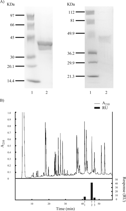
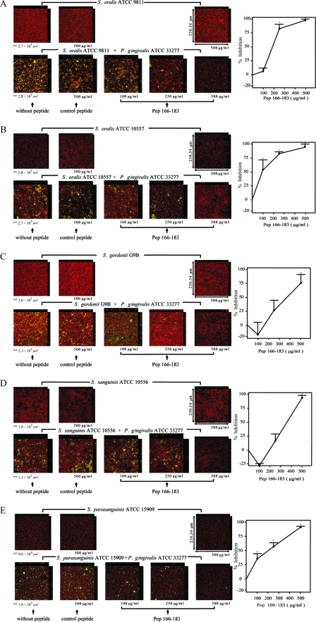
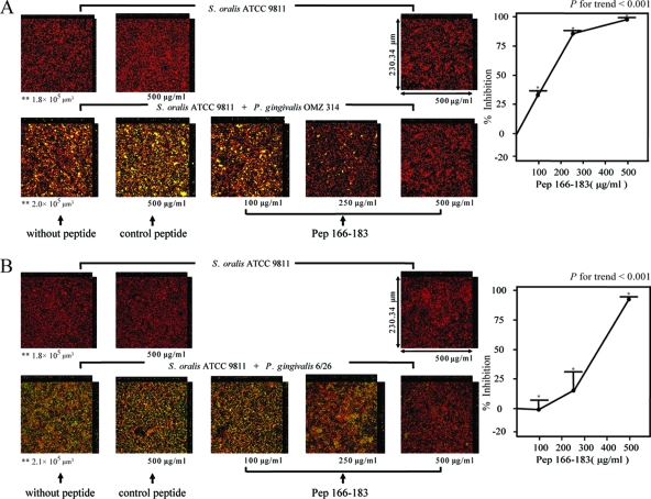
Porphyromonas gingivalis forms communities with antecedent oral biofilm constituent streptococci. P. gingivalis major fimbriae bind to glyceraldehyde-3-phosphate dehydrogenase (GAPDH) present on the streptococcal surface, and this interaction plays an important role in P. gingivalis colonization. This study identified the binding domain of Streptococcus oralis GAPDH for P. gingivalis fimbriae. S. oralis recombinant GAPDH (rGAPDH) was digested with lysyl endopeptidase. Cleaved fragments of rGAPDH were applied to a reverse-phase high-pressure liquid chromatograph equipped with a C18 column. Each peak was collected; the binding activity toward P. gingivalis recombinant fimbrillin (rFimA) was analyzed with a biomolecular interaction analysis system. The fragment displaying the strongest binding activity was further digested with various proteinases, after which the binding activity of each fragment was measured. The amino acid sequence of each fragment was determined by direct sequencing, mass spectrometric analysis, and amino acid analysis. Amino acid residues 166 to 183 of S. oralis GAPDH exhibited the strongest binding activity toward rFimA; confocal laser scanning microscopy revealed that the synthetic peptide corresponding to amino acid residues 166 to 183 of S. oralis GAPDH (pep166-183, DNFGVVEGLMTTIHAYTG) inhibits S. oralis-P. gingivalis biofilm formation in a dose-dependent manner. Moreover, pep166-183 inhibited interbacterial biofilm formation by several oral streptococci and P. gingivalis strains with different types of FimA. These results indicate that the binding domain of S. oralis GAPDH for P. gingivalis fimbriae exists within the region encompassing amino acid residues 166 to 183 of GAPDH and that pep166-183 may be a potent inhibitor of P. gingivalis colonization in the oral cavity.
Figures
Similar articles
-
Identification and characterization of Porphyromonas gingivalis client proteins that bind to Streptococcus oralis glyceraldehyde-3-phosphate dehydrogenase.Infect Immun. 2013 Mar;81(3):753-63. doi: 10.1128/IAI.00875-12. Epub 2012 Dec 21. Infect Immun. 2013. PMID: 23264054 Free PMC article.
-
Oral streptococcal glyceraldehyde-3-phosphate dehydrogenase mediates interaction with Porphyromonas gingivalis fimbriae.Microbes Infect. 2004 Nov;6(13):1163-70. doi: 10.1016/j.micinf.2004.06.005. Microbes Infect. 2004. PMID: 15488735
-
Glyceraldehyde-3-phosphate dehydrogenase of Streptococcus oralis functions as a coadhesin for Porphyromonas gingivalis major fimbriae.Infect Immun. 2004 Mar;72(3):1341-8. doi: 10.1128/IAI.72.3.1341-1348.2004. Infect Immun. 2004. PMID: 14977937 Free PMC article.
-
Molecular interaction of Porphyromonas gingivalis with host cells: implication for the microbial pathogenesis of periodontal disease.J Periodontol. 2003 Jan;74(1):90-6. doi: 10.1902/jop.2003.74.1.90. J Periodontol. 2003. PMID: 12593602 Review.
-
Post-translational Modifications in Oral Bacteria and Their Functional Impact.Front Microbiol. 2021 Dec 2;12:784923. doi: 10.3389/fmicb.2021.784923. eCollection 2021. Front Microbiol. 2021. PMID: 34925293 Free PMC article. Review.
Cited by
-
VimA-dependent modulation of the secretome in Porphyromonas gingivalis.Mol Oral Microbiol. 2012 Dec;27(6):420-35. doi: 10.1111/j.2041-1014.2012.00661.x. Epub 2012 Aug 9. Mol Oral Microbiol. 2012. PMID: 23134608 Free PMC article.
-
The pathogenic persona of community-associated oral streptococci.Mol Microbiol. 2011 Jul;81(2):305-14. doi: 10.1111/j.1365-2958.2011.07707.x. Epub 2011 Jun 3. Mol Microbiol. 2011. PMID: 21635580 Free PMC article. Review.
-
Tobacco smoke augments Porphyromonas gingivalis-Streptococcus gordonii biofilm formation.PLoS One. 2011;6(11):e27386. doi: 10.1371/journal.pone.0027386. Epub 2011 Nov 14. PLoS One. 2011. PMID: 22110637 Free PMC article.
-
Peptide-Based Inhibitors of Fimbrial Biogenesis in Porphyromonas gingivalis.Infect Immun. 2019 Feb 21;87(3):e00750-18. doi: 10.1128/IAI.00750-18. Print 2019 Mar. Infect Immun. 2019. PMID: 30642895 Free PMC article.
-
Role of vimA in cell surface biogenesis in Porphyromonas gingivalis.Microbiology (Reading). 2010 Jul;156(Pt 7):2180-2193. doi: 10.1099/mic.0.038331-0. Epub 2010 Apr 8. Microbiology (Reading). 2010. PMID: 20378652 Free PMC article.
References
-
- Abe, N., A. Baba, R. Takii, K. Nakayama, A. Kamaguchi, Y. Shibata, Y. Abiko, K. Okamoto, T. Kadowaki, and K. Yamamoto. 2004. Roles of Arg- and Lys-gingipains in coaggregation of Porphyromonas gingivalis: identification of its responsible molecules in translation products of rgpA, kgp, and hagA genes. Biol. Chem. 385:1041-1047. - PubMed
-
- Amano, A., T. Fujiwara, H. Nagata, M. Kuboniwa, A. Sharma, H. T. Sojar, R. J. Genco, and S. Shizukuishi. 1997. Porphyromonas gingivalis fimbriae mediate coaggregation with Streptococcus oralis through specific domains. J. Dent. Res. 76:852-857. - PubMed
-
- Amano, A., M. Kuboniwa, I. Nakagawa, S. Akiyama, I. Morisaki, and S. Hamada. 2000. Prevalence of specific genotypes of Porphyromonas gingivalis fimA and periodontal health status. J. Dent. Res. 79:1664-1668. - PubMed
-
- Amano, A., I. Nakagawa, N. Okahashi, and N. Hamada. 2004. Variations of Porphyromonas gingivalis fimbriae in relation to microbial pathogenesis. J. Periodontal Res. 39:136-142. - PubMed
Publication types
MeSH terms
Substances
LinkOut - more resources
Full Text Sources
Molecular Biology Databases
Research Materials
