. 2009 Oct 30;284(44):30360-71.
doi: 10.1074/jbc.M109.048322. Epub 2009 Sep 8.

Identification of the human mitochondrial linoleoyl-coenzyme A monolysocardiolipin acyltransferase (MLCL AT-1)

Affiliations

Identification of the human mitochondrial linoleoyl-coenzyme A monolysocardiolipin acyltransferase (MLCL AT-1)

William A Taylor et al. J Biol Chem. .

Abstract

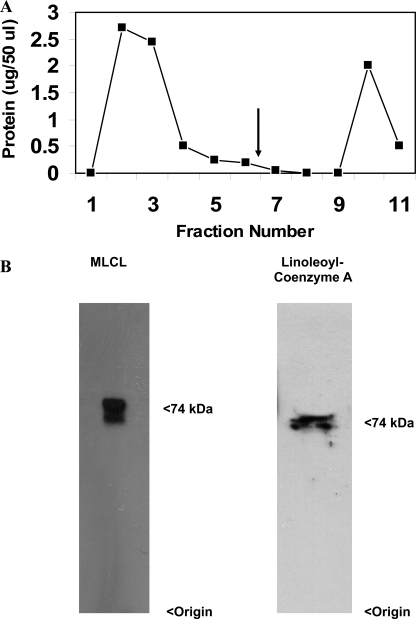
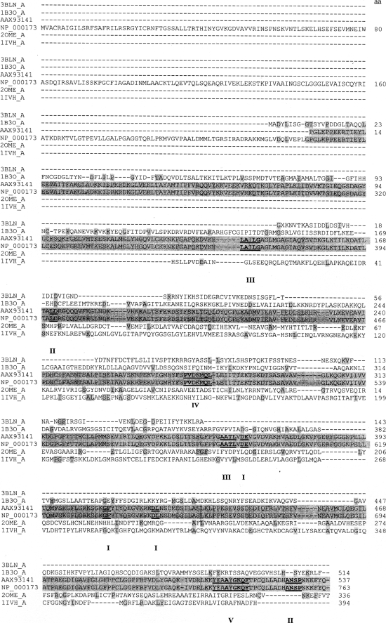
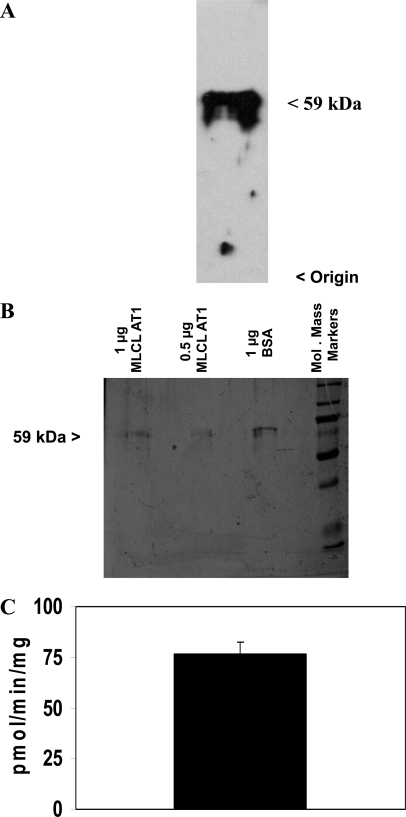
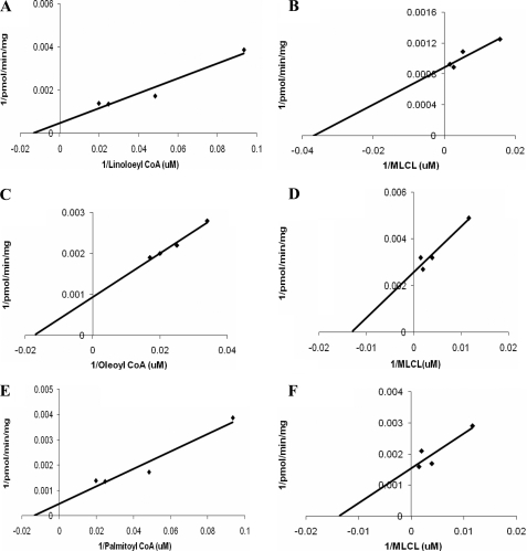
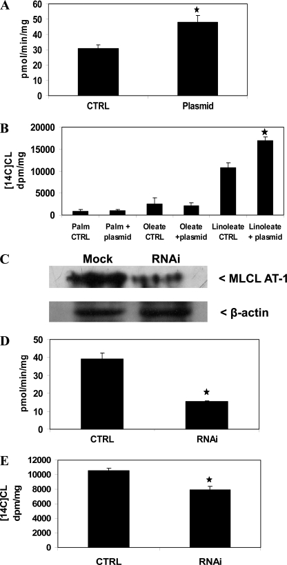
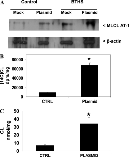
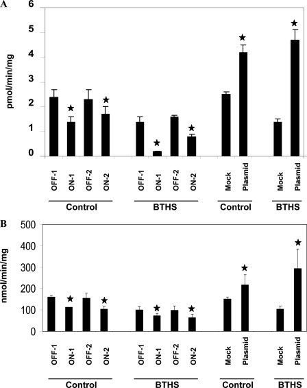
Here we report the identification of a previously uncharacterized human protein as the human monolysocardiolipin acyltransferase-1 (MLCL AT-1). Pig liver mitochondria were treated with n-butyl alcohol followed by Q-Sepharose chromatography, preparative gel electrophoresis, cytidine diphosphate-1,2-diacyl-sn-glycerol-Sepharose chromatography, and finally monolysocardiolipin-adriamycin-agarose affinity chromatography. Elution with either monolysocardiolipin or linoleoyl coenzyme A revealed a major band at 74 kDa with high specific activity (2,300 pmol/min/mg) for the acylation of monolysocardiolipin to cardiolipin using [1-(14)C]linoleoyl coenzyme A as substrate. Matrix-assisted laser desorption ionization time-of-flight-mass spectrometry analysis followed by search of the Mascot protein data base revealed peptide matches consistent with a 59-kDa protein identified as unknown human protein (GenBank(TM) protein accession number AAX93141; nucleotide accession number AC011742.3). The purified human recombinant MLCL AT-1 protein utilized linoleoyl coenzyme A > oleoyl coenzyme A > palmitoyl coenzyme A for the specific acylation of monolysocardiolipin to cardiolipin. Expression of MLCL AT-1 in HeLa cells increased mitochondrial monolysocardiolipin acyltransferase activity and [1-(14)C]linoleic acid incorporated into cardiolipin, whereas RNA interference knockdown of MLCL AT-1 in HeLa cells resulted in reduction in enzyme activity and [1-(14)C]linoleic acid incorporated into cardiolipin. In contrast, expression of MLCL AT-1 in HeLa cells did not alter [1-(14)C]oleic or [1-(14)C]palmitate incorporation into cardiolipin indicating in vivo specificity for the remodeling of cardiolipin with linoleate. Finally, expression of MLCL AT-1 in Barth syndrome lymphoblasts, which exhibit cardiolipin levels 20% that of normal lymphoblasts, increased mitochondrial monolysocardiolipin acyltransferase activity, [1-(14)C]linoleic acid incorporation into cardiolipin, cardiolipin mass, and succinate dehydrogenase (mitochondrial complex II) activity compared with mock-transfected Barth syndrome lymphoblasts. The results identify MLCL AT-1 as a human mitochondrial monolysocardiolipin acyltransferase involved in the remodeling of cardiolipin.


Figures

FIGURE 1.
FIGURE 1.
MLCL-adriamycin-agarose affinity chromatography profile and Western blot analysis. A, 4 ml of partially purified MLCL-AT was loaded onto 1 ml of adriamycin-agarose affinity gel, which was pre-equilibrated with 0.75 mg of MLCL. The gel was washed with 12 column volumes of 0.1 m borate buffer, pH 8.0, until no more protein was eluted. Finally, MLCL-AT was eluted (arrow) by the addition of 1 mm MLCL or 1 mm linoleoyl coenzyme A. B, Western blot of fraction 10 eluted with MLCL using the polyclonal antibody to MLCL AT and Western blot of fraction 10 eluted with linoleoyl coenzyme A. Origin and molecular mass are indicated on the right. Representative blots are depicted.
FIGURE 2.
FIGURE 2.
Sequence alignment of AAX93141 with proteins identified through peptide matches. Proteins (3BLN_A, 1B3O_A, 2OME_A, and 1IVH_A) obtained from the BLAST search of the peptides and human trifunctional protein (NP_000173) were analyzed by the Cobalt multialignment program. The amino acid residues of the known proteins are highlighted (gray) relative to their alignment with the human MLCL AT-1 (AAX93141) and NP_000173. Matching amino acid sequences from peptides I to V from Table 3 are indicated in boldface. Protein accession number is indicated on the left.
FIGURE 3.
FIGURE 3.
Western blot analysis, SDS-PAGE analysis, and MLCL AT activity of the human recombinant MLCL AT-1 protein. A, purified human recombinant MLCL AT-1 was prepared and Western blot analysis performed as described under “Experimental Procedures.” A representative blot is depicted. Origin and molecular mass are indicated on the right. B, SDS-PAGE of 1 μg of bovine serum albumin (BSA) or 0.5 or 1.0 μg of purified human recombinant MLCL AT-1 following imidazole elution from the nickel column. Molecular mass markers are indicated on the right. A representative blot is depicted. C, MLCL AT activity of purified human recombinant MLCL AT-1 eluted from the nickel column. Data represent the mean ± S.D. of three preparations.
FIGURE 4.
FIGURE 4.
MLCL AT activity of human recombinant MLCL AT-1 in the presence of various concentrations of MLCL or acyl-CoAs. MLCL AT enzyme activity of MLCL AT-1 was determined with varying concentrations of linoleoyl coenzyme A (A) or oleoyl coenzyme A (C) or palmitoyl coenzyme A (E) in the presence of a constant amount of MLCL or determined with varying concentrations of MLCL in the presence of a constant amount of linoleoyl coenzyme A (B) or oleoyl coenzyme A (D) or palmitoyl coenzyme A (F) as described under “Experimental Procedures.” Reciprocal plots are depicted. Data represent the mean of two experiments. The results between samples differed by less than 10%.
FIGURE 5.
FIGURE 5.
MLCT AT activity and 1-14C-labeled fatty acid incorporation into CL in HeLa cells expressing human recombinant MLCL AT-1 and MLCT AT-1 protein, activity, and 1-14C-labeled fatty acid incorporation into CL in HeLa cells transfected with MLCL AT-1 RNAi. A, HeLa cells were mock-transfected (CTRL) or transfected with human recombinant MLCL AT-1 (Plasmid) mitochondrial fractions prepared, and MLCL AT activity was determined as described under “Experimental Procedures.” B, HeLa cells were transfected as in A and incubated with 0.1 μm [1-14C]linoleate or [1-14C]oleate or [1-14C]palmitate (Palm), and radioactivity incorporated into CL was determined. CTRL, control. C, HeLa cells were mock-transfected (Mock) or transfected with MLCL AT-1 RNAi (RNAi), and Western blot analysis was performed as described under “Experimental Procedures.” A representative blot is shown for MLCL AT-1 and β-actin. D, HeLa cells were mock-transfected (CTRL) with or transfected with MLCL AT-1 RNAi (RNAi), and mitochondrial fractions were prepared, and MLCL AT activity was determined as described under “Experimental Procedures.” E, HeLa cells were transfected as in D and then incubated with 0.1 μm [1-14C]linoleate, and radioactivity incorporated into CL was determined. Data represent the mean ± S.D. of three experiments. *, p < 0.05.
FIGURE 6.
FIGURE 6.
MLCT AT-1 protein expression and [1-14C]linoleate incorporation into CL and CL mass in Barth syndrome lymphoblasts expressing human recombinant MLCL AT-1. A, age-matched control and BTHS lymphoblasts were mock-transfected (Mock) or transfected with human recombinant MLCL AT-1 plasmid (Plasmid), and Western blot analysis of MLCL AT-1 protein was determined as described under “Experimental Procedures.” A representative blot is shown for MLCL AT-1 and β-actin. BTHS lymphoblasts were mock-transfected (CTRL) or transfected with human recombinant MLCL AT-1 (Plasmid), and [1-14C]linoleate incorporation into CL (B) and CL mass (C) was determined. Data represent the mean ± S.D. of three experiments. *, p < 0.05.
FIGURE 7.
FIGURE 7.
MLCL AT-1 and succinate dehydrogenase activities in Barth syndrome lymphoblasts expressing human recombinant MLCL AT-1 or MLCL AT-1 RNAi. Control and BTHS lymphoblasts were transfected with OFF-1 or OFF-2 or ON-1 or ON-2 MLCL AT-1 RNAi plasmids or were mock-transfected (Mock) or transfected with human recombinant MLCL AT-1 plasmid (Plasmid) and MLCL AT (A) or succinate dehydrogenase (B) activities determined as described under “Experimental Procedures.” Data represent the mean ± S.D. of three experiments. *, p < 0.05.

References

    1. Hatch G. M. (2004) Biochem. Cell Biol. 82, 99–112 - PubMed
    1. Houtkooper R. H., Vaz F. M. (2008) Cell. Mol. Life Sci. 65, 2493–2506 - PMC - PubMed
    1. Schlame M. (2008) J. Lipid. Res. 49, 1607–1620 - PMC - PubMed
    1. Hostetler K. Y. (1982) Phospholipids ( Hawthorne J. N., Ansell G. B. eds) pp. 215–261, Elsevier Science Publishers B.V., Amsterdam
    1. Schlame M., Rua D., Greenberg M. L. (2000) Prog. Lipid Res. 39, 257–288 - PubMed

Publication types

Associated data