. 2009 Nov 6;284(45):31422-30.
doi: 10.1074/jbc.M109.024489. Epub 2009 Sep 8.

Interaction of organic cations with organic anion transporters

Affiliations

Interaction of organic cations with organic anion transporters

Sun-Young Ahn et al. J Biol Chem. .

Abstract

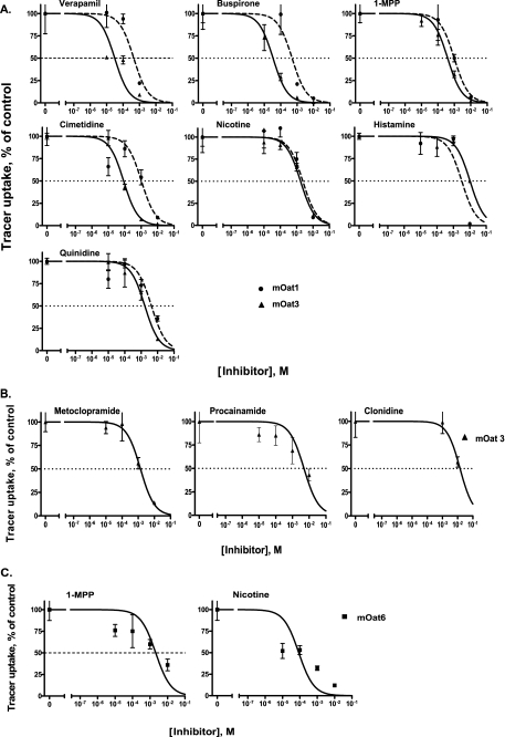
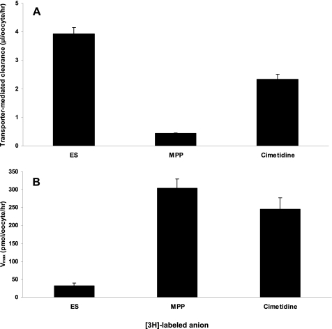
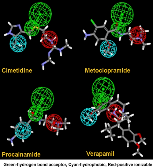
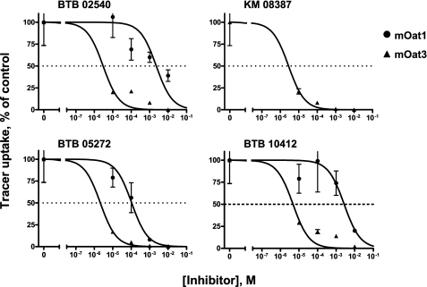
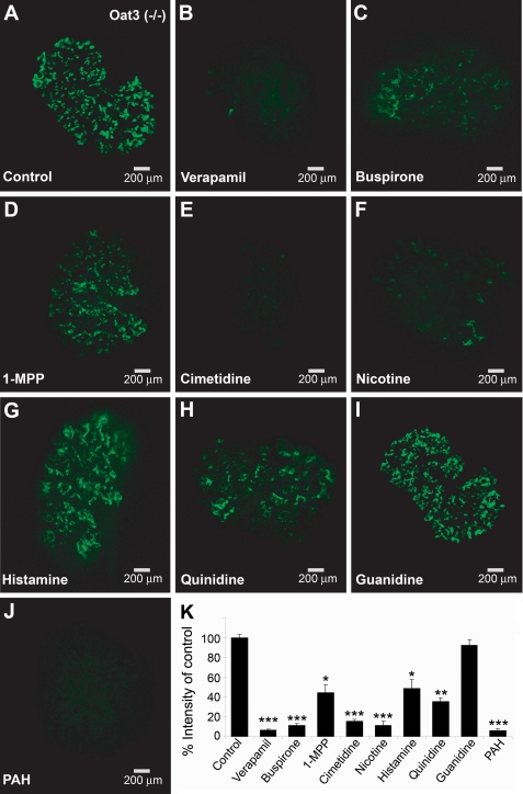
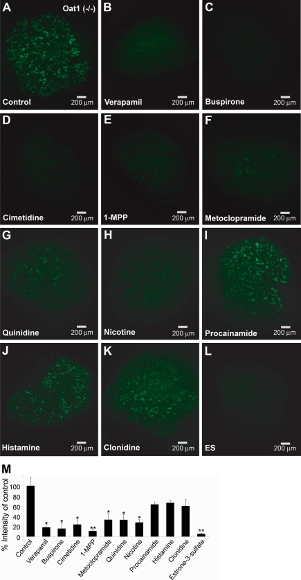
Studies of the organic anion transporters (Oats) have focused mainly on their interactions with organic anionic substrates. However, as suggested when Oat1 was originally identified as NKT (Lopez-Nieto, C. E., You, G., Bush, K. T., Barros, E. J., Beier, D. R., and Nigam, S. K. (1997) J. Biol. Chem. 272, 6471-6478), since the Oats share close homology with organic cation transporters (Octs), it is possible that Oats interact with cations as well. We now show that mouse Oat1 (mOat1) and mOat3 and, to a lesser degree, mOat6 bind a number of "prototypical" Oct substrates, including 1-methyl-4-phenylpyridinium. In addition to oocyte expression assays, we have tested binding of organic cations to Oat1 and Oat3 in ex vivo assays by analyzing interactions in kidney organ cultures deficient in Oat1 and Oat3. We also demonstrate that mOat3 transports organic cations such as 1-methyl-4-phenylpyridinium and cimetidine. A pharmacophore based on the binding affinities of the tested organic cations for Oat3 was generated. Using this pharmacophore, we screened a chemical library and were able to identify novel cationic compounds that bound to Oat1 and Oat3. These compounds bound Oat3 with an affinity higher than the highest affinity compounds in the original set of prototypical Oct substrates. Thus, whereas Oat1, Oat3, and Oat6 appear to function largely in organic anion transport, they also bind and transport some organic cations. These findings could be of clinical significance, since drugs and metabolites that under normal physiological conditions do not bind to the Oats may undergo changes in charge and become Oat substrates during pathologic conditions wherein significant variations in body fluid pH occur.


Figures

FIGURE 1.
FIGURE 1.
Interaction of organic cations with mOat1, mOat3, and mOat6. A, seven organic cations inhibited uptake of fluorescent tracers (6CF and 5CF; see “Experimental Procedures”) in both mOat1 and mOat3-expressing Xenopus oocytes. Each data point represents the mean ± S.E. values of four groups of 4–5 oocytes each. B, three cations that did not inhibit Oat1 inhibited 5CF uptake in Oat3. C, only two cations inhibited fluorescein uptake in Oat6-expressing oocytes.
FIGURE 2.
FIGURE 2.
Transport of organic cations by mOat3. A, transporter-mediated clearance of 3H-labeled estrone 3-sulfate (ES), cimetidine, and 1-MPP was calculated as the clearance in injected oocytes minus the clearance of uninjected oocytes. The measurements were performed in a single experiment using the same group of oocytes. Estrone 3-sulfate, the prototypical substrate for Oat3, was used as a positive control. B, Vmax, the maximum uptake rate, of the 3H-labeled compounds was calculated from the clearance (see “Experimental Procedures”).
FIGURE 3.
FIGURE 3.
Oat3 pharmacophore. A four-feature pharmacophore model of the interaction of organic cations with Oat3 is shown superimposed on the structures of four of the tested compounds. Pharmacophore features are color-coded as follows. Red, positive ionizable; green, hydrogen bond acceptor; cyan, hydrophobic. Compounds are color-coded as follows. Red, oxygen; blue, nitrogen; gray, carbon; white, hydrogen.
FIGURE 4.
FIGURE 4.
Compounds matching the pharmacophore interact with Oat1 and Oat3. Four cationic compounds that were identified from the Maybridge chemical library as pharmacophore matches strongly inhibited Oat3-mediated transport in Xenopus oocyte assays, whereas three of these compounds inhibited Oat1-mediated transport. Oat3 showed a higher affinity for these compounds than did Oat1.
FIGURE 5.
FIGURE 5.
Inhibition of fluorescent tracer uptake in Oat3 knock-out kidney cultures by cations. Organ cultures from Oat3 knockouts were incubated with a 2 mm concentration of the indicated compounds and 1 μm 6CF. Fluorescence was quantified, and results are presented as a percentage of control tracer uptake. J, PAH, the prototypical substrate for Oat1, was used as a positive control and showed the greatest degree of inhibition. I, guanidine, which did not show inhibition in the Xenopus oocyte assays, was the negative control. A–J, all images represent triplicate kidney cultures from the same experiment. K, values in the graph represent mean ± S.E. *, p < 0.05; **, p < 0.005; ***, p < 0.0005.
FIGURE 6.
FIGURE 6.
Inhibition of tracer uptake in Oat1 knock-out kidney culture. Organ cultures from Oat1 knockouts were coincubated with tracer (6CF; 1 μm) and the indicated compounds (2 mm), and data were analyzed as described in the legend to Fig. 5. L, estrone 3-sulfate (ES), the prototypical substrate for Oat3, served as the positive control. A–L, all images represent triplicate kidney cultures from the same experiment. M, values in the graph represent mean ± S.E. percentage of control. *, p < 0.05; **, p < 0.005; ***, p < 0.0005.

References

    1. Lopez-Nieto C. E., You G., Bush K. T., Barros E. J., Beier D. R., Nigam S. K. (1997) J. Biol. Chem. 272, 6471–6478 - PubMed
    1. Ahn S. Y., Nigam S. K. (2009) Mol. Pharmacol. 76, 481–490 - PMC - PubMed
    1. Sweet D. H., Chan L. M., Walden R., Yang X. P., Miller D. S., Pritchard J. B. (2003) Am. J. Physiol. Renal Physiol. 284, F763–F769 - PubMed
    1. Sweet D. H., Wolff N. A., Pritchard J. B. (1997) J. Biol. Chem. 272, 30088–30095 - PubMed
    1. Kaler G., Truong D. M., Sweeney D. E., Logan D. W., Nagle M., Wu W., Eraly S. A., Nigam S. K. (2006) Biochem. Biophys. Res. Commun. 351, 872–876 - PMC - PubMed

Publication types

MeSH terms