. 2009 Oct 30;284(44):30695-707.
doi: 10.1074/jbc.M109.007997. Epub 2009 Sep 8.

A conserved phosphorylation site within the forkhead domain of FoxM1B is required for its activation by cyclin-CDK1

Affiliations

A conserved phosphorylation site within the forkhead domain of FoxM1B is required for its activation by cyclin-CDK1

Yi-Ju Chen et al. J Biol Chem. .

Abstract

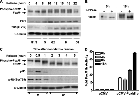
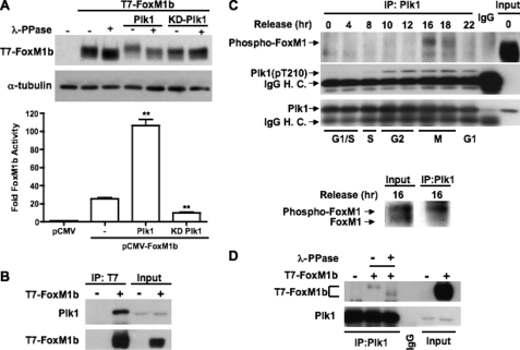
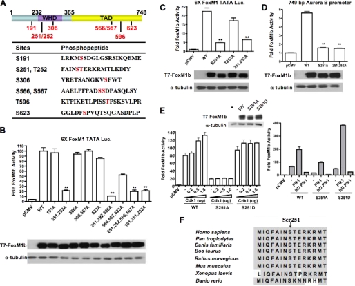
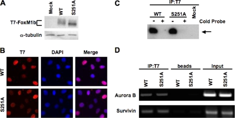
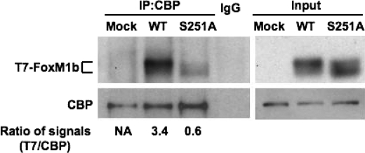
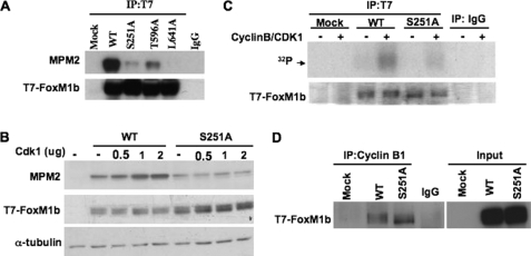
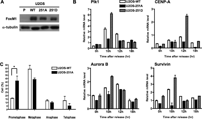
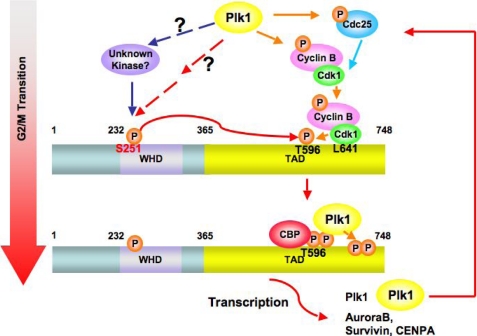
The Forkhead box M1 (FoxM1) transcription factor is critical for expression of the genes essential for G(1)/S transition and mitotic progression. To explore the cell cycle regulation of FoxM1, we examined the phosphorylation profile of FoxM1. Here, we show that the phosphorylated status and the activity of FoxM1 increase as cells progress from S to G(2)/M phases. Moreover, dephosphorylation of FoxM1 coincides with exit from mitosis. Using mass spectrometry, we have identified a new conserved phosphorylation site (Ser-251) within the forkhead domain of FoxM1. Disruption of Ser-251 inhibits phosphorylation of FoxM1 and dramatically decreases its transcriptional activity. We demonstrate that the Ser-251 residue is required for CDK1-dependent phosphorylation of FoxM1 as well as its interaction with the coactivator CREB-binding protein (CBP). Interestingly, the transcriptional activity of the S251A mutant protein remains responsive to activation by overexpressed Polo-like kinase 1 (PLK1). Cells expressing the S251A mutant exhibit reduced expression of the G(2)/M phase genes and impaired mitotic progression. Our results demonstrate that the transcriptional activity of FoxM1 is controlled in a cell cycle-dependent fashion by temporally regulated phosphorylation and dephosphorylation events, and that the phosphorylation at Ser-251 is critical for the activation of FoxM1.


Figures

FIGURE 1.
FIGURE 1.
FoxM1 is hyperphosphorylated during G2 and M phase and becomes dephosphorylated upon cells completing mitosis. A, phosphorylation profile of endogenous FoxM1 through th S-G2-M phases of the cell cycle. U2OS cells were synchronized at the G1/S boundary by a double thymidine block, released into fresh medium, and harvested at the indicated time points (hours). Cell extracts were prepared and immunoblotted for FoxM1, Plk1, active form Plk1 (pT210-Plk1), and α-tubulin. B, equal amounts of G1/S phase (0 h) or G2/M phase (16 h) cell extracts from A were immunoprecipitated using anti-FoxM1 antibody. Immunoprecipitated endogenous FoxM1 was treated with or without phosphatase (λ-PPase) followed by Western blot analysis. C, mitotic cells were collected as described under “Materials and Methods” and replated in fresh medium to allow re-entry into the cell cycle. Cell extracts were collected at various time points as indicated and immunoblotted for FoxM1, phospho-Histone H3, phospho-Rb(Ser-795), and α-tubulin. D, U2OS cells were synchronized by double thymidine block. At the release intervals, cells were cotransfected with the 6×FoxM1-TATA-luciferase reporter and a vector expressing FoxM1b. Cells were harvested at the indicated time points and analyzed by the dual luciferase assay as described under “Materials and Methods.” Results are presented as fold-induction relative to G1/S (0 h) cells transfected with a pCMV empty vector. Error bars represent S.D.
FIGURE 2.
FIGURE 2.
Plk1 hyperphosphorylates FoxM1 and stimulates its transcriptional activity. A, U2OS cells were cotransfected with vectors expressing T7-FoxM1b and either WT or kinase-dead (KD) Plk1. Upper panel, cell extracts were treated or not with λ-phosphatase (PPase) and analyzed by immunoblotting using a monoclonal antibody against T7 tag. Lower panel, cell extracts were used in a dual luciferase assay. Results are expressed as fold-induction over the pCMV control. ± SD. **, p < 0.01. B, cells were transiently transfected with pCMV-T7-FoxM1b, and cell extracts were immunoprecipitated (IP) with a monoclonal antibody recognizing T7 tag. Endogenous Plk1 in the immunoprecipitates was detected by Western blot and using a monoclonal antibody specific to Plk1. C, cell extracts prepared as described in the legend to Fig. 1A were used for coimmunoprecipitation to determine the association of endogenous FoxM1 and Plk1 during cell cycle progression. Lysates from different time points were immunoprecipitated with monoclonal antibody against Plk1 or normal mouse IgG followed by immunoblotting with anti-FoxM1, anti-pT210 Plk1, and anti-Plk1 antibody. Lower panel, Western blot of FoxM1 from the input and the immunoprecipitates from the 16-h time point extract is shown. D, cell extracts prepared as in B were subjected to coimmunoprecipitation with monoclonal Plk1 antibody or normal mouse IgG. The immunoprecipitates were treated or not with λ-phosphatase, and the T7-FoxM1b protein was detected by Western blot analysis. Detection of Plk1 demonstrated that an equal amount of Plk1 protein was pulled down in all lanes.
FIGURE 3.
FIGURE 3.
Identification of in vivo phosphorylation sites within the hyperphosphorylated FoxM1b. A, a schematic representation of the FoxM1b protein indicating the N terminus (1–232), wing-helix domain (WHD), and transcriptional activation domain (TAD). The phosphorylated serine and threonine residues as revealed by mass spectrometry are shown in red. The table lists the sequences of peptides surrounding the phosphorylated serine and/or threonine residues. B and C, Ser-251 is critical for FoxM1b transcriptional activity. Vectors expressing T7-FoxM1b WT or mutant proteins in which the indicated residues have been mutated to alanine were cotransfected into U2OS cells with the 6×FoxM1-TATA-luciferase reporter construct. Cell extracts were analyzed by dual luciferase assay and immunoblotting. Western blot shows the expression levels of T7-FoxM1b WT and the mutant proteins. D, cells were cotransfected with a vector expressing T7-FoxM1b WT, S251A, or S251A/T252A mutant proteins and a luciferase reporter construct containing −749 bp human Aurora B promoter. The Western blot shows the expression of T7-FoxM1b WT and mutant proteins. The asterisks in panels B, C, D indicate statistically significant changes, with p values calculated by the Student t test. **, p < 0.01. E, left, cells were cotransfected with a vector expressing T7-FoxM1b WT, S251A, or S251D, an increasing amount of pCMV-Cdk1 plasmid, and the 6×FoxM1-TATA luciferase reporter construct. Cell extracts were analyzed by dual luciferase assay. Right, cells were cotransfected with a vector expressing T7-FoxM1b WT, S251A, or S251D mutant, a vector expressing wild type or kinase-dead Plk1, and the 6×FoxM1-TATA luciferase reporter construct. Western blot shows the expression levels of T7-FoxM1b WT and the mutant proteins. F, comparison of the amino acid sequence surrounding Ser-251 of FoxM1b in FoxM1 orthologues.
FIGURE 4.
FIGURE 4.
Mutation in Ser-251 affects neither the nuclear localization nor the DNA binding of FoxM1b. A, mutation of Ser-251 generates mainly the hypophosphorylated form of FoxM1b. U2OS cells were transfected with a vector expressing T7-FoxM1b WT or S251A mutant proteins, and cell extracts were subjected to immunoblotting with T7-tag monoclonal antibody. B, U2OS cells constitutively expressing the T7-FoxM1b WT or S251A mutant proteins were synchronized at the G1/S boundary and released into fresh medium for 10 h to allow them reach the G2 phase. Cells were fixed and cellular localization of T7-FoxM1b WT or S251A mutant proteins was revealed by immunofluorescence staining with T7-tag antibody (red). Cells were stained also with 4′,6-diamidino-2-phenylindole (DAPI) (blue) to identify the nuclei. C, McKay assay was performed with 32P-labeled FoxM1 binding site oligonucleotide and cells extracts containing exogenous T7-FoxM1b WT or S251A mutant proteins. The T7-FoxM1b protein-DNA complex was immunoprecipitated with monoclonal antibody specific to T7 tag; the bound DNA were purified, resolved by electrophoresis on a nondenatured 5% polyacrylamide gel, and detected by autoradiography as described under “Materials and Methods.” For DNA competitions, 100-fold molar excess of cold probe was used as competitor to show binding specificity. D, promoter binding ability of the T7-FoxM1b S251A mutant determined by ChIP assay. U2OS cells constitutively expressing T7-FoxM1b WT or S251A mutant proteins were cross-linked and sonicated. The chromatin fragments were immunoprecipitated (IP) with T7 tag monoclonal antibody followed by PCR using the primers specific to human Aurora B (−1045/−741) or Survivin (−1152/−831) promoters.
FIGURE 5.
FIGURE 5.
Ser-251 of FoxM1b is required for its efficient association with the co-activator CBP. U2OS cells were transfected with T7-FoxM1b WT or S251A expression plasmids. Endogenous CBP was immunoprecipitated with a monoclonal CBP antibody and the immunocomplexes were subjected to Western blot analysis with antibodies specific to T7 tag or CBP. The relative ratio of the T7-FoxM1b signal compared with immunoprecipitated (IP) CBP signal in the immunocomplexes was determined. NA, not analyzed.
FIGURE 6.
FIGURE 6.
Ser-251 is necessary for Cdk1-dependent phosphorylation of FoxM1b. A, U2OS cells were transfected with a vector expressing T7-FoxM1b WT, S251A, T596A, or L641A mutant proteins. The exogenous T7-FoxM1b proteins were immunoprecipitated with T7 tag antibody and immunoblotted with a monoclonal antibody against the MPM2 (mitotic protein monoclonal 2) epitope or T7 tag. B, Cdk1 increases MPM2 signals on T7-FoxM1b WT but not S251A mutant protein in vivo. A vector expressing T7-FoxM1b WT or S251A was cotransfected with an increasing amount of Cdk1 plasmids into cells. Cell extracts were subjected to Western blot analysis with MPM2 antibody. C, Cdk1 phosphorylates T7-FoxM1b WT but not S251A mutant in vitro. Cells were transfected with T7-FoxM1b WT or S251A expressing plasmids, and cell extracts were immunoprecipitated with either normal mouse IgG or T7 tag antibody. The immunocomplexes were incubated with [γ-32P]ATP in the presence or absence of recombinant cyclin B-CDK1 for the kinase reaction. Following the kinase reaction, samples were subject to Coomassie Blue staining and autoradiography. D, endogenous cyclin B1 was immunoprecipitated from cell extracts prepared as in C and the immunocomplexes were analyzed by Western blot analysis using T7 tag antibody.
FIGURE 7.
FIGURE 7.
Expression of T7-FoxM1b S251A mutant protein fails to stimulate the G2/M target genes and impairs mitotic progression. A, cell extracts were prepared from parental U2OS cells (P), and cells constitutively expressing T7-FoxM1b WT (U2OS-WT), S251A (U2OS-251A), or S251D mutant proteins (U2OS-251D). The expression level of WT and mutant proteins in stable cell lines was determined by immunoblotting with FoxM1 antibody. B, U2OS-WT, U2OS-251A, and U2OS-251D cells were synchronized at the G1/S phase by double thymidine block and then released into fresh media. Following release, cells were harvested at the indicated time points for real time RT-PCR analysis. Total RNA was isolated, and the mRNA levels of G2/M target genes were analyzed by quantitative real time RT-PCR analysis using primers specific to PLK1, Aurora B, Survivin, and CENP-A. The experiments were repeated twice in triplicate. C, U2OS-251A cells exhibit mitotic delay. U2OS cells stably expressing either WT or S251A mutant FoxM1 were synchronized by nocodazole treatment. The mitotic cells were collected, replated with fresh medium on chamber slides for 40 min, fixed, and then subjected to immunostaining with α-tubulin, FoxM1, and 4′,6-diamidino-2-phenylindole. Cells in each stage of mitosis were counted and presented in the bar graph. The experiments were repeated twice in triplicate and 10 random fields were counted per sample. *, p < 0.05.
FIGURE 8.
FIGURE 8.
A model for the activation of FoxM1 regulated by sequential phosphorylation during G2 and M phase progression. First, during the G2/M transition, Ser-251 within the winged helix DNA binding domain (WHD) of FoxM1 is phosphorylated by an unknown kinase, which might be activated by Plk1. Phosphorylation at Ser-251 is required for Cdk1-mediated phosphorylation on the C terminus of FoxM1. The Cdk1-dependent phosphorylation serves as a priming factor to recruit Plk1 onto FoxM1 at M phase (33). The positive feedback loop between FoxM1 and Plk1 maintains the hyperphosphorylation status and maximal activity of FoxM1 during the progression of mitosis.

References

    1. Nigg E. A. (2001) Nat. Rev. Mol. Cell Biol. 2, 21–32 - PubMed
    1. Barr F. A., Silljé H. H., Nigg E. A. (2004) Nat. Rev. Mol. Cell Biol. 5, 429–440 - PubMed
    1. Pines J. (1999) Nat. Cell Biol. 1, E73–E79 - PubMed
    1. Hanahan D., Weinberg R. A. (2000) Cell 100, 57–70 - PubMed
    1. Takaki T., Trenz K., Costanzo V., Petronczki M. (2008) Curr. Opin. Cell Biol. 20, 650–660 - PubMed

Publication types