. 2009 Sep 15;15(18):5704-13.
doi: 10.1158/1078-0432.CCR-09-0198. Epub 2009 Sep 8.

C-Raf is associated with disease progression and cell proliferation in a subset of melanomas

Affiliations

C-Raf is associated with disease progression and cell proliferation in a subset of melanomas

Lucia B Jilaveanu et al. Clin Cancer Res. .

Abstract

Purpose: Raf-kinases include three major isoforms. Although the role of B-Raf in melanoma is well established, little is known about C-Raf. We studied effects of C-Raf knockdown in vitro and assessed expression of C-Raf in a large cohort of melanomas and nevi.

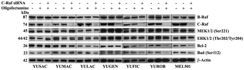
Experimental design: Using specific siRNAs, we knocked down C-Raf expression, and determined the effect on viability, MAP extracellular signal-regulated kinase (ERK)/ERK kinase signaling, and apoptosis in seven melanoma cell lines. We determined the IC(50) of the C-Raf inhibitors sorafenib and GW5074, and studied the effects of GW5074 on cell signaling. Using an automated method to measure in situ protein expression, we quantified C-Raf expression in 263 nevi and 523 melanomas.

Results: C-Raf was knocked down in three cell lines with detectable phospho-C-Raf, resulting in decreased viability in two of the three (YULAC and YUROB). This resulted in decreased Bcl-2 expression and phospho-Bad cleavage, without affecting phospho-MEK and phospho-ERK. Sensitivity to sorafenib and GW5074 varied. GW5074 inhibited mitogen-activated protein kinase signaling without Bcl-2 and phospho-Bad down-regulation. C-Raf was highly expressed in melanomas compared with nevi (P < 0.0001), and no nevi had high C-Raf expression. C-Raf expression was higher in metastatic than primary specimens (P = 0.0225).

Conclusions: C-Raf siRNA knock-down results in decreased viability of YULAC (B-Raf(V600K)) and YUROB (B-Raf(WT)) melanoma cells, likely mediated by Bcl-2 inhibition rather than mitogen-activated protein kinase inhibition. Cotargeting C-Raf and parallel pathways might be an effective therapeutic approach for melanoma. C-Raf expression is up-regulated in a subset of melanomas but not in nevi, suggesting that it might be a valuable diagnostic marker and therapeutic target.


Figures

Figure 1
Figure 1. C-Raf silencing in melanoma cell lines
YUSAC, YUMAC, YULAC, YUGEN, YUFIC, YUROB and MEL501 cells were transfected with one of two siRNA clones directed against C-Raf (lanes 3 and 4 respectively) or a non-targeting control siRNA (TS1058), (lanes 5). Following treatment, cells were lysed and proteins were extracted, electrophoresed and probed for expression of C-Raf. Equal protein gel loading was confirmed by probing for expression of β-Actin.
Figure 2
Figure 2. Effect of C-Raf silencing on cell viability
YUSAC, YUMAC, YULAC, YUGEN, YUFIC, YUROB and MEL501 cells were transfected with 75 nM of either C-Raf siRNA or a control siRNA (TS1058) for 96 hours (A), 72 hours (B) and 48 hours (C). Cell viability was determined by the CellTiter 96® Aqueous One Solution Cell Proliferation Assay. Following 96 hours treatment, 75nM C-Raf siRNA inhibited cell viability of YULAC and YUROB cell lines by 43-58% and 50-65% respectively. At 72 hours of incubation C-Raf siRNA inhibited cell proliferation of the YULAC cell line by 23-39%. Viability of the other cell lines was less affected at all time points. 75nM of the control siRNA did not affect C-Raf expression or viability.
Figure 3
Figure 3. Effect of C-Raf silencing on protein expression
YUSAC, YUMAC, YULAC, YUGEN, YUFIC, YUROB and MEL501 cells were transfected with one of the C-Raf siRNA clones at 75nM. Following 96 hour treatment, cells were lysed and proteins were extracted, electrophoresed and probed for expression of B-Raf, C-Raf, phospho-MEK1/2 (Ser 221), phospho-ERK1/2 (Thr202/Tyr204), Bcl-2, phospho-Bad (Ser112) following stripping of the blots. Equal protein gel loading was confirmed by probing for expression of β-Actin. C-Raf knock down resulted in decreased levels of Bcl-2 and phospho-Bad in YULAC and YUROB cells, while it had less or no effect at all in other cell lines.
Figure 4
Figure 4. Membranous and cytoplasmic C-Raf expression in melanoma histospots
(A) AQUA uses a cocktail of S100 and HMB45 to define the tumor mask (bottom-left), 4,6-diamidine-2-phenylindole to define the nuclear compartment (bottom-right) and Cy5 (top-right) for the target (C-Raf). The cytoplasmic compartment is generated by subtracting the nuclear compartment from the S100/HMB45 mask (top-left). C-Raf expression is then measured within the nuclear and cytoplasmic compartments within the tumor mask, and each spot is assigned a score based on pixel intensity per unit area. C-Raf expression in this example corresponds to an AQUA score of 35.922 (measured in the cytoplasmic compartment) and is shown at 10X magnification (top-right panel A) and (B) 40X magnification. (C) Regression plots for scores from the two sets of melanoma arrays for C-Raf. Each array contains histospots from the same patients, taken from different areas of the tumor.
Figure 5
Figure 5. C-Raf expression in nevi and malignant specimens
(A) Comparison of C-Raf expression (Cytoplasmic AQUA scores) in nevi, primary and metastatic specimens. AQUA scores are represented as percentages of total nevi, primaries and metastases, respectively, by deciles. 99.57% of nevi have AQUA scores between 0 and 10 while only 0.43% had scores between 10 and 20. AQUA scores higher than 20 correspond only to either primary or metastatic specimens. (B) Comparison of C-Raf expression in nevi and all malignant specimens (metastatic and primary melanomas). Unpaired t tests showed that C-Raf expression was significantly higher in malignant versus benign tissue cores. (C) Comparison between high and low C-Raf expression in primary and metastatic melanoma specimens. We defined high C-Raf expression for malignant specimens as expression that was higher than the 95th percentile score for nevi. This score was 7.537.

References

    1. Jemal A, Siegel R, Ward E, et al. Cancer statistics, 2008. CA Cancer J Clin. 2008;58:71–96. - PubMed
    1. Nashan D, Muller ML, Grabbe S, Wustlich S, Enk A. Systemic therapy of disseminated malignant melanoma: an evidence-based overview of the state-of-the-art in daily routine. J Eur Acad Dermatol Venereol. 2007;21:1305–18. - PubMed
    1. Smalley KS. A pivotal role for ERK in the oncogenic behaviour of malignant melanoma? Int J Cancer. 2003;104:527–32. - PubMed
    1. Dhawan P, Singh AB, Ellis DL, Richmond A. Constitutive activation of Akt/protein kinase B in melanoma leads to up-regulation of nuclear factor-kappaB and tumor progression. Cancer Res. 2002;62:7335–42. - PubMed
    1. Kolch W, Kotwaliwale A, Vass K, Janosch P. The role of Raf kinases in malignant transformation. Expert Rev Mol Med. 2002;4:1–18. - PubMed

Publication types

MeSH terms