. 2009 Sep 15;69(18):7224-34.
doi: 10.1158/0008-5472.CAN-09-1299. Epub 2009 Sep 8.

Tuberous sclerosis complex suppression in cerebellar development and medulloblastoma: separate regulation of mammalian target of rapamycin activity and p27 Kip1 localization

Affiliations

Tuberous sclerosis complex suppression in cerebellar development and medulloblastoma: separate regulation of mammalian target of rapamycin activity and p27 Kip1 localization

Bobby Bhatia et al. Cancer Res. .

Abstract

During development, proliferation of cerebellar granule neuron precursors (CGNP), candidate cells-of-origin for the pediatric brain tumor medulloblastoma, requires signaling by Sonic hedgehog (Shh) and insulin-like growth factor (IGF), the pathways of which are also implicated in medulloblastoma. One of the consequences of IGF signaling is inactivation of the mammalian target of rapamycin (mTOR)-suppressing tuberous sclerosis complex (TSC), comprised of TSC1 and TSC2, leading to increased mRNA translation. We show that mice, in which TSC function is impaired, display increased mTOR pathway activation, enhanced CGNP proliferation, glycogen synthase kinase-3 alpha/beta (GSK-3 alpha/beta) inactivation, and cytoplasmic localization of the cyclin-dependent kinase inhibitor p27(Kip1), which has been proposed to cause its inactivation or gain of oncogenic functions. We observed the same characteristics in wild-type primary cultures of CGNPs in which TSC1 and/or TSC2 were knocked down, and in mouse medulloblastomas induced by ectopic Shh pathway activation. Moreover, Shh-induced mouse medulloblastomas manifested Akt-mediated TSC2 inactivation, and the mutant TSC2 allele synergized with aberrant Shh signaling to increase medulloblastoma incidence in mice. Driving exogenous TSC2 expression in Shh-induced medulloblastoma cells corrected p27(Kip1) localization and reduced proliferation. GSK-3 alpha/beta inactivation in the tumors in vivo and in primary CGNP cultures was mTOR-dependent, whereas p27(Kip1) cytoplasmic localization was regulated upstream of mTOR by TSC2. These results indicate that a balance between Shh mitogenic signaling and TSC function regulating new protein synthesis and cyclin-dependent kinase inhibition is essential for the normal development and prevention of tumor formation or expansion.


Conflict of interest statement

Conflicts of Interest: None

Figures

Figure 1
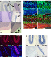
Figure 1. TSC2 inactivation leads to abnormal cerebellar development and p27 misregulation
A. TSC2 inactivation leads to abnormal cerebellar development in mice. (top row) Sagittal sections of H&E-stained post-natal day 7 wild type or TSC2-RGΔ cerebella. Arrows indicate molecular layer, compressed in the mutant mice. Carat shows expanded EGL in TSC2-RGΔ. (middle row) Sagittal sections of H&E-stained post-natal day 15 wild type and TSC2-RGΔ cerebella. Arrow indicates retained EGL in TSC2-RGΔ cerebellum. (bottom row) Sagittal sections of H&E-stained adult cerebellum. Arrow points to “rests”, clusters of cells which have failed to migrate to the IGL. B. p27Kip1 is mislocalized in TSC inactive cerebella. Immunofluorescence analysis of p27Kip1 levels and localization in post natal day 7 wild-type and TSC2-RGΔ sagittal sections shows that wild-type mice have nuclear p27Kip1 (green) in post-mitotic region EGLb whereas TSC2-RGΔ mice have cytoplasmic-localization of p27Kip1 in throughout the EGL (top row). PCNA (red) shows proliferating cells in EGLa. Nuclei co-stained with DAPI (blue). White line indicates the entire EGL (TSC-RGΔ) and EGLa/EGLb (wild-type). 80x magnification was used. C. The expanded and retained EGL in TSC2-RGΔ cerebella contains proliferating cells as indicated by immunofluorescence staining for the proliferation marker Ki67 (red). Ki67 staining is localized in the EGL of post-natal day 7 and 15 cerebella. TSC2-RGΔ mice have increased Ki67 (right column) compared to wild-type (left column) in post-natal day 7 (top row) and post-natal day 15 (bottom row). Ki67 (red) was co-stained with DAPI (blue). 40x magnification was used. D. The expanded and retained EGL in TSC2-RGΔ cerebella contains proliferating cells as indicated by in situ hybridization for cyclin D2. Cyclin D2 can be detected in the EGL of wild type and TSC2-RGΔ mice at PN 7, but by PN 15 (bottom), only the mutant mice still have cyclin D2-expressing cells in the EGL. 40x magnification was used. Bars: A (top and middle row), C–D, 16 µm; A (bottom row), 32µm; B, 8 µm.
Figure 2
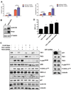
Figure 2. TSC inactivation increases the basal proliferative capacity of CGNPs through increased mTOR signaling and inactivating phosphorylation of p27 and GSK3α/β
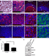
A. (Left) Quantification of BrdU incorporation in wild-type and TSC2-RGΔ CGNPs treated with Shh or Shh vehicle alone. Vehicle-treated mutant CGNPs have significantly higher levels of BrdU incorporation than wild type CGNPs (* P value = 0.0004). In the presence of Shh, TSC2-RGΔ CGNP BrdU incorporation is significantly greater than that of Shh-treated wild-type CGNPs (** P value = 0.0245). Two-tailed t-tests were used to test significance. (Right) Quantification of staining for the mitotic marker phosphorylated Histone H3 in wild-type and TSC2-RGΔ CGNPs treated with Shh or Shh vehicle alone. TSC2-RGΔ CGNPs have increased levels of phospho-Histone H3 in comparison with wild-type CGNPs even in the absence of Shh (* P value = 0.0021). Levels of phospho-Histone H3 are significantly increased in mutant CGNPs in the presence of Shh (** P value = 0.0108). Two-tailed t-tests were used to test significance. B. Western blot shows TSC2-RGΔ CGNPs have higher endogenous levels of cyclin D2, N-myc, and phospho-GSK3α/β Ser21/9 in comparison to wild-type freshly isolated CGNPs. β-tubulin was used as loading control. Representative data shown from four wild-type post-natal day 4/5 mice and four TSC2-RGΔ post-natal day 4/5 mice. C. Western blot analysis of wild type Shh-treated CGNPs infected with TSC shRNA lentiviruses. Pooled TSC shRNA lentiviruses increased phosphorylation of rp S6, phosphorylation GSK3α/β, mis-localized p27Kip1, and proliferation due to TSC knockdown. Short-term treatment with mTOR inhibitor rapamycin blocks S6 phosphorylation but does not significantly affect phosphorylated-p27Kip1 and cyclin D2 levels. GFP shRNA lentiviruses were used as control to knock down GFP in CGNPs derived from Math-1-GFP mice. Protein levels from TSC1, TSC2, phospho-rpS6, and phospho-GSK3α/β protein levels were unaffected. D. Knockdown of TSC significantly increases CGNP proliferation (* P value < 0.0001). Ki67 staining was quantified using automated detection and scoring. One-way ANOVA test was used to test significance.
Figure 3
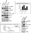
Figure 3. TSC2 inactivation in mice leads to p27Kip1 mislocalization, GSK3α/β destabilization, and decreased survival latency
A. Protein lysates extracted from age-matched post-natal day 4/5 wild-type and TSC2-RGΔ CGNPs, and medulloblastomas from Patched heterozygote (Ptc +/−) and NeuroD2-Smoothened mutant (SmoA1) mice were used for Western Blot analyses of the indicated proteins. Data representative of four wild-type mice, four TSC2-RGΔ mice, three Ptc+/− medulloblastomas, and six SmoA1 medulloblastomas. B. Graph indicating medulloblastoma incidence in NeuroD2-SmoA1 and Ptc +/− mice in the presence or absence of TSC2-RGΔ allele. Increased tumor incidence suggests that inactivation of the TSC can synergize with oncogenic Shh signaling. C. Kaplan-Meier survival curve indicates survival latency in SmoA1 tumor-bearing mice in the presence or absence of TSC2-RGΔ allele. TSC2-RGΔ mice were crossed into the SmoA1 line. Tumors arising in the SmoA1/TSC2-RGΔ mice (red) have shorter latency than SmoA1 alone (blue). Survival curves are significantly different using Gehan-Breslow-Wilcoxon (P value 0.0084) and Mantel-Cox (P value 0.0063) tests. D. TSC2 inactivation in SmoA1 medulloblastoma enhances mTOR signaling, GSK3α/β inactivation, and p27Kip1 phosphorylation. Protein lysates extracted from tumors of SmoA1 and SmoA1/TSC2-RGΔ mice were used for Western Blot analyses of the indicated proteins. Data representative of three SmoA1 medulloblastomas and three SmoA1/TSC2-RGΔ medulloblastomas.
Figure 4
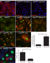
Figure 4. TSC inactivation and p27 dys-regulation in medulloblastoma cells are associated with increased proliferation
A. Under normal conditions, Pzp53med cells have phosphorylated ribosomal S6 protein. Cells treated with 10 nM PI3-kinase inhibitor wortmannin or 10 nM mTOR inhibitor rapamycin for 8 hours shut down levels of phosphorylated S6. Cells were immunostained for phospho-rp S6 (red) and co-stained for DAPI (blue). 40x magnification was used. B. Under normal conditions, Pzp53med cells have barely detectable TSC2 and p27 (left panel). Akt inhibition by 10 nM wortmannin for 8 hours rescued TSC2 and nuclear-localized p27Kip1 (middle panel). However, mTOR inhibition by 10 nM rapamycin for 8 hours did not rescue either of these two proteins (right panel). TSC2 (red), p27Kip1 (green) were co-stained. 40x magnification was used. C. Ectopic TSC2 expression retains nuclear p27Kip1 in medulloblastoma cells. Nuclear p27Kip1 can be easily detected in cells transfected with exogenous TSC2 (left and middle panel). Hi-power image shows striking contrast in p27Kip1 localization between a TSC2-transfected cell and an untransfected neighboring cell (middle panel). 40x (left panel) and 63x (right panel) magnification were used. TSC2 (red), p27 (green) were co-stained. Right panel: automated quantification of TSC2 and p27Kip1 immunostaining. The vast majority of TSC2-positive cells have nuclear p27 (* P value < 0.0001). Two-tailed t-tests were used to test significance. D. TSC2-expressing cells have reduced immunostaining for BrdU incorporation (left panel). TSC2 (red) positive cells are Brdu (green) negative. Cells were co-stained with DAPI (blue). Right panel shows results from automated quantification of TSC2 and BrdU immunostaining. TSC2-positive cells have significantly reduced proliferation (* P value = 0.0003). Two-tailed t-tests were used to test significance.
Figure 5
Figure 5. TSC2 phosphorylation, p27 localization, and GSK3α/β phosphorylation in Shh-mediated medulloblastoma
A. SmoA1 medulloblastoma tumor cells have robust Bmi 1 (DAB staining), phospho-S6 protein (red) and little GFAP (green). Only Purkinje neurons (PL) of wild-type adult cerebella have phosphorylated rp S6. Representative image from a wild-type mouse treated with mTOR inhibitor CCI-779 shows loss of phospho-rp S6 in Purkinje neurons, indicating that this drug crosses the blood-brain barrier. 40x magnification was used. B. SmoA1 medulloblastoma untreated (top row) or treated (bottom row) with CCI-779 for 10 days. Immunostaining for phosphorylated S6 (red)(first column) serves as a read-out for mTOR activity. Phosphorylated GSK3α/β (red)(second column) is affected by mTOR inhibition. Inactivating Akt-mediated phosphorylation of TSC2 (red)(third column) and complete cytoplasmic localization of p27Kip1 (green)(fourth column) in murine SmoA1 medulloblastoma are not altered by mTOR inhibition. All cells were co-stained with DAPI (blue). 63x (first and second column) and 120x (third and fourth column) magnification were used. C. Quantification of staining for the mitotic marker phosphorylated Histone H3 in SmoA1 medulloblastoma treated with or without CCI-779 for 10 days. mTOR inhibition reduced levels of phospho-Histone H3 (* P value = 0.0052). Two-tailed t-tests were used to test significance. D. CCI-779 treatment reduces levels of cell cycle regulators in SmoA1 tumor cells. SmoA1 medulloblastomas lose N-myc, cyclin D2, and VEGF protein upon mTOR inhibition for 10 days. Wild-type mice were used as control. Bars: A, B (first and second column), 16µm; B (third and fourth column), 8 µm.
Figure 6
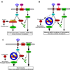
Figure 6. Model for how TSC activity is integrated with Shh proliferative signaling during cerebellar development and in cancer
A. Under normal conditions in the developing brain, CGNPs are exposed to Shh and IGF. Normal levels of Shh signaling promote CGNP proliferation and cerebellar development, with appropriate levels of mTOR activity and p27 localization. B. When the TSC is inactivated, p27Kip1 moves to the cytoplasm where it can be phosphorylated. mTOR is released from inhibition, increasing mRNA translation. MTOR may also act through S6 kinase to further inhibit GSK-3α/β. C. In medulloblastomas, loss of Ptc function or aberrant activation of Smo drives oncogenic Shh activity which cooperates with enhanced signaling by growth factors such as PDGF or IGF, leading TSC inactivation. p27Kip1 moves to the cytoplasm where it may be inactivated or gain new oncogenic functions. mTOR activity is increased, contributing to the transformed phenotype. TSC inactivation by mutation/deletion also leads to mTOR activation, p27Kip1 misregulation and enhanced activity of Shh pathway effectors.

References

    1. Rubin JB, Rowitch DH. Medulloblastoma: a problem of developmental biology. Cancer Cell. 2002;2:7–8. - PubMed
    1. Knoepfler PS, Kenney AM. Neural precursor cycling at sonic speed: N-Myc pedals, GSK-3 brakes. Cell Cycle. 2006;5:47–52. - PubMed
    1. Wetmore C. Sonic hedgehog in normal and neoplastic proliferation: insight gained from human tumors and animal models. Curr Opin Genet Dev. 2003;13:34–42. - PubMed
    1. Pietsch T, Waha A, Koch A, et al. Medulloblastomas of the desmoplastic variant carry mutations of the human homologue of Drosophila patched. Cancer Res. 1997;57:2085–2088. - PubMed
    1. Raffel C, Jenkins RB, Frederick L, et al. Sporadic medulloblastomas contain PTCH mutations. Cancer Res. 1997;57:842–845. - PubMed

Publication types

MeSH terms

Substances