. 2009 Oct 1;69(19):7538-47.
doi: 10.1158/0008-5472.CAN-08-4626. Epub 2009 Sep 8.

Melanoma proteoglycan modifies gene expression to stimulate tumor cell motility, growth, and epithelial-to-mesenchymal transition

Affiliations

Melanoma proteoglycan modifies gene expression to stimulate tumor cell motility, growth, and epithelial-to-mesenchymal transition

Jianbo Yang et al. Cancer Res. .

Abstract

Melanoma chondroitin sulfate proteoglycan (MCSP) is a plasma membrane-associated proteoglycan that facilitates the growth, motility, and invasion of tumor cells. MCSP expression in melanoma cells enhances integrin function and constitutive activation of Erk1,2. The current studies were performed to determine the mechanism by which MCSP expression promotes tumor growth and motility. The results show that MCSP expression in radial growth phase, vertical growth phase, or metastatic cell lines causes sustained activation of Erk1,2, enhanced growth, and motility which all require the cytoplasmic domain of the MCSP core protein. MCSP expression in a radial growth phase cell line also promotes an epithelial-to-mesenchymal transition based on changes in cell morphology and the expression of several epithelial-to-mesenchymal transition markers. Finally, MCSP enhances the expression of c-Met and hepatocyte growth factor, and inhibiting c-Met expression or activation limits the increased growth and motility of multiple melanoma cell lines. The studies collectively show the importance of MCSP in promoting progression by an epigenetic mechanism and they indicate that MCSP could be targeted to delay or inhibit tumor progression in patients.


Figures

Figure 1
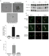
Figure 1. Expression of MCSP stimulates tumor formation, anchorage independent growth and constitutive activation of ERK 1, 2 in melanoma cells
(a) 2×10⁶ cells were injected into the flank of NOD.SCID mice. Tumors were excised at 42 days post injection and weighed to determine the tumor mass. *p<0.001 by students two-tailed t test, n = 25. (b)Pictures of representative colonies from cells plated in 0.2% agarose for 17 days (scale bar = 20μm). Colonies were counted as described in Methods. Bars represent the average number of cells per 5 fields in triplicate wells +/− s.e.m. (*p<0.0001 by student’s two tailed t test), from one representative experiment. (c)Indicated melanoma cell lines were transfected with MCSP specific- or negative-control siRNA or then serum starved for 24 hours prior to harvesting the cells for western analysis. (d)Melanoma cell lines were transfected with MCSP specific siRNA for 24 hours and plated in the agarose 3-dimensional growth assay for 14 days. Bars represent the average number of cells per 5 fields in triplicate wells +/− s.e.m. (ANOVA p<0.001, *p<0.001 vs. control siRNA treated cells by Bonferroni’s adjusted t-test), from one representative experiment.
Figure 2
Figure 2. Full length MCSP, but not MCSPΔCD, promotes growth, tumor formation and Erk 1,2 activation
(a) WM1552C parental and transfected cells were plated in 0.2% agarose for 17 days. Pictures are of representative colonies (Bar = 20μm). Colonies were counted as in 1b. Bars represent the average number of cells per 5 fields in triplicate wells +/− s.e.m. (ANOVA p<0.001, *p<0.001 vs. WM1552C/Mock cells by Bonferroni’s adjusted t-test), from one representative experiment. (b) 2×10⁶ cells were injected into the flank of NOD.SCID mice. Tumors were excised at 42 days post injection and weighed to determine the tumor mass. *p<0.001 by students two-tailed t-test, n = 10. (c) Serum starved cells were suspended in serum free 1.6% methylcellulose medium for the indicated time, and harvested for western blot analysis (1: WM1552C/Mock, 2: WM1552C/MCSP, 3: WM1552C/MCSPΔCD). (d) Cells were cultured on glass coverslips in serum free medium overnight, and the cells fixed and stained with antibody against phosphorylated Erk 1,2 (pERK 1,2, green) and Propidium Iodide (P.I., red) to visualize the nucleus. The merged image (yellow) demonstrates localization of phosphorylated Erk 1, 2 to the nucleus. Scale bar = 20 μm.
Figure 3
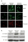
Figure 3. WM1552C/MCSP cells show increased expression of mesenchymal cell markers
(a) Indicated cell lines were grown on glass coverslips, serum starved for 48 hours, then fixed and stained with antibodies against the indicated proteins. Bar = 20 μm. (b) Cells were serum starved 24 hours and lysates subjected to western blot analysis.
Figure 4
Figure 4. MCSP stimulates an autocrine loop of HGF/c-Met that activates c-Met
(a) Mock(1), MCSP (2) and MCSPΔCD (3) transfected WM1552C cells were serum starved for 24 hours, stimulated with 2.5 ng/ml rHGF for various times and cell lysates analyzed by western blot. Densitometry analysis normalized to tubulin (not shown) demonstrates that the addition of HGF to MCSP expressing cells causes approximately a 2 fold increase in c-Met activation within thirty minutes compared to the Mock or MCSPΔCD cells.(b)WM1552C transfected cells were serum starved for 24 hours followed by transfection with nothing (1), negative control siRNA (2) or MCSP specific siRNA (3) for 48 hours, and cell lysates evaluated for HGF expression. (c)WM1552C/MCSP cells were serum starved for 24 hours prior to transfection with nothing (1), negative control siRNA (2) or HGF specific siRNA (3) for 48 hours and cell lysates analyzed by western blot. (d) Scratch wound migration assay of the indicated cell lines over 72 hours in serum free medium, +/− 2.5ng/ml rHGF, as indicated (Scale bar = 100 μm).
Figure 5
Figure 5. WM1552C/MCSP cell migration and anchorage-independent growth is dependent upon the expression and activation of C-Met
(a) WM1552C/MCSP cells transfected with the indicated siRNAs were plated in 6 well plates, serum starved, and subjected to a scratch wound migration assay 24 hours post transfection. Bars represent the percent of open area in the wound at 48 hours, +/− s.e.m., from triplicate wells, from one representative experiment (ANOVA p<0.001, *p<0.001 vs. neg. control by Bonferroni’s adjusted t-test). (b)SiRNA transfected WM1225C/MCSP cells were harvested as for ‘a’ plated in soft agar and counted after 17 days as in 1b. Bars represent the average number of cells per 5 fields in triplicate wells +/− s.e.m. (ANOVA p<0.001, *p<0.0001 vs. negative control by Bonferroni’s adjusted t-test), from one representative experiment. (c) Confluent monolayers of WM1552C/MCSP cells were serum starved for 24hrs, wounded and incubated for 72hrs in the presence of C-Met inhibitor SU11274 (4.0uM) or DMSO. Pictures from one representative experiment are shown. Scale bar = 100 μm. (d)Indicated melanoma cell lines were transfected with MCSP siRNA, then serum starved for 24 hours prior to harvesting for western analysis.
Figure 6
Figure 6. Erk 1,2 mediates enhanced c-Met expression through MITF
(a) WM1341D and WM1552C/MCSP transfected cells were incubated with 2.4 μM U0126 or control compound U0124 overnight in serum free medium. Lysates were evaluated by western blot using the indicated antibodies. (b) WM1552C/MCSP cells were transfected with MITF specific or control siRNA for 48 hours and assayed for expression of C-Met, MITF, and pErk 1, 2 by western blot. (c) WM1552C/MCSP cells were transfected with c-Met specific or control siRNA for 48 hours and assayed for expression of C-Met, MITF and pErk 1,2 and MCSP by western blot.

References

    1. Campoli MR, Chang CC, Kageshita T, Wang X, McCarthy JB, Ferrone S. Human high molecular weight-melanoma-associated antigen (HMW-MAA): a melanoma cell surface chondroitin sulfate proteoglycan (MSCP) with biological and clinical significance. Crit Rev Immunol. 2004;24:267–96. - PubMed
    1. Chekenya M, Rooprai HK, Davies D, Levine JM, Butt AM, Pilkington GJ. The NG2 chondroitin sulfate proteoglycan: role in malignant progression of human brain tumours. Int J Dev Neurosci. 1999;17:421–35. - PubMed
    1. Stallcup WB. The NG2 proteoglycan: past insights and future prospects. J Neurocytol. 2002;31:423–35. - PubMed
    1. Kageshita T, Nakamura T, Yamada M, Kuriya N, Arao T, Ferrone S. Differential expression of melanoma associated antigens in acral lentiginous melanoma and in nodular melanoma lesions. Cancer Res. 1991;51:1726–32. - PubMed
    1. Sorensen PH, Chen CS, Smith FO, et al. Molecular rearrangements of the MLL gene are present in most cases of infant acute myeloid leukemia and are strongly correlated with monocytic or myelomonocytic phenotypes. J Clin Invest. 1994;93:429–37. - PMC - PubMed

Publication types

MeSH terms

Substances