. 2010 Aug;43(2):142-51.
doi: 10.1165/rcmb.2009-0113OC. Epub 2009 Sep 8.

Bacterial lipopolysaccharide enhances PDGF signaling and pulmonary fibrosis in rats exposed to carbon nanotubes

Affiliations

Bacterial lipopolysaccharide enhances PDGF signaling and pulmonary fibrosis in rats exposed to carbon nanotubes

Mark F Cesta et al. Am J Respir Cell Mol Biol. 2010 Aug.

Abstract

Engineered multi-walled carbon nanotubes (MWCNT) represent a possible health risk for pulmonary fibrosis due to their fiber-like shape and potential for persistence in the lung. We postulated that bacterial lipopolysaccharide (LPS), a ubiquitous agent in the environment that causes lung inflammation, would enhance fibrosis caused by MWCNT. Rats were exposed to LPS and then intratracheally instilled with MWCNT or carbon black (CB) nanoparticles 24 hours later. Pulmonary fibrosis was observed 21 days after MWCNT exposure, but not with CB. LPS alone caused no fibrosis but enhanced MWCNT-induced fibrosis. LPS plus CB did not significantly increase fibrosis. MWCNT increased platelet-derived growth factor-AA (PDGF-AA), a major mediator of fibrosis. PDGF-AA production in response to MWCNT, but not CB, was synergistically enhanced by LPS. Immunostaining showed PDGF-AA in bronchiolar epithelial cells and macrophages. Since macrophages engulfed MWCNT, were positive for PDGF-AA, and mediate fibroblast responses, experiments were performed with rat lung macrophages (NR8383 cells) and rat lung fibroblasts in vitro. LPS exposure increased PDGF-A mRNA levels in NR8383 cells and enhanced MWCNT-induced PDGF-A mRNA levels. Moreover, LPS increased MWCNT- or CB-induced PDGF receptor-alpha (PDGF-Ralpha) mRNA in fibroblasts. Our data suggest that LPS exacerbates MWCNT-induced lung fibrosis by amplifying production of PDGF-AA in macrophages and epithelial cells, and by increasing PDGF-Ralpha on pulmonary fibroblasts. Our findings also suggest that individuals with pre-existing pulmonary inflammation are at greater risk for the potential adverse effects of MWCNT.


Figures
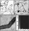
Figure 1.
Figure 1.
Transmission electron microscopic (TEM) characterization of bulk multi-walled carbon nanotubes (MWCNT). (A) Low magnification photomicrograph of MWCNT (bar = 2 μm). (B) Intermediate magnification TEM image of MWCNT (bar = 2 μm). (C) High-magnification TEM photomicrograph of MWCNT (bar = 20 nm). (D) Energy dispersive X-ray (EDX) analysis of MWCNT.
Figure 2.
Figure 2.
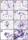
Lung histopathology in rats 21 days after exposure (blue staining indicates collagen). (A) Saline control. (B) Lipopolysaccharide (LPS) alone. (C) Carbon black (CB) nanoparticle alone. (D) CB nanoparticles with LPS pre-exposure. (E) MWCNT alone. (F) MWCNT with LPS pre-exposure. (G) Higher magnification of inset from E. (H) Higher magnification of inset from F. Original magnification: ×10 for all panels (except G and H, original magnification ×40).
Figure 3.
Figure 3.
Lung pathology scoring in rats 21 days after exposure. The lungs were scored for the amount of collagen present (based on Masson's trichrome–stained sections), the thickness of the alveolar walls, and the number of fibroblasts associated with the particle-associated lesions. These average scores were then divided by the relative scores for the amount of nanoparticles present in the lungs to give an adjusted average score (see Materials and Methods). *P < 0.05 or **P < 0.01 compared with control or LPS as determined by one-way ANOVA with post Dunnett's multiple comparison test.
Figure 4.
Figure 4.
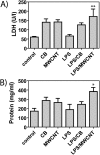
Markers of lung injury. (A) Lactate dehydrogenase (LDH) and (B) total protein in the lungs of rats 24 hours after exposure to MWCNT or CB nanoparticles, with or without a 24-hour LPS pre-exposure. *P < 0.05 or **P < 0.01 compared with control or LPS as determined by one-way ANOVA with post Tukey's test or Bonferroni's test.
Figure 5.
Figure 5.
PDGF-AA protein levels in bronchoalveolar lavage (BAL) fluid collected from rats 1 day or 21 days after exposure to 4 mg/kg MWCNT or CB nanoparticles, with or without a 24-hour LPS pre-exposure. PDGF-AA was measured by ELISA. *P < 0.05 or ***P < 0.001 compared with control as determined by one-way ANOVA with post Tukey's test or Bonferroni's test.
Figure 6.
Figure 6.
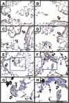
Immunohistochemistry for PDGF-AA at 24 hours after exposure. Brown staining indicates PDGF-AA protein. Open arrows indicate PDGF-positive epithelium. Solid arrows indicate PDGF-positive macrophages, some containing CB or MWCNT. (A) Saline control. (B) LPS alone. (C) CB nanoparticle alone. (D) CB nanoparticles with LPS pre-exposure. (E) MWCNT alone. (F) MWCNT with LPS pre-exposure. (G) Higher magnification of inset from E. (H) Higher magnification of inset from F. Hematoxylin counterstain (light blue). Original magnification: ×20 for all panels (except G and H, original magnification ×40). Bars = 100 μm.
Figure 7.
Figure 7.
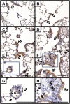
Immunohistochemistry for PDGF-AA at 21 days after exposure. Brown staining indicates PDGF-AA protein. Open arrows indicate PDGF-positive epithelium. Solid arrows indicate PDGF-positive macrophages, some containing CB or MWCNT. (A) Saline control. (B) LPS alone. (C) CB nanoparticle alone. (D) CB nanoparticles with LPS pre-exposure. (E) MWCNT alone. (F) MWCNT with LPS pre-exposure. (G) Higher magnification of inset from E. (H) Higher magnification of inset from F. Hematoxylin counterstain (light blue). Original magnification: ×20 for all panels (except G and H, original magnification ×40). Bars = 100 μm.
Figure 8.
Figure 8.
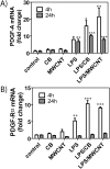
(A) PDGF-A and (B) PDGF-Rα mRNA levels in rat alveolar macrophages (NR8383 cells) exposed to MWCNT for 4 or 24 hours with or without 1 hour of LPS pre-exposure. Nonadherent cultures of NR8383 cells were grown to near confluence in 10% fetal bovine serum–Dulbecco's Modified Eagle's Medium, then washed and rendered quiescent in serum-free defined medium, and treated with 10 μg/cm2 MWCNT or CB for 4 or 24 hours before collecting RNA. PDGF-A and PDGF-Rα mRNA levels were measured by real-time RT-PCR as described in Materials and Methods. *P < 0.05, **P < 0.01, or ***P < 0.001 compared with control as determined by one-way ANOVA with post Tukey's test or Bonferroni's test.
Figure 9.
Figure 9.
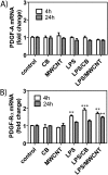
(A) PDGF-A and (B) PDGF-Rα mRNA levels in early passage rat lung fibroblasts (RLF) exposed to MWCNT for 4 or 24 hours with or without 1 hour of LPS pre-exposure. Confluent, quiescent cultures of RLF were treated with 10 μg/cm2 MWCNT or CB for 4 or 24 hours before collecting RNA. PDGF-A and PDGF-Rα mRNA levels were measured by Taqman quantitative real-time RT-PCR as described in Materials and Methods. **P < 0.01, or ***P < 0.001 compared with control as determined by one-way ANOVA with post Tukey's test or Bonferroni's test.

References

    1. Baughman RH, Zakhidov AA, de Heer WA. Carbon nanotubes – the route toward applications. Science 2002;297:787–792. - PubMed
    1. Avouris P, Chen Z, Perebeinos V. Carbon-based electronics. Nat Nanotechnol 2007;2:605–615. - PubMed
    1. Bianco A, Kostarelos K, Prato M. Applications of carbon nanotubes in drug delivery. Curr Opin Chem Biol 2005;9:674–679. - PubMed
    1. Donaldson K, Aitken R, Tran L, Stone V, Duffin R, Forrest G, Alexander A. Carbon nanotubes: a review of their properties in relation to pulmonary toxicology and workplace safety. Toxicol Sci 2006;92:5–22. - PubMed
    1. Card JW, Zeldin DC, Bonner JC, Nestmann ER. Pulmonary applications and toxicity of engineered nanoparticles. Am J Physiol Lung Cell Mol Physiol 2008;295:L400–L411. - PMC - PubMed

Publication types