. 2009 Oct 23;284(43):29383-90.
doi: 10.1074/jbc.M109.024745. Epub 2009 Sep 8.

RNA-destabilizing factor tristetraprolin negatively regulates NF-kappaB signaling

Affiliations

RNA-destabilizing factor tristetraprolin negatively regulates NF-kappaB signaling

Jian Liang et al. J Biol Chem. .

Abstract

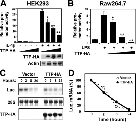
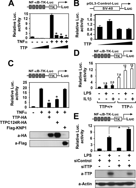
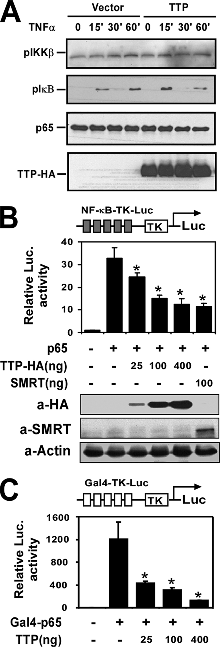
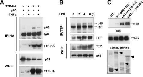
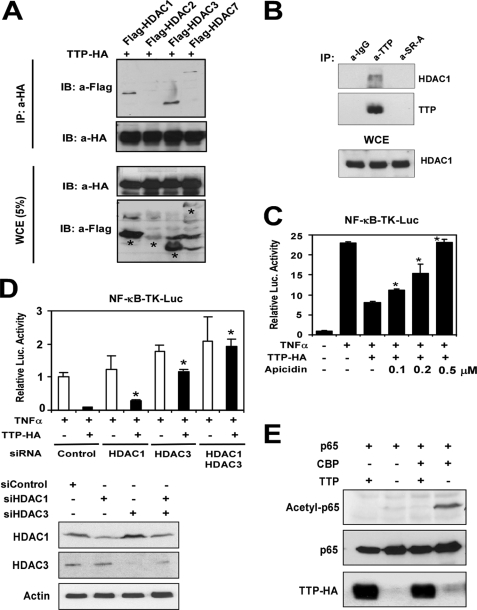
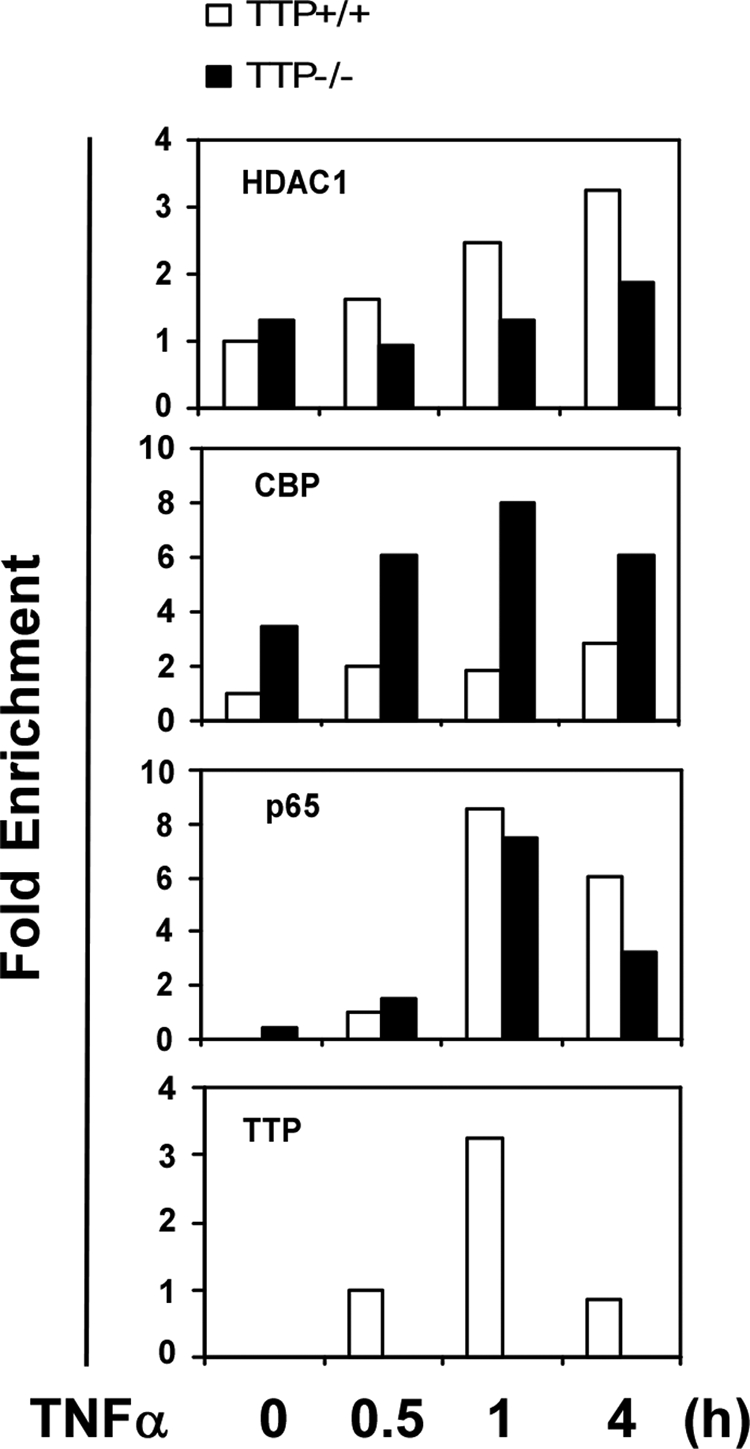
Tristetraprolin (TTP) is a CCCH zinc finger-containing protein that destabilizes mRNA by binding to an AU-rich element. Mice deficient in TTP develop a severe inflammatory syndrome mainly because of overproduction of tumor necrosis factor alpha. We report here that TTP also negatively regulates NF-kappaB signaling at the transcriptional corepressor level, by which it may repress inflammatory gene transcription. TTP expression inhibited NF-kappaB-dependent transcription. However, overexpression of TTP did not affect reporter mRNA stability. Instead, TTP functioned as a corepressor of p65/NF-kappaB. In support of this concept, we found that TTP physically interacted with the p65 subunit of NF-kappaB and was also associated with HDAC1, -3, and -7 in vivo. Treatment with histone deacetylase inhibitors or small interfering RNA induced HDAC1 or HDAC3 knockdown completely or partly abolished the inhibitory activity of TTP on NF-kappaB reporter activation. Consistently, chromatin immunoprecipitation showed decreased recruitment of HDAC1 and increased recruitment of CREB-binding protein on the Mcp-1 promoter in TTP(-/-) cells compared with wild-type cells. Moreover, overexpression of TTP blocked CREB-binding protein-induced acetylation of p65/NF-kappaB. Taken together, these data suggest that TTP may also function in vivo as a modulator in suppressing the transcriptional activity of NF-kappaB.


Figures

FIGURE 1.
FIGURE 1.
TTP expression inhibits TNFα promoter activity. A and B, HEK293 or RAW264.7 cells were cotransfected with a luciferase reporter plasmid under the transcriptional control of the mouse TNFα promoter and increasing amounts of TTP-HA (0, 25, 100, and 400 ng/well). After being quiescent for 24 h, cells were treated with or without IL-1β (10 ng/ml) or LPS (0.1 μg/ml) as shown and collected for analysis of reporter gene activity 24 h later. Data represent means ± S.D. from three independent experiments. *, p < 0.05; **, p < 0.01 versus IL-1β stimulation without the TTP-HA-transfected group. The gels under the graph in A are the protein levels of TTP-HA and actin in the transfected cell lysates detected by Western blotting. C, HEK293 cells were transiently transfected with the pcDNA3.1 or pTTP-HA expression construct. After 24 h, the cells were quiescent for 18 h and then treated with IL-1β (10 ng/ml) for 8 h, and actinomycin D (10 μg/ml) was added to stop transcription. RNA was harvested after different time points as indicated. The luciferase (Luc.) mRNA level was examined by Northern blotting. D, the intensity of the bands in B was quantified using an AlphaImager 2200 system (Alpha Innotech). The value of the 0 point was set as 100%, and the values of the other points were transformed according to the 0 point. The data from three independent experiments were averaged and plotted.
FIGURE 2.
FIGURE 2.
TTP represses NF-κB-dependent gene expression. A and B, HEK293 cells were cotransfected with the NF-κB-TK-Luc (A) or pGL3-Control (B) vector as indicated with increasing amounts of pTTP-HA (0, 25, 100, and 400 ng/well) by the calcium phosphate method. After being quiescent for 16 h, cells were treated with or without TNFα (10 ng/ml) (A) and collected for analysis of reporter gene activity 24 h later. *, p < 0.01 versus stimulation without the TTP-HA-transfected group. C, HEK293 cells were cotransfected with NF-κB-TK-Luc with or without expression plasmids as indicated and subjected to the same treatment as described above. The proper protein expression is shown in the lower panels. D, NF-κB-Luc was transfected into TTP+/+ or TTP−/− MEFs by FuGENE 6. 24 h later, the cells were stimulated with LPS (0.1 μg/ml) or IL-1β (10 ng/ml) for another 24 h. The luciferase (Luc.) activity was measured and normalized by β-galactosidase activity. The number on each bar represents -fold over the untreated group in TTP+/+ cells. E, scrambled control (siControl) or TTP (siTTP) siRNA was cotransfected with NF-κB-Luc into RAW264.7 cells by electroporation (Amaxa). 24 h later, the cells were quiescent for 12 h and then stimulated with LPS (0.1 μg/ml) for another 24 h. The luciferase activity was measured and normalized by β-galactosidase activity. The TTP and β-actin protein levels in the transfected cells were analyzed by Western blotting and are shown in the lower panels.
FIGURE 3.
FIGURE 3.
TTP represses transactivation of p65. A, HEK293 cells were transfected with pcDNA3.1 or pTTP-HA. After 24 h, the transfected cells were quiescent for 16 h and then stimulated with TNFα (10 ng/ml) for the indicated times. Whole cell extracts were isolated and subjected to Western blot analysis using anti-phospho-IKKβ, anti-phospho-IκBα, anti-p65, or anti-HA antibody as indicated. B and C, HEK293 cells were transfected with NF-κB-TK-Luc, p65, and increasing amounts of pTTP-HA as indicated or SMRT (B) or with Gal4-TK-Luc with an expression construct encoding the Gal4-p65(TAD) fusion protein and increasing amounts of pTTP-HA (C) as indicated. 36 h later, the cells were collected for analysis of reporter gene activity and protein expression. *, p < 0.01 versus stimulation without the TTP-HA-transfected group. Luc., luciferase.
FIGURE 4.
FIGURE 4.
TTP physically interacts with p65. A, HEK293 cells were transiently cotransfected with p65 with or without the pTTP-HA expression plasmid. The transfected cells were stimulated with or without TNFα for 30 min. The whole cell lysates (WCE) were subjected to co-immunoprecipitation (IP) with anti-HA antibody followed by Western blotting with anti-p65 or anti-HA antibody. The same lysates were subjected to SDS-PAGE and probed with anti-p65 or anti-HA antibody for proper protein expression. B, RAW264.7 cells were treated with LPS (0.1 μg/ml) for 0, 2, 4, and 8 h. The cell extracts were subjected to immunoprecipitation with anti-TTP antibody. Both whole cell extracts and immunoprecipitates were analyzed by Western blotting with anti-TTP and p65 antibodies. C, HEK293 cells were transfected with the pTTP-HA plasmid. After 24 h of transfection, the cells were treated with TNFα for 30 min. Aliquots of cell lysates were subjected to purification with glutathione-Sepharose beads already bound to GST, GST-p65-(1–305), GST-p65-(245–355), or GST-p65-(345–551). Proteins co-precipitated with GST beads were analyzed by Western blotting with anti-HA antibody. Whole cell extracts were also analyzed by Western blot analysis with anti-HA antibody to monitor the expression of transfected DNA. Coomassie (Comas.) Brilliant Blue stain showed the expression of GST and GST fusion proteins.
FIGURE 5.
FIGURE 5.
Involvement of HDACs in the TTP-mediated inhibitory effect on NF-κB. A, HEK293 cells were cotransfected with pTTP-HA and the FLAG-HDAC1, FLAG-HDAC2, FLAG-HDAC3, or FLAG-HDAC7 expression plasmid. After 24 h of transfection, the whole cell lysates (WCE) were subjected to co-immunoprecipitation (IP) with anti-HA antibody followed by Western blotting (immunoblotting (IB)) with anti-FLAG or anti-HA antibody as indicated. The same lysates were subjected to SDS-PAGE and probed with anti-FLAG or anti-HA antibody for proper protein expression. B, RAW264.7 cells were treated with LPS for 8 h. The cell extracts were subjected to immunoprecipitation by anti-TTP antibody, no specific IgG, or an unrelated antibody followed by Western blot analysis with anti-TTP or HDAC1 antibody. The HDAC1 protein level in whole cell extracts was also probed by anti-HDAC1 antibody. C, HEK293 cells were cotransfected with NF-κB-TK-Luc and TTP-HA or the empty vector as indicated. The transfected cells were treated with or without HDAC inhibitor (apicidin at 0.1, 0.2, and 0.5 μm) for 2 h and then stimulated with TNFα (10 ng/ml) for 24 h. Cells were collected for analysis of reporter gene activity. The data are from three independent experiments (means ± S.D.). *, p < 0.01 versus TTP-HA-transfected without the apicidin treatment group. D, HEK293 cells were transfected with scrambled control siRNA (siControl), HDAC1 siRNA (siHDAC1), or HDAC3 siRNA (siHDAC3) together with NF-κB-Luc and the TTP-HA plasmid as indicated. 24 h later, the cells were quiescent for 12 h and then stimulated with TNFα for 24 h. The reporter activity was analyzed. The HDAC1 and HDAC3 protein levels in the cell lysates were detected by Western blot analysis. *, p < 0.01 versus the scrambled control siRNA- and TTP-HA-transfected group. E, HEK293 cells were cotransfected with p65, CBP, and pTTP-HA as indicated. Cell lysates were extracted and analyzed by Western blotting with anti-acetyl-p65, anti-p65, and anti-HA antibodies.
FIGURE 6.
FIGURE 6.
ChIP analysis of the Mcp-1 promoter in TTP−/− MEFs. ChIP assays were performed with MEFs from wild-type (+/+) or TTP null (−/−) littermates untreated or treated with TNFα (10 ng/ml). Protein-DNA complexes were immunoprecipitated with the indicated antibodies, and immunoprecipitated DNA was analyzed by quantitative PCR with specific primers against the IκB site in the murine Mcp-1 promoter. The values were normalized by input DNA. The experiment was independently repeated twice with similar results.

References

    1. O'Neill L. A. (2006) Nat. Rev. Drug Discov. 5, 549–563 - PubMed
    1. Pfitzner E., Kliem S., Baus D., Litterst C. M. (2004) Curr. Pharm. Des. 10, 2839–2850 - PubMed
    1. Natoli G., Chiocca S. (2008) Sci. Signal. 1, pe1. - PubMed
    1. Ogawa S., Lozach J., Jepsen K., Sawka-Verhelle D., Perissi V., Sasik R., Rose D. W., Johnson R. S., Rosenfeld M. G., Glass C. K. (2004) Proc. Natl. Acad. Sci. U.S.A. 101, 14461–14466 - PMC - PubMed
    1. Wormald S., Hilton D. J. (2004) J. Biol. Chem. 279, 821–824 - PubMed

Publication types

MeSH terms

LinkOut - more resources