Review
. 2010 Jan;30(1):2-14.
doi: 10.1038/jcbfm.2009.188. Epub 2009 Sep 9.

Cerebral blood flow response to functional activation

Affiliations
Review

Cerebral blood flow response to functional activation

Olaf B Paulson et al. J Cereb Blood Flow Metab. 2010 Jan.

Abstract

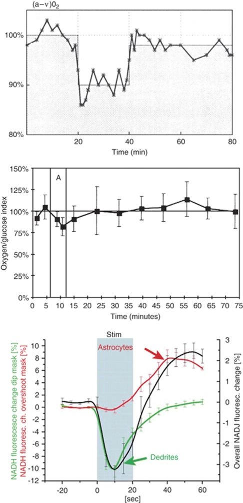
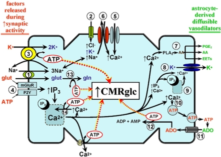
Cerebral blood flow (CBF) and cerebral metabolic rate are normally coupled, that is an increase in metabolic demand will lead to an increase in flow. However, during functional activation, CBF and glucose metabolism remain coupled as they increase in proportion, whereas oxygen metabolism only increases to a minor degree-the so-called uncoupling of CBF and oxidative metabolism. Several studies have dealt with these issues, and theories have been forwarded regarding the underlying mechanisms. Some reports have speculated about the existence of a potentially deficient oxygen supply to the tissue most distant from the capillaries, whereas other studies point to a shift toward a higher degree of non-oxidative glucose consumption during activation. In this review, we argue that the key mechanism responsible for the regional CBF (rCBF) increase during functional activation is a tight coupling between rCBF and glucose metabolism. We assert that uncoupling of rCBF and oxidative metabolism is a consequence of a less pronounced increase in oxygen consumption. On the basis of earlier studies, we take into consideration the functional recruitment of capillaries and attempt to accommodate the cerebral tissue's increased demand for glucose supply during neural activation with recent evidence supporting a key function for astrocytes in rCBF regulation.


Figures

Figure 1
Figure 1
(A) Experiment in humans with a global maximal activation period of 10 mins (from 20–30 mins) induced by the Wisconsin card sorting test. Values are expressed as the percentage of the baseline measurements. Note the continuous resetting of the oxygen to glucose ratio, which may be accounted for by a prolonged phase of non-oxidative glucose metabolism. Measurements were performed as arterio-venous (jugular) differences of glucose and oxygen combined with CBF measurement by the Kety–Schmidt method (Madsen et al, 1995). (B) The cerebral oxygen-glucose index before, during, and after maximal global activation of rats (duration 6 mins, period ‘A') induced by opening the sheltering box for 6 m. Values represent the mean of global cerebral arterio-venous determinations in a group of six animals. Vertical bars represent s.d. Note the decreased oxygen to glucose uptake ratio, signifying non-oxidative glucose consumption during and immediately after activation, and the reversal of that ratio after 35 mins, in which more oxygen than glucose is taken up by the brain (Madsen et al, 1998). A and B reprinted with permission from NPG. (C) Metabolic changes in hippocampal slice preparations after brief 20 secs stimulation. The biphasic NADH response represent early oxidative metabolism in neuronal dendrites followed by late activation of glycolysis in astrocytes (Kasischke et al, 2004). Reprinted with permission from AAAS.
Figure 2
Figure 2
A generalized depiction of the energy cost of neurovascular coupling and its impact on astrocytic glucose utilization (CMRglc). After enhanced synaptic activity, increases in extracellular levels of ATP, glutamate (glut), and K+ occur (shown in red on the left hand margin of the figure). It has been suggested that these factors, paricularly glutamate, arise from pre-synaptic neuronal elements, at least at low to moderate increases in synaptic activity. However, at higher levels of activity, post-synaptic contributions may come into play (Petzold et al, 2008). Glutamate enters the astrocyte through the glut-3Na+ cotransporter [1], whereas key K+ routes of entry include the Na+-K+-2Cl cotransporter [2], and, in exchange for the increased Na+ from [1] and [2], the Na-K ATPase [3]. Glutamate and ATP can activate their respective metabotropic receptors [4] leading to IP3-mediated Ca2+ release from the ER. Depletion of ER Ca2+ promotes Ca2+ entry through store-operated Ca2+ channels [5]. In addition, the elevated intracellular Na+ may be exchanged for extracellular Ca2+, through the plasma membrane Na-Ca exchanger operating in reverse mode [6], contributing further to the elevation in intracellular Ca2+. The Ca2+ rise provides a proximal stimulus for a variety of vasodilating pathways. Some of the major paracrine vasodilating factors arising from these pathways are shown in green on the right hand margin of the figure. This includes arachidonic acid (AA) derived from Ca2+-activated phospholipase A2 (PLA2) [7]. The AA can be further metabolized, through cyclooxygenase-1 (COX1), to vasodilating prostanoids, primarily PGE2, and/or converted to epoxyeicosatrienoic acids (EETs) by a cytochrome P450 epoxygenase (Takano et al, 2006; Shi et al, 2008). Arachidonic acid itself can act as a direct vasodilator (Bryan et al, 2006). An increase in K+ levels (up to 15–20 mmol/L) in the extracellular milieu provides a potent vasodilating stimulus through acting on smooth muscle inward rectifier K+ channels (Straub and Nelson, 2007). A key conduit for astrocytic release of K+ is the large-conductance Ca2+-operated K+ (BKCa) channel [8]. Those channels are well expressed in astrocyte foot processes (Price et al, 2002), along with IP3-sensitive Ca2+ storage and release sites [9] (Straub et al, 2006; Straub and Nelson, 2007). The latter is thought to provide the localized elevations in [Ca2+] necessary for channel activation. Increased cytosolic Ca2+ may also promote ATP exocytosis (Blum et al, 2008) [10], although Ca2+-independent and plasma membrane hemichannel routes may also exist (Coco et al, 2003). The released ATP is rapidly hydrolyzed to adenosine (ADO) by astrocytic ecto-nucleotidases (Xu and Pelligrino, 2007). The ADO is returned to the cell through nucleoside transporters [11] to enter the adenosine kinase-mediated adenine nucleotide salvage pathway [12]. There are multiple sites of ATP consumption that, in turn, may trigger increased CMRglc. Some of the key sites that can be directly linked to astrocyte-derived vasodilating factors are shown. Principal among these is the plasma membrane Na-K ATPase [3]. Other ATP-consuming sites depicted in the figure are glutamine (gln) synthase-mediated conversion of glut to gln [13], the ER Ca-ATPase [14], the plasma membrane Ca-ATPase [15], and adenosine kinase-mediated formation of ADP and AMP [12].

References

    1. Ances BM, Buerk DG, Greenberg JH, Detre JA. Temporal dynamics of the partial pressure of brain tissue oxygen during functional forepaw stimulation in rats. Neurosci Lett. 2001;306:106–110. - PubMed
    1. Anderson AW, Heptulla RA, Driesen N, Flanagan D, Goldberg PA, Jones TW, Rife F, Sarofin H, Tamborlane W, Sherwin R, Gore JC. Effects of hypoglycemia on human brain activation measured with fMRI. Magn Reson Imaging. 2006;24:693–697. - PubMed
    1. Aubert A, Pellerin L, Magistretti PJ, Costalat R. A coherent neurobiological framework for functional neuroimaging provided by a model integrating compartmentalized energy metabolism. Proc Natl Acad Sci USA. 2007;104:4188–4193. - PMC - PubMed
    1. Attwell D, Laughlin SB. An energy budget for signaling in the grey matter of the brain. J Cereb Blood Flow Metab. 2001;21:1133–1145. - PubMed
    1. Barros LF, Porras OH, Bittner CX. Why glucose transport in the brain matters for PET. Trends Neurosci. 2005;28:117–119. - PubMed

LinkOut - more resources