. 2009 Nov;7(11):1886-96.
doi: 10.1111/j.1538-7836.2009.03606.x. Epub 2009 Sep 9.

Endothelial cell specific adhesion molecule (ESAM) localizes to platelet-platelet contacts and regulates thrombus formation in vivo

Affiliations

Endothelial cell specific adhesion molecule (ESAM) localizes to platelet-platelet contacts and regulates thrombus formation in vivo

T J Stalker et al. J Thromb Haemost. 2009 Nov.

Abstract

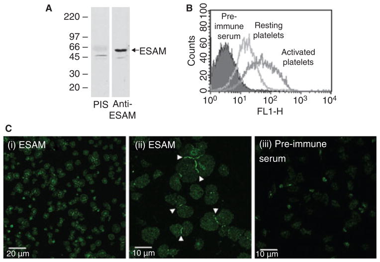
Background: In resting platelets, endothelial cell specific adhesion molecule (ESAM) is located in alpha granules, increasing its cell surface expression following platelet activation. However, the function of ESAM on platelets is unknown.

Objective: To determine whether ESAM has a role in thrombus formation.

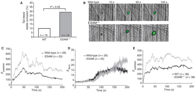
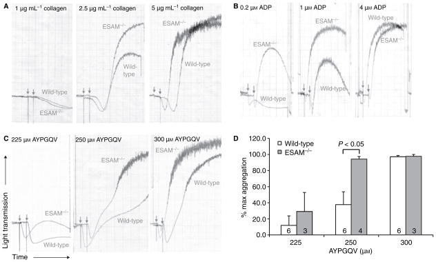
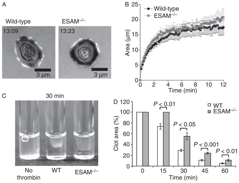
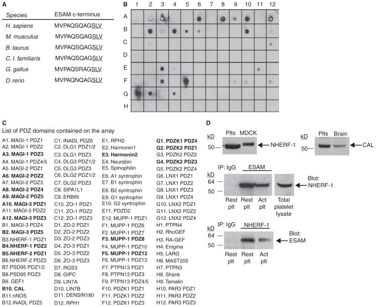
Methods and results: We found that following platelet activation ESAM localizes to the junctions between adjacent platelets, suggesting a role for this protein in contact-dependent events that regulate thrombus formation. To test this hypothesis we examined the effect of ESAM deletion on platelet function. In vivo, ESAM(-/-) mice achieved more stable hemostasis than wild-type mice following tail transection, and developed larger thrombi following laser injury of cremaster muscle arterioles. In vitro, ESAM(-/-) platelets aggregated at lower concentrations of G protein-dependent agonists than wild-type platelets, and were more resistant to disaggregation. In contrast, agonist-induced calcium mobilization, alpha(IIb)beta(3) activation, alpha-granule secretion and platelet spreading, were normal in ESAM-deficient platelets. To understand the molecular mechanism by which ESAM regulates platelet activity, we utilized a PDZ domain array to identify the scaffold protein NHERF-1 as an ESAM binding protein, and further demonstrated that it associates with ESAM in both resting and activated platelets.

Conclusions: These findings support a model in which ESAM localizes to platelet contacts following platelet activation in order to limit thrombus growth and stability so that the optimal hemostatic response occurs following vascular injury.


Conflict of interest statement

Disclosure of Conflict of Interests

The authors state that they have no conflict of interest.

Figures

Fig. 1
Fig. 1
ESAM localizes to platelet–platelet contacts following platelet activation. (A) Human gel-filtered platelets were lysed and proteins separated by SDS–PAGE. An immunoblot was performed using rabbit anti-ESAM or rabbit pre-immune serum (PIS). (B) Flow cytometric analysis of resting or activated (10 μM SFLLRN) human platelets stained with anti-ESAM or pre-immune serum followed by fluorescently labeled anti-rabbit secondary antibody. (C) Human platelets were activated with 10 μM SFLLRN and allowed to spread on immobilized fibrinogen. The platelets were stained with either rabbit anti-ESAM (panels i and ii) or pre-immune serum (panel iii), as described in Methods. Scale bars: (i) 20 μm, (ii, iii) 10 μm. Arrowheads indicate intense ESAM staining at platelet–platelet contacts. The photomicrographs are representative of at least three independent experiments.
Fig. 2
Fig. 2
ESAM-deficient mice form larger, more stable thrombi than wild-type mice in vivo. (A) A bleeding time assay was performed by cutting the distal 5 mm of the mouse tail. The percentage of mice that formed stable hemostatic plugs (i.e. did not re-bleed) up to 10 min after initial occlusion is shown. Statistical analysis was performed using Fisher’s exact test. (B) Photomicrographs show representative time-lapse images of laser-induced thrombus formation in cremaster muscle arterioles of wild-type and ESAM−/− mice. Platelets are labeled green. (C, D) Median integrated fluorescence intensity is reported (arbitrary units) as a quantitative measure of platelet (C) and fibrin accumulation (D) in cremaster muscle arterioles following laser injury. Statistical analysis was performed using the Mann–Whitney test (n = 26 thrombi from 4 wild-type mice and n = 25 thrombi from 5 ESAM−/− mice). (E) Laser-induced thrombus formation in radiation chimera mice expressing ESAM on their endothelium with or without ESAM expression on their platelets (n = 36 thrombi from 4 mice with wild-type platelets and n = 38 thrombi from four mice with ESAM-deficient platelets).
Fig. 3
Fig. 3
Lack of ESAM increases platelet aggregation. Platelet aggregation was performed using platelets from wild-type or ESAM−/− mice. (A–C) Representative aggregation tracings for the indicated concentrations of (A) collagen, (B) ADP and (C) the PAR-4 agonist peptide AYPGQV. Arrows indicate addition of agonist. Tracings are representative of at least three independent experiments performed for each condition. (D) Quantitative analysis of aggregation in response to AYPGQV. Data are reported as the extent of aggregation at each agonist concentration normalized to the maximum response in each experiment (max = 100%). Values are mean ± SEM and the numbers at the base of the columns indicate the number of mice in each group.
Fig. 4
Fig. 4
Lack of ESAM attenuates platelet disaggregation. Platelet aggregation was performed using platelets from wild-type or ESAM−/− mice with ADP (2 μM) as the agonist. Following peak aggregation, either apyrase (0.2 U mL−1, left) or tirofiban (0.5 μM, right) was added to induce disaggregation at the time point indicated by the arrowheads. Arrows indicate addition of ADP.
Fig. 5
Fig. 5
ESAM does not regulate calcium mobilization or αIIbβ3 activation in isolated mouse platelets. (A) Calcium mobilization in response to ADP (left panel) and the PAR4 agonist AYPGKF (right panel) were measured using the fluorescent calcium indicator Fura-2 as described in Methods. Values are mean ± SD for two independent experiments. (B) Flow cytometry was used to evaluate αIIbβ3 activation by stimulating platelets from wild-type and ESAM−/− mice with AYPGQV (500 μM) in the presence of either FITC-fibrinogen (right panel) or phycoerythrin-labeled JON/A antibody (left panel). Solid histogram is resting wild-type platelets, solid line is activated wild-type platelets and the dotted line is activated ESAM−/− platelets. Histograms are representative of three independent experiments.
Fig. 6
Fig. 6
Clot retraction is delayed in the absence of ESAM. (A, B) Wild-type and ESAM−/− platelets were spread on immobilized fibrinogen in the presence of the PAR-4 agonist AYPGKF (200 μM). The time stamp indicates time after initial contact for the platelet shown. The area (mean ± SEM) of platelets over time in two independent experiments is reported in (B). Black circles represent wild-type and gray circles represent ESAM−/− platelets. (C, D) Clot retraction was induced by adding 10 U mL−1 thrombin to either wild-type or ESAM−/− PRP at 37 °C. Image is representative of difference 30 min after the addition of thrombin. Clot retraction was quantified by determining the two-dimensional area occupied by the clot as described in Methods. Values are mean ± SEM for six mice in each group.
Fig. 7
Fig. 7
ESAM associates with the scaffold protein NHERF-1 in human platelets. (A) Amino acid sequence of the ESAM cytoplasmic tail c-terminus in multiple species. The PDZ domain binding motif is underlined. (B) A PDZ domain array was probed using a GST-ESAM cytoplasmic tail fusion protein as described in Methods. There were several positive hits, including PDZ domain 2 of NHERF-1. (C) A complete list of PDZ domains present on the array. Strongly positive hits are in bold. (D) Top panel: Western blot results demonstrating NHERF-1 and CAL expression in human platelets. MDCK cells expressing human NHERF-1 and human brain lysate were used as positive controls. Bottom panel: Co-immunoprecipitation of ESAM and NHERF-1 in human platelets. Human platelet lysates were subjected to immunoprecipitation with anti-ESAM, anti-NHERF-1 or a non-specific IgG control antibody. The membranes were immunoblotted using an anti-NHERF-1 antibody (for ESAM IP) or an anti-ESAM antibody (NHERF-1 IP). Blots are representative of three independent experiments.

References

    1. Law DA, DeGuzman FR, Heiser P, Ministri-Madrid K, Killeen N, Phillips DR. Integrin cytoplasmic tyrosine motif is required for outside-in alphaIIbbeta3 signalling and platelet function. Nature. 1999;401:808–11. - PubMed
    1. Cosemans JM, Munnix IC, Wetzker R, Heller R, Jackson SP, Heemskerk JW. Continuous signaling via PI3K isoforms beta and gamma is required for platelet ADP receptor function in dynamic thrombus stabilization. Blood. 2006;108:3045–52. - PubMed
    1. Goto S, Tamura N, Ishida H, Ruggeri ZM. Dependence of platelet thrombus stability on sustained glycoprotein IIb/IIIa activation through adenosine 5′-diphosphate receptor stimulation and cyclic calcium signaling. J Am Coll Cardiol. 2006;47:155–62. - PubMed
    1. Andre P, Prasad KS, Denis CV, He M, Papalia JM, Hynes RO, Phillips DR, Wagner DD. CD40L stabilizes arterial thrombi by a beta3 integrin–dependent mechanism. Nat Med. 2002;8:247–52. - PubMed
    1. Angelillo-Scherrer A, Burnier L, Flores N, Savi P, DeMol M, Schaeffer P, Herbert JM, Lemke G, Goff SP, Matsushima GK, Earp HS, Vesin C, Hoylaerts MF, Plaisance S, Collen D, Conway EM, Wehrle-Haller B, Carmeliet P. Role of Gas6 receptors in platelet signaling during thrombus stabilization and implications for antithrombotic therapy. J Clin Invest. 2005;115:237–46. - PMC - PubMed

Publication types

Substances