. 2009 Sep;128(1 Suppl):e237-49.
doi: 10.1111/j.1365-2567.2008.02954.x. Epub 2008 Oct 21.

Hypoxia skews dendritic cells to a T helper type 2-stimulating phenotype and promotes tumour cell migration by dendritic cell-derived osteopontin

Affiliations

Hypoxia skews dendritic cells to a T helper type 2-stimulating phenotype and promotes tumour cell migration by dendritic cell-derived osteopontin

Meixiang Yang et al. Immunology. 2009 Sep.

Abstract

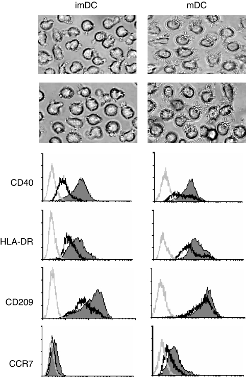
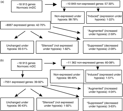
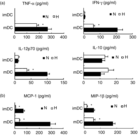
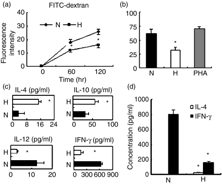
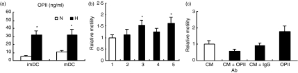
It is well recognized that tissue microenvironments are involved in regulating the development and function of dendritic cells (DC). Oxygen supply, which varies in different tissues, has been accepted as an important microenvironmental factor in regulating the biological functions of several immune cells and as being involved in tumour progression and metastasis. However, little is known about the effect of hypoxia on the biological functions of DC and the effect of these hypoxia-conditioned DC on tumour metastasis. In this study, we analysed the transcriptional profiles of human monocyte-derived immature DC (imDC) and mature DC (mDC) cultured under normoxia and hypoxia by microarray, and found a body of potential targets regulating the functions of DC during hypoxia. In addition, the phagocytic ability of hypoxic imDC markedly decreased compared with that of normoxic imDC. Importantly, hypoxic DC poorly induced the proliferation of allogeneic T cells, but polarized allogeneic CD4(+) naive T cells into a T helper type 2 (Th2) response. Moreover, hypoxic DC secreted large amounts of osteopontin, which were responsible for the enhanced migration of tumour cells. Therefore, our study provides new insights into the biological functions of DC under hypoxic conditions and one of mechanisms underlying tumour immune escape during hypoxia.


Figures

Figure 1
Figure 1
Morphology and phenotypes of human monocyte-derived dendritic cells (DC) under normoxia or hypoxia. Human monocyte-derived DC were cultured under normoxic (N) or hypoxic (H) conditions for 5 days in the presence of granulocyte–macrophage colony-stimulating factor and interleukin-4 and induced for further maturation by the treatment with lipopolysaccharide for 48 hr, as described in the Materials and methods section. (a) Morphology of immature DC (imDC) and mature DC (mDC) under normoxic or hypoxic condition (200× magnification). (b) Surface marker expression on immature and mature DC under normoxia (thin and filled line) and hypoxia (bold and open line). The grey lines represent staining with a control isotype-matched antibody. Results are representative of four separate experiments.
Figure 2
Figure 2
Gene expression profiles in dendritic cells (DC) modulated by hypoxia environment. This schematic illustrates gene expression profiles in normoxic DC and hypoxic DC. Based on a greater than twofold difference in expression in hypoxic DC relative to normoxic DC, genes significantly altered were categorized as ‘silenced’, ‘downregulated’, ‘upregulated’ and ‘induced’. (a) The gene expression profiles in immature DC (imDC) cultured under normoxia or hypoxia. (b) The gene expression profiles in mature DC (mDC) cultured under normoxia or hypoxia.
Figure 3
Figure 3
Cytokine secretion by dendritic cells (DC). The DC were induced under hypoxia (H) or normoxia (N). The supernatants were analysed by Bio-Plex Protein Array system for the secretion of tumour necrosis factor-α (TNF-α), interferon-γ (IFN-γ), interleukin-12 (IL-12) p70, IL-10 (a), and monocyte chemotactic protein 1 (MCP-1), macrophage inflammatory protein 1β (MIP-1β) (b). The results are shown as the mean ± SD. *P< 0·05.
Figure 4
Figure 4
Hypoxia inhibited the phagocytic ability of immature dendritic cells (imDC) and skewed mature dendritic cells (mDC) to mediate a T helper type 2 (Th2)-polarizing response. (a) The imDC were induced under hypoxia (H) or normoxia (N). The cells endocytic ability was evaluated by the uptake of 1 mg/ml fluorescein isothiocyanate (FITC)-dextran measured using flow cytometry. Results were expressed as the mean ± SD of the fluorescence intensity. (b) The mixed lymphocyte reaction (MLR) test was performed with allogeneic T cells and hypoxic mDC (H) or normoxic mDC (N). [3H]Thymidine was added 16 hr before cells were collected and proliferation was assessed. (c) The supernatants were collected after 72 hr of culture and analysed for the production of interleukin-4 (IL-4), IL-10, IL-12p70 and interferon-γ (IFN-γ). (d) Naive CD4+ CD45RA+ T cells isolated by immunomagnetic negative depletion were polarized by culture with hypoxic or normoxic mDC. Production of the cytokines IL-4 and IFN-γ was induced by adding phorbol 12-myristate 13-acetate and ionomycin on day 9. The supernatant was collected after 24 hr and analysed using the Bio-Plex Protein Array system as described in the Materials and methods section. Data are expressed as the mean ± SD of four independent experiments. *P< 0·05.
Figure 5
Figure 5
Hypoxic dendritic cells (DC) promoted the motility of the breast tumour cells MDA-MD-231 by producing osteopontin (OPN). (a) The migratory activity of tumour cells was analysed in Transwell chambers in vitro as described in the Materials and methods section. Tumour cells were preincubated with or without DC-conditioned media collected from normoxic immature DC (imDC) (2), hypoxic imDC (3), normoxic mature DC (mDC) (4) or hypoxic mDC (5) for 4 hr. Complete RPMI medium served as a control (1). The number of cells migrating through the Matrigel-coated membranes was counted. (b) OPN production by imDC and mDC induced under normoxia (N) or hypoxia (H). Human CD14+ monocytes were incubated for 5 days and matured using lipopolysaccharide for another 2 days. OPN in the culture supernatant was quantified with using an enzyme-linked immunosorbent assay kit. (c) Tumour cells preincubated with imDC-conditioned medium with or without the OPN neutralizing antibody. (CM) hypoxic DC-conditioned medium; (IgG) normal goat immunoglobulin G served as isotype-matched control (10 μg/ml); (OPN Ab) OPN neutralizing antibody (10 μg/ml); (OPN) recombinant human OPN served as positive control (5 μg/ml). Data are the mean ± SD of five independent experiments. *P< 0·05.

References

    1. Caldwell CC, Kojima H, Lukashev D, Armstrong J, Farber M, Apasov SG, Sitkovsky MV. Differential effects of physiologically relevant hypoxic conditions on T lymphocyte development and effector functions. J Immunol. 2001;167:6140–9. - PubMed
    1. Vaupel P, Kallinowski F, Okunieff P. Blood flow, oxygen and nutrient supply, and metabolic microenvironment of human tumors: a review. Cancer Res. 1989;49:6449–65. - PubMed
    1. Brahimi-Horn MC, Pouyssegur J. The hypoxia-inducible factor and tumor progression along the angiogenic pathway. Int Rev Cytol. 2005;242:157–213. - PubMed
    1. Vaupel P, Briest S, Hockel M. Hypoxia in breast cancer: pathogenesis, characterization and biological/therapeutic implications. Wien Med Wochenschr. 2002;152:334–42. - PubMed
    1. Okunieff P, Fenton B, Chen Y. Past, present, and future of oxygen in cancer research. Adv Exp Med Biol. 2005;566:213–22. - PubMed

Publication types

MeSH terms