Superantigen-presentation by rat major histocompatibility complex class II molecules RT1.Bl and RT1.Dl
- PMID: 19740318
- PMCID: PMC2753945
- DOI: 10.1111/j.1365-2567.2008.03033.x
Superantigen-presentation by rat major histocompatibility complex class II molecules RT1.Bl and RT1.Dl
Abstract
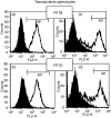
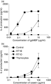
Rat major histocompatibility complex (MHC) class II molecules RT1.B(l) (DQ-like) and RT1.D(l) (DR-like) were cloned from the LEW strain using reverse transcription-polymerase chain reaction and expressed in mouse L929 cells. The transduced lines bound MHC class II-specific monoclonal antibodies in an MHC-isotype-specific manner and presented peptide antigens and superantigens to T-cell hybridomas. The T-cell-hybridomas responded well to all superantigens presented by human MHC class II, whereas the response varied considerably with rat MHC class II-transduced lines as presenters. The T-cell hybridomas responded to the pyrogenic superantigens Staphylococcus enterotoxin B (SEB), SEC1, SEC2 and SEC3 only at high concentrations with RT1.B(l)-transduced and RT1.D(l)-transduced cells as presenters. The same was true for streptococcal pyrogenic exotoxin A (SPEA), but this was presented only by RT1.B(l) and not by RT1.D(l). SPEC was recognized only if presented by human MHC class II. Presentation of Yersinia pseudotuberculosis superantigen (YPM) showed no MHC isotype preference, while Mycoplasma arthritidis superantigen (MAS or MAM) was presented by RT1.D(l) but not by RT1.B(l). Interestingly, and in contrast to RT1.B(l), the RT1.D(l) completely failed to present SEA and toxic shock syndrome toxin 1 even after transduction of invariant chain (CD74) or expression in other cell types such as the surface MHC class II-negative mouse B-cell lymphoma (M12.4.1.C3). We discuss the idea that a lack of SEA presentation may not be a general feature of RT1.D molecules but could be a consequence of RT1.D(l)beta-chain allele-specific substitutions (arginine 80 to lysine, asparagine 82 to aspartic acid) in the extremely conserved region flanking the Zn(2+)-binding histidine 81, which is crucial for high-affinity SEA-binding.
Figures


Similar articles
-
The Forgotten: Identification and Functional Characterization of MHC Class II Molecules H2-Eb2 and RT1-Db2.J Immunol. 2016 Feb 1;196(3):988-99. doi: 10.4049/jimmunol.1403070. Epub 2016 Jan 6. J Immunol. 2016. PMID: 26740108
-
Polymorphic class II sequences linked to the rat major histocompatibility complex (RT1) homologous to human DR and DQ sequences.J Immunol. 1985 Aug;135(2):1450-5. J Immunol. 1985. PMID: 2989367
-
Allelic polymorphisms at the H-2A and HLA-DQ loci influence the response of murine lymphocytes to the Mycoplasma arthritidis superantigen MAM.Infect Immun. 1997 Oct;65(10):4190-8. doi: 10.1128/iai.65.10.4190-4198.1997. Infect Immun. 1997. PMID: 9317026 Free PMC article.
-
Structure-function studies of T-cell receptor-superantigen interactions.Immunol Rev. 1998 Jun;163:177-86. doi: 10.1111/j.1600-065x.1998.tb01196.x. Immunol Rev. 1998. PMID: 9700510 Review.
-
MHC class II-dependent peptide antigen versus superantigen presentation to T cells.Hum Immunol. 1997 May;54(2):194-201. doi: 10.1016/s0198-8859(97)00074-8. Hum Immunol. 1997. PMID: 9297538 Review.
Cited by
-
Toxic Shock Syndrome: A Literature Review.Antibiotics (Basel). 2024 Jan 18;13(1):96. doi: 10.3390/antibiotics13010096. Antibiotics (Basel). 2024. PMID: 38247655 Free PMC article. Review.
References
-
- Sundberg EJ, Li Y, Mariuzza RA. So many ways of getting in the way: diversity in the molecular architecture of superantigen-dependent T-cell signaling complexes. Curr Opin Immunol. 2002;14:36–44. - PubMed
-
- Kreiss M, Asmuss A, Krejci K, et al. Contrasting contributions of complementarity-determining region 2 and hypervariable region 4 of rat BV8S2+ (Vβ8.2) TCR to the recognition of myelin basic protein and different types of bacterial superantigens. Int Immunol. 2004;16:655–63. - PubMed
-
- Sellins KS, Bellgrau D, Gold DP. Specificity of rat T cell receptor Vβ chain usage in proliferative responses to staphylococcal enterotoxin B. Eur J Immunol. 1992;22:1931–4. - PubMed
Publication types
MeSH terms
Substances
LinkOut - more resources
Full Text Sources
Research Materials
Miscellaneous

