. 2009 Sep;128(1 Suppl):e746-57.
doi: 10.1111/j.1365-2567.2009.03077.x. Epub 2009 Mar 26.

Impact of human immunodeficiency virus 1 infection and inflammation on the composition and yield of cervical mononuclear cells in the female genital tract

Affiliations

Impact of human immunodeficiency virus 1 infection and inflammation on the composition and yield of cervical mononuclear cells in the female genital tract

Nonhlanhla N Nkwanyana et al. Immunology. 2009 Sep.

Abstract

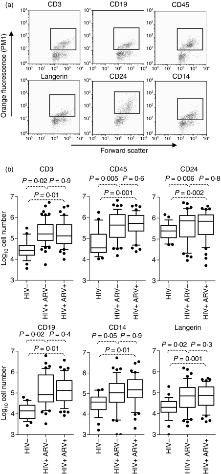
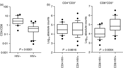
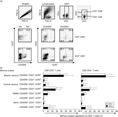
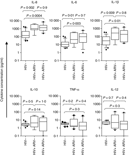
Cervical cytobrush sampling is a relatively non-invasive method for obtaining mucosal cells from the female genital tract. To define mucosal immune cells sampled by cervical cytobrushing and to validate this approach for local immunity studies, we investigated the impact of human immunodeficiency virus (HIV) status and inflammation on the yield and composition of cervical cytobrush specimens. Cervical cytobrush samples were obtained from 89 chronically HIV-infected and 46 HIV-negative women. The HIV-infected women had significantly higher yields of CD3(+), CD45(+), CD19(+), CD14(+), Langerin(+) and CD24(+) cells than the uninfected women. While cytobrush-derived T cells from uninfected women were predominantly CD4(+) (4.2 CD4 : 1 CD8), CD8(+) T cells were predominant in HIV-infected women (0.6 CD4 : 1 CD8). The majority of CD4(+) and CD8(+) T cells from HIV-infected and uninfected women were of the effector memory (CD45RA(-) CCR7(-) CD27(-)) phenotype. HIV-infected women had significantly elevated levels of interleukin (IL)-1beta, IL-6 and IL-8 in cervical supernatants compared with uninfected women. We observed a significant positive correlation between T-cell counts and IL-1beta, tumour necrosis factor (TNF)-alpha and IL-12 concentrations. Neutrophil counts correlated significantly with cervical concentrations of IL-1beta, TNF-alpha, IL-8, IL-6 and IL-10. Antigen-presenting cell numbers correlated significantly with TNF-alpha and IL-12 concentrations. HIV-infected women on antiretroviral therapy had similar levels of cervical lymphocyte infiltration and inflammation to women naïve to therapy. In conclusion, we suggest that inflammation at the cervix and HIV infection are likely to be key determinants in the absolute number of mucosal immune cells recovered by cervical cytobrushing.


Figures

Figure 1
Figure 1
Impact of human immunodeficiency virus (HIV) infection on ex vivo cervical cytobrush yields. (a) Representative plots of ex vivo cervical CD3+, CD19+, CD45+, Langerin+, CD24+, and CD14+ populations quantified using a Guava automated cell counter. Red dots indicate cells that were considered positive for each phenotypic marker while green dots represent cells considered negative. (b) Summary of absolute immune cell counts isolated from ex vivo cervical cytobrushes determined using a Guava automated cell counter from 46 HIV-negative women (HIV−), 48 therapy-naive chronically HIV-infected women (HIV+ ARV−) and 41 HIV-infected women on ARV (HIV+ ARV+). Box and whisker plots indicate the median and the 25th and 75th percentiles. Dots represent outliers. The Mann–Whitney U-test was applied to compare cervical counts in HIV-negative versus HIV-positive women. P-values ≤0·05 were considered significant.
Figure 2
Figure 2
CD4:CD8 ratio and absolute CD4 and CD8 T-cell counts in cervical cytobrush samples from uninfected and human immunodeficiency virus (HIV)-1-infected women. (a) CD4:CD8 T-cell ratios were determined by flow cytometry from ex vivo derived cervical cytobrush samples stained with CD3, CD4 and CD8. (b) Absolute CD4 and CD8 T-cell counts were determined using ratios inferred by flow cytometry and absolute CD3 counts were determined using Guava automated cell counting of CD3+ cells. Box and whisker plots indicate the median and 25th and 75th percentiles. Dots represent outliers. The Mann–Whitney U-test was applied to compare cervical counts in HIV-negative versus HIV-positive women. P-values ≤0·05 were considered significant.
Figure 3
Figure 3
Memory subset composition of ex vivo cervical cytobrush-derived T cells. (a) Representative plots of staining combinations used to differentiate memory subsets of cervical T cells based on differential expression of CD3, CD8, CD45RA, CCR7 and CD27. (b) CD45RA+ CD27+ CCR7+ (naïve), CD45RA+ CD27 CCR7+, CD45RA+ CD27+ CCR7, CD45RA+ CD27 CCR7 (effector memory cells expressing RA (EMRA)), CD45RA CD27+ CCR7, CD45RA CD27+ CCR7+ (central memory), CD45RA CD27 CCR7+ and CD45RA CD27 CCR7 (effector memory) CD8+ (left panel) and CD8 (right panel) T-cell frequencies in cervical cytobrush samples derived from HIV-infected (black bars) and uninfected (clear bars) women. Each bar represents the mean percentage memory marker expression by CD8 and CD8+ T cells (± standard deviation). The unpaired Student’s t-test was applied to compare cervical counts in HIV-negative versus HIV-positive women. P-values ≤0·05 were considered significant.
Figure 4
Figure 4
Relationship between HIV infection and inflammatory cytokine concentrations in the female genital tract. Interleukin (IL)-8, IL-6, IL-1β, IL-10, tumour necrosis factor (TNF)-α and IL-12 p70 concentrations at the cervix of uninfected women (HIV−), therapy-naïve HIV-infected women (HIV+ ARV−) and HIV-infected women on antiretroviral (ARV) therapy (HIV+ ARV+) were measured. The Mann–Whitney U-test was applied to compare cervical cytokine concentrations in HIV-negative versus HIV-positive women. P-values ≤0·05 were considered significant.

References

    1. UNAIDS . AIDS epidemic update. 2007. URL http://data.unaids.org/pub/FactSheet/2008/(epi07_fs_regionalsummary_subs... [accessed on 08 April 2009]
    1. Johansson EL, Rudin A, Wassen L, Holmgren J. Distribution of lymphocytes and adhesion molecules in human cervix and vagina. Immunology. 1999;96:272–277. - PMC - PubMed
    1. Musey L, Ding Y, Cao J, Lee J, Galloway C, Yuen A, Jerome KR, McElrath MJ. Ontogeny and specificities of mucosal and blood human immunodeficiency virus type 1-specific CD8(+) cytotoxic T lymphocytes. J Virol. 2003;77:291–300. - PMC - PubMed
    1. Anton PA, Elliott J, Poles MA, et al. Enhanced levels of functional HIV-1 co-receptors on human mucosal T cells demonstrated using intestinal biopsy tissue. AIDS. 2000;14:1761–1765. - PubMed
    1. Guadalupe M, Reay E, Sankaran S, Prindiville T, Flamm J, McNeil A, Dandekar S. Severe CD4+ T-cell depletion in gut lymphoid tissue during primary human immunodeficiency virus type 1 infection and substantial delay in restoration following highly active antiretroviral therapy. J Virol. 2003;77:11708–11717. - PMC - PubMed

Publication types

MeSH terms