Correlation between recombinase activating gene 1 ubiquitin ligase activity and V(D)J recombination
- PMID: 19740377
- PMCID: PMC2767310
- DOI: 10.1111/j.1365-2567.2009.03101.x
Correlation between recombinase activating gene 1 ubiquitin ligase activity and V(D)J recombination
Abstract
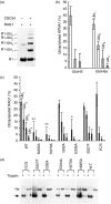
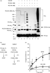
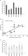
The really interesting new gene (RING) finger ubiquitin ligase domain of the recombinase activating gene 1 (RAG1) V(D)J recombinase protein adopts a standard cross-brace architecture but co-ordinates three zinc ions as opposed to the canonical two. We demonstrated previously that disruption of the conserved zinc co-ordination sites resulted in loss of structural integrity and ubiquitin ligase (E3) activity and interfered with the ability of full-length RAG1 to support recombination. Here we present evidence that amino acids surrounding the third, non-canonical site also contribute to functional interaction with the ubiquitin conjugating (E2) enzyme CDC34, while certain residues on the RING domain's surface important for interaction between other E2-E3 pairs are less critical to the functional RAG1-CDC34 interaction in this assay. Partial reduction of ubiquitin ligase activity was significantly correlated with reduction in the ability of RAG1 to support recombination of extra-chromosomal substrates (r = 0.805, P = 0.009). While poly-ubiquitin chains could be generated, RAG1 did not promote rapid chain extension following mono-ubiquitylation of substrate, regardless of the E2 enzyme used. No single ubiquitin lysine mutant disrupted the ability of CDC34 to form ubiquitin chains on RAG1, and mass spectrometric analysis of the poly-ubiquitylated products indicated ubiquitin chain linkages through lysines 48 and 11. These data suggest that RAG1 promotes a mono-ubiquitylation reaction that is required for optimal levels of V(D)J recombination.
Figures


Similar articles
-
Autoubiquitylation of the V(D)J recombinase protein RAG1.Proc Natl Acad Sci U S A. 2003 Dec 23;100(26):15446-51. doi: 10.1073/pnas.2637012100. Epub 2003 Dec 11. Proc Natl Acad Sci U S A. 2003. PMID: 14671314 Free PMC article.
-
The RAG1 V(D)J recombinase/ubiquitin ligase promotes ubiquitylation of acetylated, phosphorylated histone 3.3.Immunol Lett. 2011 May;136(2):156-62. doi: 10.1016/j.imlet.2011.01.005. Epub 2011 Jan 20. Immunol Lett. 2011. PMID: 21256161
-
Herpes simplex virus 1 mutant in which the ICP0 HUL-1 E3 ubiquitin ligase site is disrupted stabilizes cdc34 but degrades D-type cyclins and exhibits diminished neurotoxicity.J Virol. 2003 Dec;77(24):13194-202. doi: 10.1128/jvi.77.24.13194-13202.2003. J Virol. 2003. PMID: 14645576 Free PMC article.
-
The roles of the RAG1 and RAG2 "non-core" regions in V(D)J recombination and lymphocyte development.Arch Immunol Ther Exp (Warsz). 2009 Mar-Apr;57(2):105-16. doi: 10.1007/s00005-009-0011-3. Epub 2009 Mar 31. Arch Immunol Ther Exp (Warsz). 2009. PMID: 19333736 Review.
-
Regulation of RAG transposition.Adv Exp Med Biol. 2009;650:16-31. doi: 10.1007/978-1-4419-0296-2_2. Adv Exp Med Biol. 2009. PMID: 19731798 Review.
Cited by
-
Autoinhibition of DNA cleavage mediated by RAG1 and RAG2 is overcome by an epigenetic signal in V(D)J recombination.Proc Natl Acad Sci U S A. 2010 Dec 28;107(52):22487-92. doi: 10.1073/pnas.1014958107. Epub 2010 Dec 13. Proc Natl Acad Sci U S A. 2010. PMID: 21149691 Free PMC article.
-
Requirement for ubiquitin conjugation and 26S proteasome activity at an early stage in V(D)J recombination.Mol Immunol. 2010 Mar;47(6):1173-80. doi: 10.1016/j.molimm.2010.01.004. Epub 2010 Feb 8. Mol Immunol. 2010. PMID: 20116856 Free PMC article.
-
The nucleosome binding protein HMGN1 interacts with PCNA and facilitates its binding to chromatin.Mol Cell Biol. 2012 May;32(10):1844-54. doi: 10.1128/MCB.06429-11. Epub 2012 Mar 5. Mol Cell Biol. 2012. PMID: 22393258 Free PMC article.
-
Cellular strategies for making monoubiquitin signals.Crit Rev Biochem Mol Biol. 2012 Jan-Feb;47(1):17-28. doi: 10.3109/10409238.2011.620943. Epub 2011 Oct 8. Crit Rev Biochem Mol Biol. 2012. PMID: 21981143 Free PMC article. Review.
-
An IKKα-nucleophosmin axis utilizes inflammatory signaling to promote genome integrity.Cell Rep. 2013 Dec 12;5(5):1243-55. doi: 10.1016/j.celrep.2013.10.046. Epub 2013 Nov 27. Cell Rep. 2013. PMID: 24290756 Free PMC article.
References
-
- Roman CA, Cherry SR, Baltimore D. Complementation of V(D)J recombination deficiency in RAG-1(−/−) B cells reveals a requirement for novel elements in the N-terminus of RAG-1. Immunity. 1997;7:13–24. - PubMed
Publication types
MeSH terms
Substances
Grants and funding
LinkOut - more resources
Full Text Sources
Research Materials

