. 2009 Oct;128(2):260-70.
doi: 10.1111/j.1365-2567.2009.03115.x.

Investigation of the mechanism of action of alemtuzumab in a human CD52 transgenic mouse model

Affiliations

Investigation of the mechanism of action of alemtuzumab in a human CD52 transgenic mouse model

Yanping Hu et al. Immunology. 2009 Oct.

Abstract

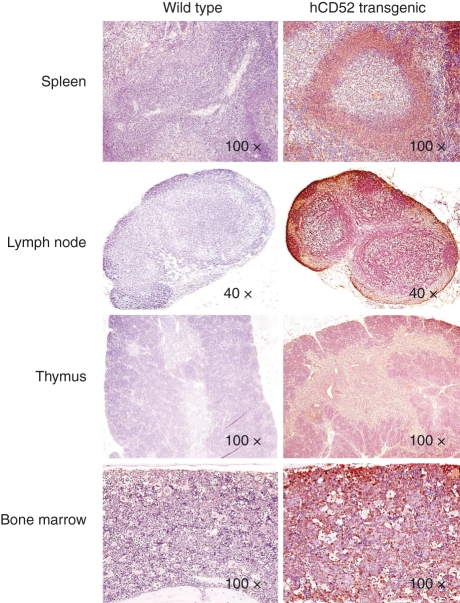
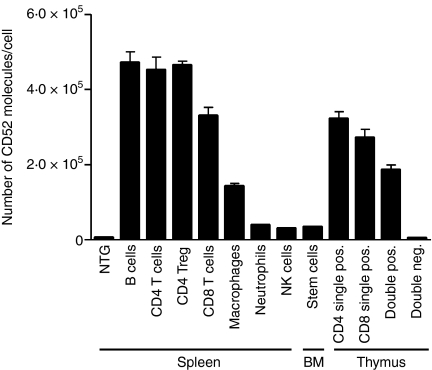
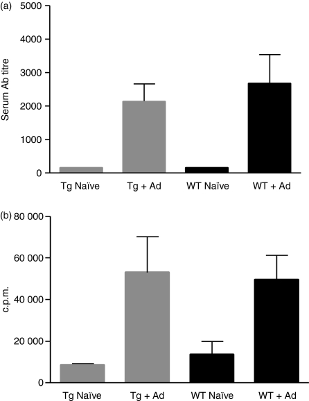
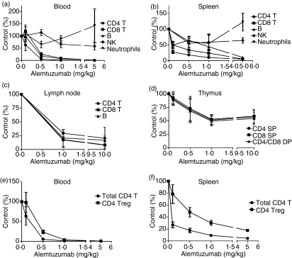
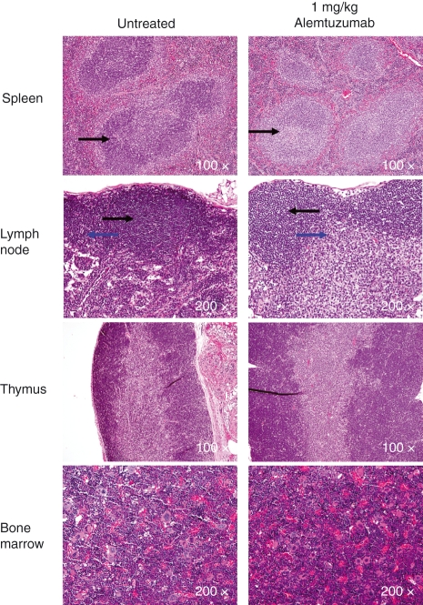
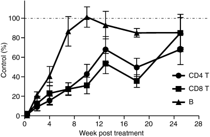
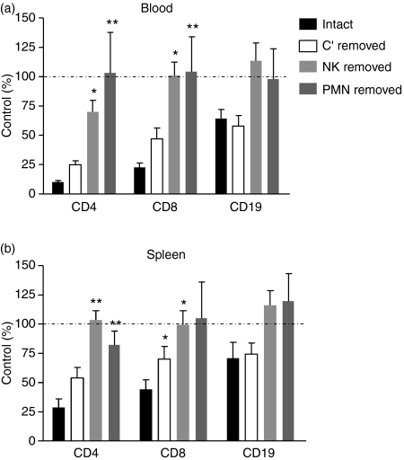
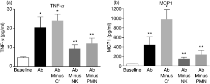
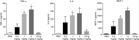
Alemtuzumab is a humanized monoclonal antibody against CD52, an antigen found on the surface of normal and malignant lymphocytes. It is approved for the treatment of B-cell chronic lymphocytic leukaemia and is undergoing Phase III clinical trials for the treatment of multiple sclerosis. The exact mechanism by which alemtuzumab mediates its biological effects in vivo is not clearly defined and mechanism of action studies have been hampered by the lack of cross-reactivity between human and mouse CD52. To address this issue, a transgenic mouse expressing human CD52 (hCD52) was created. Transgenic mice did not display any phenotypic abnormalities and were able to mount normal immune responses. The tissue distribution of hCD52 and the level of expression by various immune cell populations were comparable to those seen in humans. Treatment with alemtuzumab replicated the transient increase in serum cytokines and depletion of peripheral blood lymphocytes observed in humans. Lymphocyte depletion was not as profound in lymphoid organs, providing a possible explanation for the relatively low incidence of infection in alemtuzumab-treated patients. Interestingly, both lymphocyte depletion and cytokine induction by alemtuzumab were largely independent of complement and appeared to be mediated by neutrophils and natural killer cells because removal of these populations with antibodies to Gr-1 or asialo-GM-1, respectively, strongly inhibited the activity of alemtuzumab whereas removal of complement by treatment with cobra venom factor had no impact. The hCD52 transgenic mouse appears to be a useful model and has provided evidence for the previously uncharacterized involvement of neutrophils in the activity of alemtuzumab.


Figures

Figure 1
Figure 1
Expression of hCD52 in lymphoid tissues. Representative images of immunohistochemical staining for human CD52 in the spleen, lymph nodes, thymus and bone marrow of wild-type CD1 mice (left panels) and hCD52 transgenic mice (right panels) are shown. Human CD52 positivity is indicated by brown staining. As expected, no hCD52 expression was detected in any of the tissues from wild-type mice while hCD52 positivity was prominent in the lymphoid organs of transgenic mice. There was diffuse CD52 staining in the spleen with intensely positive lymphoid cells and macrophages in the white pulp and positive mononuclear cells in the sinusoids (red pulp). The lymph nodes also stained diffusely positive for hCD52 with positive cells in the B-cell and T-cell areas. In the thymus, there was diffuse CD52 staining of lymphocytes in both the cortex and the medulla. In the bone marrow, approximately 50% of cells were positive and the vast majority exhibited mononuclear cell morphology.
Figure 2
Figure 2
Level of CD52 expression on immune cell populations. Human CD52 expression was quantified on the indicated cell populations from the spleen, bone marrow (BM) and thymus of hCD52 transgenic mice. Using multi-parameter flow cytometry, hCD52 mean fluorescence intensities were quantified and used to calculate the number of hCD52 molecules/cell as described in the Materials and methods section. The cell populations examined included B220+ B cells, CD4+ T cells, CD4+ CD25+ FoxP3+ T cells (CD4 Treg), CD8+ T cells, CD11b+ CD11c macrophages, Gr-1+ neutrophils, NK1.1+ CD49b+ mature NK cells, c-kit+ Sca+ CD45 bone marrow stem cells, CD4 and CD8 single-positive thymocytes, double-positive and double-negative thymocytes. Non-transgenic (NTG) B220+ B cells are shown as a representative population to demonstrate the level of background staining for all NTG cell populations. Error bars indicate the SEM of six animals/group.
Figure 3
Figure 3
Immune status of hCD52 transgenic mice. The humoral and cellular immune responses of hCD52 transgenic (Tg) CD-1 mice immunized with adenovirus (Ad) were compared with those of wild type (WT) CD-1 mice; (a) Mean Ad-specific antibody titres ± SEM of serum samples from individual naïve or immunized mice. (b) Mean Ad-induced proliferation ± SEM of spleen cells from individual naïve or immunized mice (n = 3). There were no significant differences between the responses of wild-type and hCD52 transgenic mice (P> 0.05).
Figure 4
Figure 4
Immune cell depletion after treatment with alemtuzumab. Absolute numbers of immune cell populations remaining at 72 hr after the administration of various intravenous doses of alemtuzumab were assessed as described in the Materials and methods section. Results shown are the mean ± SEM of individual mice (n = 5) and are expressed as the per cent of cells remaining after treatment relative to the number of cells present in vehicle-treated control mice (% Control). The organs examined included the blood (a), spleen (b), inguinal lymph nodes (c) and thymus (d). The cell populations analysed consisted of CD4+ T cells, CD8+ T cells, single-positive (SP) and double-positive (DP) thymocytes, B220+ B cells, NK1.1+ CD49b+ NK cells and Gr-1+ neutrophils. Analysis of remaining numbers of CD4+ CD25+ FoxP3+ Tregs compared with total CD4+ T cells was also performed for the blood (e) and spleen (f).
Figure 5
Figure 5
Histology of lymphoid organs after treatment with alemtuzumab. Lymphoid tissues collected from hCD52 transgenic mice left untreated or treated intraperitoneally with 1 mg/kg alemtuzumab (72 hr post-dosing) were stained with haematoxylin & eosin for histological examination. There was mild lymphoid depletion in the spleen (periarteriolar lymphoid sheath shown by arrow) and in the paracortex (bottom arrow) and, to a lesser extent, the cortex (top arrow) of inguinal lymph nodes. There were no notable differences in the cellularity of the thymus or bone marrow in treated versus untreated mice. Depletion of bone marrow stem cells was undetectable by fluorescence-activated cell sorting up to a dose of 10 mg/kg (not shown).
Figure 6
Figure 6
Pattern of lymphocyte repopulation after treatment with alemtuzumab. Blood samples were collected at various time-points following the intraperitoneal administration of 10 mg/kg alemtuzumab and the absolute numbers of CD4+ T cells, CD8+ T cells and CD19+ B cells were assessed as described in the Materials and methods section. Results shown are the mean ± SEM of individual mice (n = 8) and are expressed as the per cent of cells remaining after treatment relative to the number of cells present in untreated, age-matched control mice (% Control).
Figure 7
Figure 7
Mechanism of lymphocyte depletion by alemtuzumab. Immune effector arms were selectively inactivated to study the impact on the lymphocyte-depleting activity of alemtuzumab. Mice were either left untreated (intact) or were treated with cobra venom factor to remove complement (C′ removed), anti-asialo-GM1 to remove natural killer (NK) cells (NK removed) or anti-Gr-1 to remove neutrophils (PMN removed) before the administration of alemtuzumab (0.1 mg/kg, intraperitoneally). Absolute numbers of CD4+ T cells, CD8+ T cells and CD19+ B cells remaining in the blood (a) and spleen (b) at 72 hr post-alemtuzumab were assessed as described in the Materials and methods. Results shown are the mean ± SEM of individual mice (n = 7) and are expressed as the per cent of cells remaining after treatment relative to the number of cells present in untreated, control mice (% Control). *P < 0.05, **P < 0.01 versus intact mice.
Figure 9
Figure 9
Mechanism of cytokine induction by alemtuzumab. Immune effector arms were selectively inactivated to study the impact on the cytokine-inducing activity of alemtuzumab. Serum levels of tumour necrosis factor-α (TNF-α) and monocyte chemoattractant protein 1 (MCP-1) at 2 hr post-alemtuzumab (0.1 mg/kg, intraperitoneally) are shown for mice that were either left untreated (Ab) or were treated with cobra venom factor to remove complement (Ab minus C′), anti-asialo-GM1 to remove natural killer (NK) cells (Ab minus NK) or anti-Gr-1 to remove neutrophils (Ab minus PMN) before the administration of alemtuzumab. Results shown are the mean ± SEM of individual mice (n = 5). Background (baseline) levels of serum cytokines in untreated mice are also shown. *P < 0.01, **P > 0.05 versus baseline.
Figure 8
Figure 8
Induction of serum cytokines by alemtuzumab. Mice were injected with various doses of alemtuzumab intraperitoneally (0.5, 1 or 5 mg/kg) or with phosphate-buffered saline (PBS) or Remicade® as an isotype control (Ctl Ig, 5 mg/kg). Remicade® is a human immunoglobulin G1 specific for human tumour necrosis factor-α (TNF-α) and does not cross-react with murine TNF-α. Serum samples were collected at 1, 2, 4, 24 hr post-dosing with alemtuzumab and cytokine levels measured with a multiplex mouse cytokine assay kit. Data are shown for the 2-hr cytokine peak only and represent the mean ± SEM of individual mice (n = 5) for TNF-α, interleukin-6 (IL-6) and monocyte chemoattractant protein 1 (MCP-1). *P < 0.01 versus PBS.

References

    1. Hale G, Xia MQ, Tighe HP, Dyer MJ, Waldmann H. The CAMPATH-1antigen (CDw52) Tissue Antigens. 1990;35:118–27. - PubMed
    1. Hale G. CD52 (Campath-1) J Biol Regul Homeost Agents. 2001;15:386–91. - PubMed
    1. Huh Y, Kantarjian H, Pierce SM, et al. Expression of human CD52 in human hematopoietic malignancies. Blood. 1998;92 Abstract 4199.
    1. Elsner J, Hochstetter R, Spiekermann K, Kapp A. Surface and mRNA expression of the CD52 antigen by human eosinophils but not by neutrophils. Blood. 1996;88:4684–93. - PubMed
    1. Gilleece MH, Dexter TM. Effect of Campath-1H antibody on human hematopoietic progenitors in vitro. Blood. 1993;82:807–12. - PubMed

MeSH terms

LinkOut - more resources