

Myoepithelioma within the carpal tunnel: a case report and review of the literature
- PMID: 19740441
- PMCID: PMC2748076
- DOI: 10.1186/1477-7800-6-15
Myoepithelioma within the carpal tunnel: a case report and review of the literature
Abstract
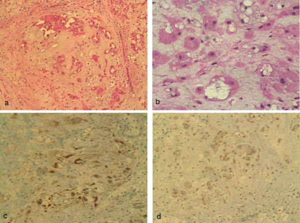
Myoepitheliomas of the extremity are rare and usually benign, while a minority display malignant features. This case demonstrates the diagnosis and management of myoepithelioma within the carpal tunnel. Clinical and radiological tumour features were evaluated. Hematoxylin and eosin stained tumour sections were examined, and immunohistochemistry was performed. Histology revealed a nodular mass of epithelioid cells in clusters within a myxoid/chondroid stroma. No mitoses were noted. Cytokeratins, neuron-specific enolase, synaptophysin, glial fibrillary acidic protein, and S100 were positive on immunohistochemistry. A literature review revealed very few prior reports of myoepithelioma in the wrist, and limited data concerning any relationship between recurrence and quality of surgical margins. In this case, wide local excision would have significantly compromised dominant hand function, and therefore a marginal excision was deemed appropriate in the context of bland histological features. Surgical margins noted in future case reports will aid clinical decision making.
Figures


Similar articles
-
[Soft tissue myoepithelioma, a rare tumor. A case report].Ann Pathol. 2003 Feb;23(1):55-8. Ann Pathol. 2003. PMID: 12743501 French.
-
Cutaneous myoepithelioma: a clinicopathologic and immunohistochemical study of 14 cases.Hum Pathol. 2004 Jan;35(1):14-24. doi: 10.1016/j.humpath.2003.08.016. Hum Pathol. 2004. PMID: 14745720
-
Salivary gland myoepithelioma variants. Histological, ultrastructural, and immunocytological features.Virchows Arch A Pathol Anat Histopathol. 1989;416(1):25-42. doi: 10.1007/BF01606467. Virchows Arch A Pathol Anat Histopathol. 1989. PMID: 2479165
-
Malignant myoepithelioma of the breast. Case report with immunohistochemical study.Arch Anat Cytol Pathol. 1996;44(4):193-8. Arch Anat Cytol Pathol. 1996. PMID: 9157829 Review.
-
Malignant myoepithelioma of minor salivary gland origin.Acta Pathol Jpn. 1992 Jul;42(7):518-22. doi: 10.1111/j.1440-1827.1992.tb03099.x. Acta Pathol Jpn. 1992. PMID: 1329434 Review.
References
-
- Fletcher CDM, Unni KK, Mertens F, eds . World Health Organization Classification of Tumours: Pathology and Genetics of Tumours of Soft Tissue and Bone. Lyon, France: IARC Press; 2002.
-
- Colombat M, Lesourd A, Moughabghab M, Coindre JM, Carton S. [Soft tissue myoepithelioma, a rare tumor. A case report] Ann Pathol. 2003;23:55–8. - PubMed
LinkOut - more resources
Full Text Sources
