

Changes in resting state effective connectivity in the motor network following rehabilitation of upper extremity poststroke paresis
- PMID: 19740732
- PMCID: PMC3595191
- DOI: 10.1310/tsr1604-270
Changes in resting state effective connectivity in the motor network following rehabilitation of upper extremity poststroke paresis
Abstract
Background: A promising paradigm in human neuroimaging is the study of slow (<0.1 Hz) spontaneous fluctuations in the hemodynamic response measured by functional magnetic resonance imaging (fMRI). Spontaneous activity (i.e., resting state) refers to activity that cannot be attributed to specific inputs or outputs, that is, activity intrinsically generated by the brain.
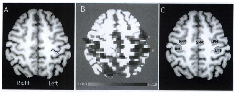
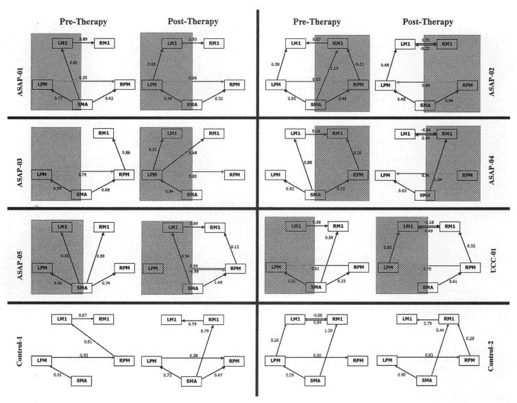
Method: This article presents pilot data examining neural connectivity in patients with poststroke hemiparesis before and after 3 weeks of upper extremity rehabilitation in the Accelerated Skill Acquisition Program (ASAP). Resting-state fMRI data acquired pre and post therapy were analyzed using an exploratory adaptation of structural equation modeling (SEM) to evaluate therapy-related changes in motor network effective connectivity.
Results: Each ASAP patient showed behavioral improvement. ASAP patients also showed increased influence of the affected hemisphere premotor cortex (a-PM) upon the unaffected hemisphere premotor cortex (u-PM) following therapy. The influence of a-PM on affected hemisphere primary motor cortex (a-M1) also increased with therapy for 3 of 5 patients, including those with greatest behavioral improvement.
Conclusions: Our findings suggest that network analyses of resting-state fMRI constitute promising tools for functional characterization of functional brain disorders, for intergroup comparisons, and potentially for assessing effective connectivity within single subjects; all of which have important implications for stroke rehabilitation.
Conflict of interest statement
All other authors report no conflicts of interest.
Figures
References
-
- Rosamond W, Flegal K, Friday G, et al. Heart disease and stroke statistics–2007 update: a report from the American Heart Association Statistics Committee and Stroke Statistics Subcommittee. Circulation. 2007;115(5):e69–171. - PubMed
-
- Catalan MJ, Honda M, Weeks RA, Cohen LG, Hallett M. The functional neuroanatomy of simple and complex sequential finger movements: a PET study. Brain. 1998;121(Pt 2):25364. - PubMed
-
- Deiber MP, Passingham RE, Colebatch JG, Friston KJ, Nixon PD, Frackowiak RS. Cortical areas and the selection of movement: a study with positron emission tomography. Exp Brain Res. 1991;84(2):393–402. - PubMed
Publication types
MeSH terms
Grants and funding
LinkOut - more resources
Full Text Sources
Medical