. 2009 Oct 30;284(44):30288-97.
doi: 10.1074/jbc.M109.045104. Epub 2009 Sep 9.

Uptake of oxidized low density lipoprotein by CD36 occurs by an actin-dependent pathway distinct from macropinocytosis

Affiliations

Uptake of oxidized low density lipoprotein by CD36 occurs by an actin-dependent pathway distinct from macropinocytosis

Richard F Collins et al. J Biol Chem. .

Abstract

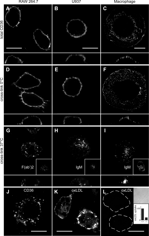
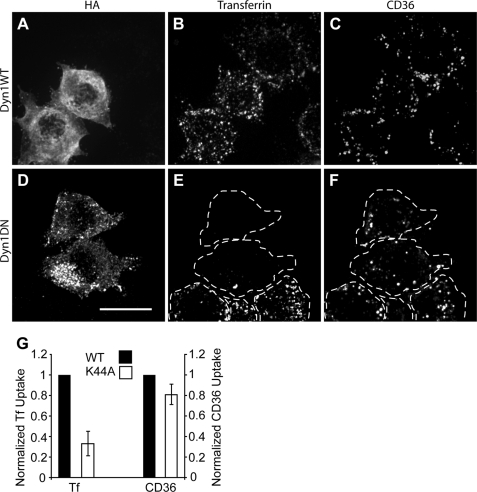
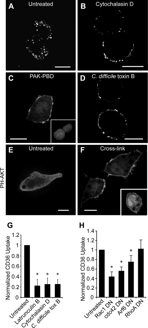
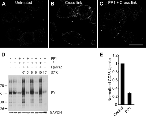
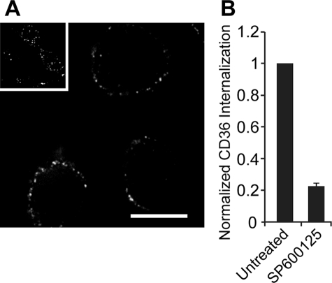
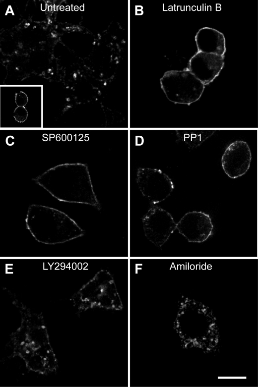
The class B scavenger receptor CD36 has numerous ligands that include modified forms of low density lipoprotein, fibrillar amyloid, apoptotic cells, and Plasmodium falciparum-infected red blood cells, linking this molecule to atherosclerosis, Alzheimer disease, malaria, and other diseases. We studied the signaling events that follow receptor engagement and lead to CD36 and ligand internalization. We show that oxidized low density lipoprotein or antibody-induced clustering of CD36 triggers macropinocytosis and internalization of the receptor-ligand complex. Remarkably, however, CD36 internalization is independent of macropinocytosis and occurs by a novel endocytic mechanism that depends on actin, but not dynamin. This actin-driven endocytosis requires the activation Src family kinases, JNK, and Rho family GTPases, but, unlike macropinocytosis, it is not affected by inhibitors of phosphatidylinositol 3-kinase or Na/H exchange. Manipulation of this unique mode of internalization may prove helpful in the prevention and management of the wide range of diseases in which CD36 is implicated.


Figures

FIGURE 1.
FIGURE 1.
Distribution of endogenous CD36 following antibody and oxLDL cross-linking. A–C, distribution of CD36 on permeabilized resting RAW 264.7 cells (A), U937 cells (B), or human primary macrophages (C). D–F, cross-linking antibodies at 0 °C did not alter distribution of CD36 on RAW cells (D), U937 cells (E), or primary human macrophages (F). G–I, warming of cross-linked cells to 37 °C resulted in internalization of CD36. G, cross-linking in murine RAW cells involved an IgA primary antibody or a F(ab′)2 fragment (inset). H and I, cross-linking of human U937 cells or primary macrophages involved IgG (clone 131.2) or IgM antibodies (insets). J, oxLDL also triggered CD36 internalization in RAW cells. K and L, CD36 is the major receptor for oxLDL. Wild-type mouse peritoneal macrophages (K) or CD36−/− knock-out mouse peritoneal macrophages (L) were incubated with DiI-labeled oxidized LDL, and noninternalized oxLDL was removed by acid wash. Because the cells in L bind little oxLDL and are not readily visible by fluorescence microscopy, they are outlined in the main panels and shown by differential interference contrast image in the top inset. The lower inset shows quantitation of oxLDL uptake in murine wild-type (+/+) and CD36-deficient (−/−) macrophages, quantified from experiments like K and L. Data are means of three experiments, each counting at least 50 cells of each type. To facilitate comparison between experiments, data were normalized to wild type. In images A–I, the x-z plane is shown below. Scale bars, 10 μm.
FIGURE 2.
FIGURE 2.
Effect of dynamin1 K44A dominant negative mutant on CD36 internalization. RAW 264.7 cells were transfected with hemagglutinin (HA)-tagged wild-type (WT) dynamin1 (Dyn1; A–C) or dynamin1 K44A (D–F), cross-linked in the cold with anti-CD36 IgA antibody plus labeled secondary antibody, incubated with transferrin-Alexa Fluor 647, fixed, and stained for hemagglutinin (A and D) and imaged for transferrin (B and E) and CD36 (C and F). DN, dominant negative. Dashed lines indicate the outline of the cells. Scale bar, 10 μm. G, for quantification, the same assay was done with the addition of two acid washes prior to fixation. Data are means ± S.E. of three experiments with at least 100 cells counted in each case. Transferrin (Tf) and internalized CD36 data are normalized to their respective wild-type controls (black bars).
FIGURE 3.
FIGURE 3.
Actin inhibitors block internalization of CD36. RAW 264.7 cells were pretreated as follows: no treatment (A), cytochalasin D (B), or C. difficile toxin B (D). CD36 receptors were then cross-linked, and cells were warmed, fixed, and imaged. Acid wash was omitted to preserve surface labeling. C, Rac1 activation was demonstrated by recruitment of PAK1-PBD-YFP to the membrane. RAW 264.7 cells transfected with PAK-PBD-YFP were imaged following CD36 receptor cross-linking with (C) or without (C, inset) anti-CD36 antibodies. E and F, cross-linking activates PI3K. RAW 264.7 cells transiently expressing PH-AKT-GFP were treated without (E) or with (F) CD36 cross-linking antibody and warming. F, inset, cross-linking was done in the presence of LY294002. To quantify CD36 internalization (G), the same assay was performed with the addition of two acid washes. Cells were warmed to 37 °C, and internalized CD36 was imaged live on a spinning disc microscope. H, to demonstrate dependence of CD36 internalization on Rho or Arf family small GTPases, RAW 264.7 cells were transfected with dominant negative (DN) DNA constructs of Rac1, Cdc42 Arf6 or RhoA, and internalized CD36 receptors were quantified as above. Scale bars, 10 μm. *, p < 0.05 compared with untreated control.
FIGURE 4.
FIGURE 4.
Macropinocytosis is induced by cross-linking CD36, but CD36 internalization is not sensitive to inhibitors of macropinocytosis. A, untreated RAW 264.7 cells take up little sulforhodamine B by macropinocytosis, but treatment with M-CSF (B) or cross-linking with antibody (C) stimulates macropinocytosis. Sulforhodamine is indicated in red. Scale bar, 10 μm. D, inhibitors of macropinocytosis block sulforhodamine uptake. RAW 264.7 cells were pretreated with no inhibitor, LY294002, amiloride, or the amiloride analog HOE-694 followed by cross-linking with antibody or mouse M-CSF followed by warming with sulforhodamine. Data are means ± S.E. of three experiments with at least 500 cells counted in each case. E, internalization of CD36 after cross-linking is not blocked by inhibitors of macropinocytosis. RAW 264.7 cells were left untreated or were pretreated with wortmannin, LY294002, amiloride, or HOE-694 prior to cross-linking CD36 with antibody in the cold, and warming in the presence of appropriate inhibitors. Finally cells were treated with two acid washes and fixed. Data are means ± S.E. of three experiments with at least 100 cells counted in each case. Data are normalized to the untreated control.
FIGURE 5.
FIGURE 5.
Cross-linking of CD36 receptors causes tyrosine phosphorylation. A–C, RAW 264.7 cells untreated (A), cross-linked with anti-CD36 (B), or cross-linked after pretreatment with the Src family tyrosine kinase inhibitor PP1 (C) were fixed and stained for phosphotyrosine. Scale bar, 10 μm. D, immunoblot of tyrosine phosphorylation subsequent to CD36 cross-linking and internalization. RAW 264.7 cells were pretreated without or with PP1 followed by cross-linking of CD36 with primary IgA antibody in the cold, secondary F(ab′)2 antibody and warming for 0 min, 5 min, or 10 min. Immunoblot was probed for phosphotyrosine (4G10), stripped, and reprobed for GAPDH. E, dependence of CD36 internalization on tyrosine phosphorylation. RAW cells were pretreated without (control) or with PP1 prior to cross-linking of CD36 receptors and warming in the presence of PP1. Cells were acid-washed twice before fixation and imaging on a spinning disc confocal microscope. Data are normalized to the control.
FIGURE 6.
FIGURE 6.
JNK inhibitor SP600125 blocks internalization of cross-linked CD36. A, RAW cells were treated without (inset) or with (main panel) SP600125 prior to cross-linking of CD36 and warmed with or without SP600125 present, and then fixed and imaged on a spinning disc confocal microscope. Scale bar, 10 μm. B, quantification of A. As in A, but with the addition of two acid wash steps before fixation. Images were acquired and quantified. Data are normalized to the untreated control and represent means ± S.E. of three experiments with at least 100 cells counted in each case.
FIGURE 7.
FIGURE 7.
Inhibitors that block internalization of cross-linked CD36 also block oxidized LDL-dependent internalization of CD36. A, untreated RAW cells have internalized CD36 following incubation with nonlabeled oxLDL. Inset, no oxLDL added. B–F, RAW cells were pretreated with latrunculin B (B), SP600125 (C), PP1 (D), LY294002 (E), or amiloride (F) before incubation with nonlabeled oxLDL and stained for total CD36. Scale bar, 10 μm.

References

    1. Endemann G., Stanton L. W., Madden K. S., Bryant C. M., White R. T., Protter A. A. (1993) J. Biol. Chem. 268, 11811–11816 - PubMed
    1. Nozaki S., Kashiwagi H., Yamashita S., Nakagawa T., Kostner B., Tomiyama Y., Nakata A., Ishigami M., Miyagawa J., Kameda-Takemura K. (1995) J. Clin. Invest. 96, 1859–1865 - PMC - PubMed
    1. Kunjathoor V. V., Febbraio M., Podrez E. A., Moore K. J., Andersson L., Koehn S., Rhee J. S., Silverstein R., Hoff H. F., Freeman M. W. (2002) J. Biol. Chem. 277, 49982–49988 - PubMed
    1. Graf G. A., Connell P. M., van der Westhuyzen D. R., Smart E. J. (1999) J. Biol. Chem. 274, 12043–12048 - PubMed
    1. Thorne R. F., Mhaidat N. M., Ralston K. J., Burns G. F. (2007) FEBS Lett. 581, 1227–1232 - PubMed

Publication types