. 2009 Oct 30;284(44):30016-23.
doi: 10.1074/jbc.M109.035568. Epub 2009 Sep 9.

Insulin-stimulated phosphorylation of the Rab GTPase-activating protein TBC1D1 regulates GLUT4 translocation

Affiliations

Insulin-stimulated phosphorylation of the Rab GTPase-activating protein TBC1D1 regulates GLUT4 translocation

Grantley R Peck et al. J Biol Chem. .

Abstract

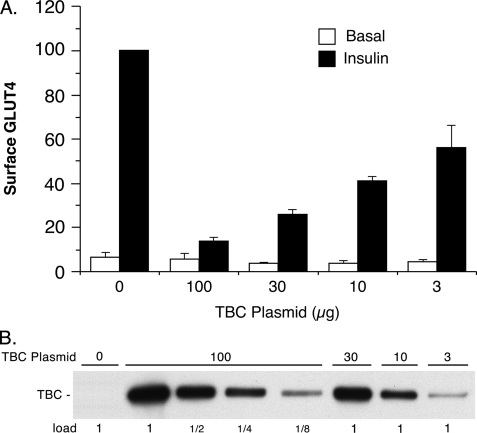
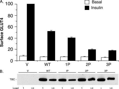
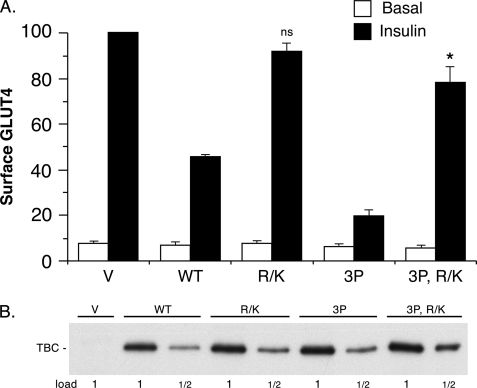
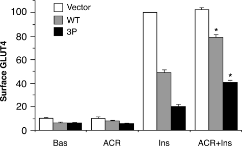
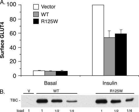
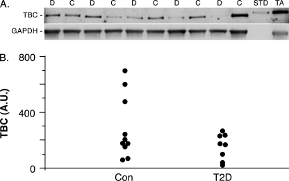
Insulin stimulates the translocation of the glucose transporter GLUT4 from intracellular locations to the plasma membrane in adipose and muscle cells. Prior studies have shown that Akt phosphorylation of the Rab GTPase-activating protein, AS160 (160-kDa Akt substrate; also known as TBC1D4), triggers GLUT4 translocation, most likely by suppressing its Rab GTPase-activating protein activity. However, the regulation of a very similar protein, TBC1D1 (TBC domain family, member 1), which is mainly found in muscle, in insulin-stimulated GLUT4 translocation has been unclear. In the present study, we have identified likely Akt sites of insulin-stimulated phosphorylation of TBC1D1 in C2C12 myotubes. We show that a mutant of TBC1D1, in which several Akt sites have been converted to alanine, is considerably more inhibitory to insulin-stimulated GLUT4 translocation than wild-type TBC1D1. This result thus indicates that similar to AS160, Akt phosphorylation of TBC1D1 enables GLUT4 translocation. We also show that in addition to Akt activation, activation of the AMP-dependent protein kinase partially relieves the inhibition of GLUT4 translocation by TBC1D1. Finally, we show that the R125W variant of TBC1D1, which has been genetically associated with obesity, is equally inhibitory to insulin-stimulated GLUT4 translocation, as is wild-type TBC1D1, and that healthy and type 2 diabetic individuals express approximately the same level of TBC1D1 in biopsies of vastus lateralis muscle. In conclusion, phosphorylation of TBC1D1 is required for GLUT4 translocation. Thus, the regulation of TBC1D1 resembles that of its paralog, AS160.


Figures

FIGURE 1.
FIGURE 1.
TBC1D1 expression during C2C12 differentiation. SDS samples of C2C12 myoblasts (day 0) and myotubes at different stages of differentiation (days 2, 4, 6, and 8) were immunoblotted for TBC1D1 (TBC) and myogenin (Myo). The 1× load of protein was 90 μg.
FIGURE 2.
FIGURE 2.
Effect of TBC1D1 expression level on GLUT4 translocation. A, relative amounts of surface GLUT4 in basal and insulin-stimulated 3T3-L1 adipocytes transfected with HA-GLUT4-GFP plasmid, together with varying amounts of wild-type TBC1D1 (TBC) plasmid and control vector plasmid, such that the total of the two was 100 μg of plasmid. Values are the mean ± S.E. for each condition from two (100, 30, and 3 μg TBC1D1 plasmid) or three (0 and 10 μg of TBC1D1 plasmid) replicate experiments. B, SDS lysates of cells in A were immunoblotted for TBC1D1 with anti-FLAG antibody. The 1× load was 10 μg.
FIGURE 3.
FIGURE 3.
Inhibition of GLUT4 translocation by TBC1D1 phosphorylation mutants. A, relative amounts of surface GLUT4 in basal and insulin-stimulated 3T3-L1 adipocytes transfected with HA-GLUT4-GFP plasmid, together with the control vector plasmid alone (V) or with the plasmids for wild-type (WT) and phosphorylation site mutants (1P, 2P, and 3P) of TBC1D1 (TBC). The values are the mean ± S.E. from two replicate experiments. Figs. 4A and 5A show six additional experiments in which TBC1D1 wild-type and 3P mutant were among those examined. The lower amount of GLUT4 at the cell surface in the insulin state for the 3P mutant compared with wild-type TBC1D1 for these eight experiments was significant, with p < 0.05. B, SDS lysates of cells in A were immunoblotted for TBC1D1 with anti-FLAG antibody. The 1× load was 10 μg. A representative immunoblot is shown.
FIGURE 4.
FIGURE 4.
Effect of the TBC1D1 GAP activity on GLUT4 translocation. A, relative amounts of surface GLUT4 in basal and insulin-stimulated 3T3-L1 adipocytes transfected with HA-GLUT4-GFP plasmid, together with the control vector plasmid (V) alone or with the plasmids for wild-type TBC1D1 (WT), GAP-inactive TBC1D1 (R/K), the 3P mutant of TBC1D1, and the GAP-inactive mutant thereof (3P, R/K). Values are mean ± S.E. from three replicate experiments. ns, not statistically different from the value for vector insulin; *, p < 0.05 for comparison to the value for vector insulin. B, SDS lysates of cells in A were immunoblotted for TBC1D1 (TBC) with anti-FLAG antibody. The 1× load was 10 μg. A representative immunoblot is shown.
FIGURE 5.
FIGURE 5.
Reduction in TBC1D1 inhibition of GLUT4 translocation by AICAR. Relative amounts of surface GLUT4 in 3T3-L1 adipocytes transfected HA-GLUT4-GFP plasmid, together with the control vector plasmid alone or with the plasmids for wild-type TBC1D1 (WT) or the 3P mutant of TBC1D1. Cells were untreated (Bas) or treated with AICAR (ACR) or insulin (Ins) or both (ACR+Ins). Values are mean ± S.E. from three replicate experiments. *, p < 0.05 for comparison with insulin alone for the same plasmid.
FIGURE 6.
FIGURE 6.
Effect of TBC1D1 on cell surface transferrin receptor. Relative amounts of surface transferrin receptor (TfR) in basal and insulin-stimulated 3T3-L1 adipocytes transfected with the HA-GLUT4-GFP plasmid, together with the control vector plasmid (V) alone or with the plasmids for wild-type TBC1D1 (WT) or the 3P mutant thereof. Values are mean ± S.E. from three replicate experiments.
FIGURE 7.
FIGURE 7.
Effect of TBC1d1 R125W on GLUT4 translocation. A, relative amounts of surface GLUT4 in basal and insulin-stimulated 3T3-L1 adipocytes transfected with the HA-GLUT4-GFP plasmid, together with the control vector plasmid (V) alone or with the plasmids for wild-type TBC1D1 (WT) or the R125W mutant thereof. Values are mean ± S.E. from three replicate experiments. B, SDS lysates of cells in A were immunoblotted for TBC1D1 (TBC) with anti-FLAG antibody. The 1× load was 10 μg. A representative immunoblot is shown.
FIGURE 8.
FIGURE 8.
TBC1D1 expression in control and diabetic human skeletal muscle. A, representative immunoblot of SDS lysates of vastus lateralis biopsies from control (C) and type 2 diabetic (D) subjects, immunoblotted for TBC1D1 or GAPDH as a loading control. Each lane was loaded with 75 μg protein. One ng of recombinant mouse FLAG-tagged TBC1D1 isolated from transfected human embryonic kidney 293 cells (STD) and 50 μg of mouse tibialis anterior muscle protein (TA) were included as positive controls. B, dot plot of individual values for control (Con) and type 2 diabetic (T2D) subjects expressed as arbitrary units (A.U.) for the TBC1D1 signal normalized to the GAPDH signal.

References

    1. Huang S., Czech M. P. (2007) Cell. Metab. 5, 237–252 - PubMed
    1. Zaid H., Antonescu C. N., Randhawa V. K., Klip A. (2008) Biochem. J. 413, 201–215 - PubMed
    1. Sakamoto K., Holman G. D. (2008) Am. J. Physiol. Endocrinol. Metab. 295, E29–E37 - PMC - PubMed
    1. Roach W. G., Chavez J. A., Mîinea C. P., Lienhard G. E. (2007) Biochem. J. 403, 353–358 - PMC - PubMed
    1. Chavez J. A., Roach W. G., Keller S. R., Lane W. S., Lienhard G. E. (2008) J. Biol. Chem. 283, 9187–9195 - PMC - PubMed

Publication types

MeSH terms