. 2009 Oct 30;284(44):30067-75.
doi: 10.1074/jbc.M109.044800. Epub 2009 Sep 8.

Dual role of DNA in regulating ATP hydrolysis by the SopA partition protein

Affiliations

Dual role of DNA in regulating ATP hydrolysis by the SopA partition protein

Yoan Ah-Seng et al. J Biol Chem. .

Abstract

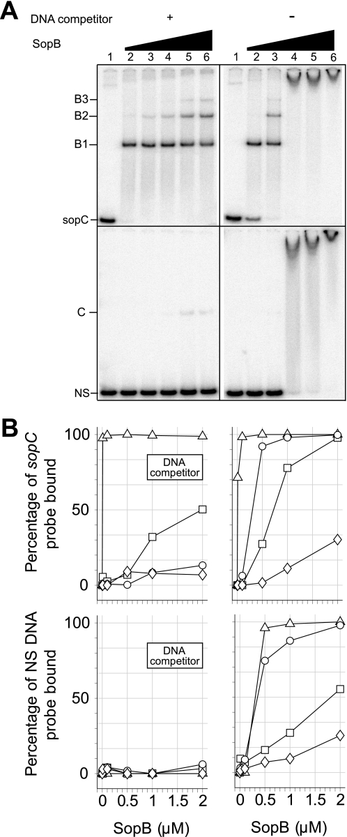
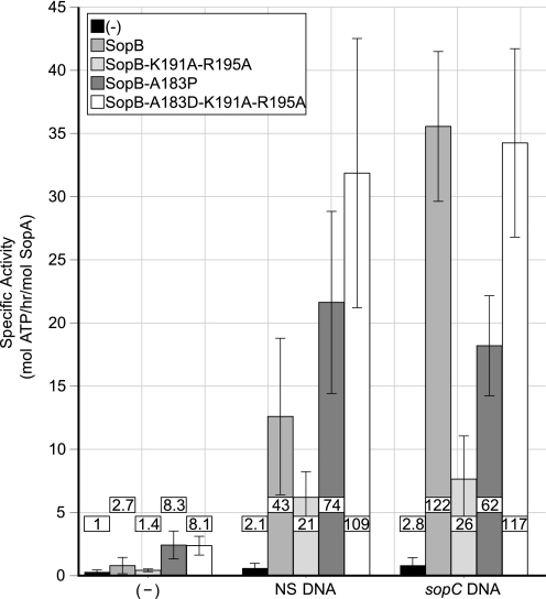
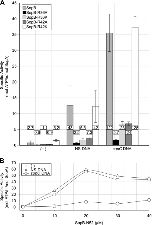
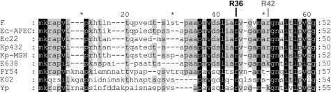
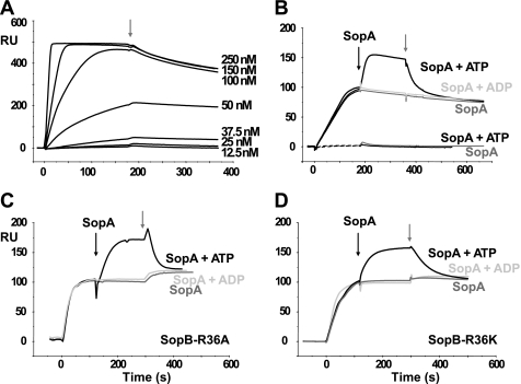
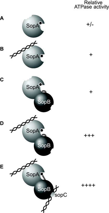
In bacteria, mitotic stability of plasmids and many chromosomes depends on replicon-specific systems, which comprise a centromere, a centromere-binding protein and an ATPase. Dynamic self-assembly of the ATPase appears to enable active partition of replicon copies into cell-halves, but for Walker-box partition ATPases the molecular mechanism is unknown. ATPase activity appears to be essential for this process. DNA and centromere-binding proteins are known to stimulate the ATPase activity but molecular details of the stimulation mechanism have not been reported. We have investigated the interactions which stimulate ATP hydrolysis by the SopA partition ATPase of plasmid F. By using SopA and SopB proteins deficient in DNA binding, we have found that the intrinsic ability of SopA to hydrolyze ATP requires direct DNA binding by SopA but not by SopB. Our results show that two independent interactions of SopA act in synergy to stimulate its ATPase. SopA must interact with (i) DNA, through its ATP-dependent nonspecific DNA binding domain and (ii) SopB, which we show here to provide an arginine-finger motif. In addition, the latter interaction stimulates ATPase maximally when SopB is part of the partition complex. Hence, our data demonstrate that DNA acts on SopA in two ways, directly as nonspecific DNA and through SopB as centromeric DNA, to fully activate SopA ATP hydrolysis.


Figures

FIGURE 1.
FIGURE 1.
Characterization of SopA ATPase activity. Reaction mixtures contained 2 μm SopA or SopAK340A and, except as indicated, 2 μm SopB and 60 μg·ml−1 nonspecific (NS; pBSKS) or sopC-containing (pJYB57) plasmid DNA. For each value the corresponding control value without SopA has been subtracted. A, ATP hydrolysis by SopA proteins. SopA (left panel) or SopAK340A (right panel) were incubated alone (black bars) or with SopB (white bars) in the absence of DNA (−) or the presence of NS or sopC-containing DNA. Stimulation factors, normalized to 1 for SopA or SopAK340A alone, are shown inside or above bars. Error bars for are standard deviations from at least 20 (SopA) and 3 (SopAK340A) independent experiments. B, DNA concentration dependence of SopA ATPase activity. Reaction mixtures contained SopA in the presence of SopB and increasing concentrations of NS (triangle) or sopC-containing DNA (square). C, SopB concentration dependence of SopA ATPase activity. Reaction mixtures contained SopA and NS (triangle) or sopC-containing DNA (square) and increasing concentrations of SopB.
FIGURE 2.
FIGURE 2.
DNA binding properties of wild-type SopB and hth mutants. A, retardation of SopB-DNA complexes. 32P-labeled sopC (top panels) or NS (bottom panels) DNA probes were incubated alone (lane 1) or with increasing concentrations of SopB (lanes 2–6; 0.01, 0.1, 0.5, 1, 2 μm) in the presence (+; left panels) or absence (−; right panels) of 100 μg·ml−1 competitor DNA. Reaction mixtures were analyzed by electrophoresis on polyacrylamide gels. Positions of DNA probes and protein complexes are indicated on the left. B1, B2, and B3 denote specific complexes. C denotes a nonspecific complex. B, DNA binding properties of SopB wt and hth mutants. EMSAs were performed as in A. After electrophoresis, bands were quantitated and percentage of total probe bound was calculated. WT SopB (triangle), SopB*1 (square), SopB*2 (circle), or SopB*3 (diamond) were incubated with sopC (top panels) or NS (bottom panels) DNA probes in the presence (left panels) or absence (right panels) of 100 μg·ml−1 competitor DNA.
FIGURE 3.
FIGURE 3.
Activation of SopA ATPase activity by SopB hth mutants. Reaction mixtures contained 2 μm SopA, 2 μm SopB (defined in inset), and, either DNA (−) or 60 μg·ml−1 NS DNA (pBSKS) or sopC-containing DNA (pJYB57). For each value the corresponding control value without SopA has been subtracted. Error bars are standard deviations from at least three independent experiments. Stimulation factors, normalized to 1 for SopA alone, are shown in boxes inside or above bars.
FIGURE 4.
FIGURE 4.
Activation of SopA ATPase activity by (A) SopB arginine mutants (inset) and (B) a SopB N-terminal peptide. Assay conditions and data treatment are as described in the legend to Fig. 3.
FIGURE 5.
FIGURE 5.
Alignment of partial amino acid sequences of ParB homologs. ParB members very closely related to SopB of plasmid F were found in the data base using the Blast algorithm. Nine of them, present on plasmids or phages, having >30% amino acid identity in the N terminus (>45% in the whole sequence), display an arginine corresponding to SopB-R36. SopB homologs of identical N-terminal sequences were not included. In the aligned sequences, conserved residues and conservative substitutions (V/I/L, T/S, and R/K) are shaded in black if present in all sequences or in gray if present in five or more sequences. The arginine residues denoted R36 and R42 refer to SopB of F. Plasmid partitioning proteins are from E. coli APEC O1 (Ec-APEC), E. coli K12 (F), E. coli E22 (Ec22), Klebsiella pneumoniae 342 (Kp342), K. pneumoniae MGH (Kp-MGH), Enterobacter sp. 638 (E638), Yersinia pestis bv Microtus (Yp). Phage partitioning proteins homologous to SopB are from Yersinia phage PY54 (PY54) and from Klebsiella phage phiKO2 (KO2).
FIGURE 6.
FIGURE 6.
Surface plasmon resonance analysis of SopA binding to SopB immobilized on sopC DNA probes. Results are expressed as the difference in RU between the signals obtained with a specific (sopC) 136-bp probe and a nonspecific 136-bp probe, and are plotted as a function of time (seconds). A, SopB dose-response on sopC DNA. WT SopB at concentrations ranging from 12.5 to 250 nm was injected at time 0 over immobilized DNA probes. After binding, complexes were washed with the same buffer without SopB at the time indicated by the gray arrow. B, SopA binding to SopB/sopC complex. Buffer containing WT SopB at 37.5 nm (black lines) or not (black dashed lines) was passed over immobilized DNA probes at time 0. After 180 s, buffer with SopA (250 nm) in the absence (dark gray line) or presence of ATP (black line) or ADP (light gray line) was injected (black arrow). Complexes were washed with buffer without proteins at the time indicated by the gray arrow. C–D, as in B except that 250 nm SopB-R36A and SopB-R36K, respectively, were injected.
FIGURE 7.
FIGURE 7.
Model for SopA ATPase activation by nonspecific DNA and the partition complex. SopA ATPase (light gray sphere) binds ATP (black circle), nonspecific DNA (intertwined helices), and SopB (dark gray sphere) and these interactions stimulate ATP hydrolyis by SopA to various extents, as indicated on the right. Both SopA and SopB form dimers but are represented as unique spheres for simplicity. A, SopA alone exhibits very low intrinsic ATPase activity. B, in the presence of NS DNA, SopA ATPase activity is modestly activated (2-fold). C, interaction with SopB alone stimulates ATP hydrolysis about 3-fold. This activation requires the R-finger motif (curved gray extension entering the SopA catalytic pocket) in the N terminus of SopB. D, when both NS DNA and SopB contact SopA, ATPase activity is highly stimulated (40-fold). These two independent interactions act in synergy. SopA bound to NS DNA could adopt a slightly different conformation that renders the R-finger motif of SopB more efficient for the neutralization of the negative charges generated during phosphoryl transfer. E, in the presence of NS DNA and the partition complex, i.e. SopB bound to sopC-containing DNA (intertwined helices containing a light gray ovoid box), SopA ATPase activity is strongly stimulated (120-fold). As in D, the two independent interactions act in synergy and the 3-fold supplementary stimulation could be provided by a conformational change that would occur upon specific binding to sopC centromere. The R-finger motif of SopB would then adopt an optimal conformation (straight gray extension) in the SopA catalytic pocket for maximal stimulatory activity.

Similar articles

Cited by

References

    1. Ebersbach G., Gerdes K. (2005) Annu. Rev. Genet. 39, 453–479 - PubMed
    1. Hayakawa Y., Murotsu T., Matsubara K. (1985) J. Bacteriol. 163, 349–354 - PMC - PubMed
    1. Funnell B. E., Gagnier L. (1993) J. Biol. Chem. 268, 3616–3624 - PubMed
    1. Bouet J. Y., Surtees J. A., Funnell B. E. (2000) J. Biol. Chem. 275, 8213–8219 - PubMed
    1. Schumacher M. A., Funnell B. E. (2005) Nature 438, 516–519 - PubMed

Publication types

LinkOut - more resources